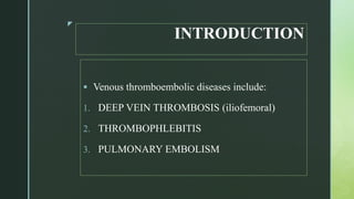
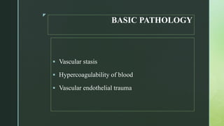
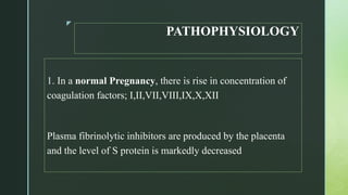
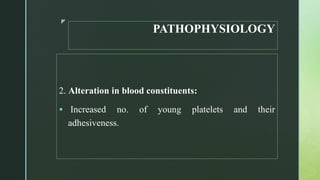
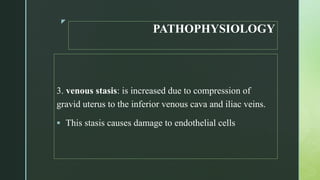
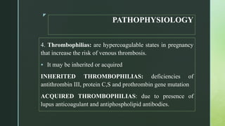
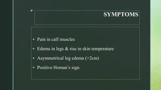
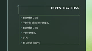
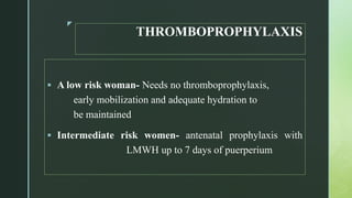
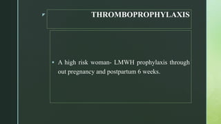
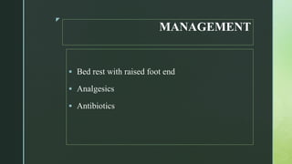
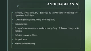
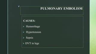
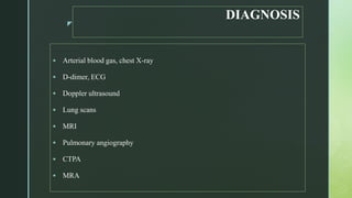
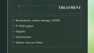
z
PUERPERAL VENOUS
THROMBOSIS
&
PULMONARY
EMBOLISM (PE)
NIKITA SHARMA
NURSING TUTOR
BECON, JAMMU
z
INTRODUCTION
๏‚ง Venous thromboembolic diseases include:
1. DEEP VEIN THROMBOSIS (iliofemoral)
2. THROMBOPHLEBITIS
3. PULMONARY EMBOLISM
z
BASIC PATHOLOGY
๏‚ง Vascular stasis
๏‚ง Hypercoagulability of blood
๏‚ง Vascular endothelial trauma
z
PATHOPHYSIOLOGY
1. In a normal Pregnancy, there is rise in concentration of
coagulation factors; I,II,VII,VIII,IX,X,XII
Plasma fibrinolytic inhibitors are produced by the placenta
and the level of S protein is markedly decreased
z
PATHOPHYSIOLOGY
2. Alteration in blood constituents:
๏‚ง Increased no. of young platelets and their
adhesiveness.
z
PATHOPHYSIOLOGY
3. venous stasis: is increased due to compression of
gravid uterus to the inferior venous cava and iliac veins.
๏‚ง This stasis causes damage to endothelial cells
z
PATHOPHYSIOLOGY
4. Thrombophilias: are hypercoagulable states in pregnancy
that increase the risk of venous thrombosis.
๏‚ง It may be inherited or acquired
INHERITED THROMBOPHILIAS: deficiencies of
antithrombin III, protein C,S and prothrombin gene mutation
ACQUIRED THROMBOPHILIAS: due to presence of
lupus anticoagulant and antiphospholipid antibodies.
z
RISK FACTORS
HIGH RISK:
๏ƒ˜ Previous VTE
๏ƒ˜ Thrombophilias
INTERMEDIATE RISK:
๏ƒ˜ Heart disease
๏ƒ˜ SLE
๏ƒ˜ Surgical procedures (LSCS)
z
RISK FACTORS
๏‚ง LOW RISK
๏ƒ˜ Presence of < 3 from any f these risk factors:
1. Age > 35 yrs
2. Obesity (BMI>35)
3. Parity >3
4. Immobility
5. Dehydration
6. Hyperemesis
7. Multiple pregnancy
z
DVT
DIAGNOSIS
๏‚ง Clinical diagnosis is unreliable
๏‚ง Majority, It remains asymptomatic
z
SYMPTOMS
๏‚ง Pain in calf muscles
๏‚ง Edema in legs & rise in skin temperature
๏‚ง Asymmetrical leg edema (>2cm)
๏‚ง Positive Homanโ€™s sign
z
INVESTIGATIONS
๏‚ง Doppler USG
๏‚ง Venous ultrasonography
๏‚ง Doppler USG
๏‚ง Venography
๏‚ง MRI
๏‚ง D-dimer assays
z
PELVIC THROMBOPHLEITIS
๏‚ง It originates in the thrombosed veins at the placental
site by organisms such as streptococci and
bacteroides
๏‚ง When localized in the pelvis, it is called pelvic
thrombophlebitis
z
CLINICAL FEATURES
๏‚ง There is no specific clinical feature of this, but it
should be suspected in cases where the pyrexia
continues for > a week in spite of antibiotic therapy.
z
EXTRAPELVIC SPREAD
๏‚ง Lungs
๏‚ง Left kidney
๏‚ง Iliofemoral veins (Phlegmasia alba dolens or
white leg)
z
PHLEGMASIAALBA DOLENS OR WHITE
LEG
๏‚ง It is a clinicopathological condition usually caused
by retrograde extension of pelvic thrombophlebitis
to involve the iliofemoral vein.
z CLINICAL FEATURES
๏‚ง Develops on the 2nd week of puerperium
๏‚ง Mild pyrexia with chills and rigors
๏‚ง Headache
๏‚ง Malaise and rising pulse rate
๏‚ง Features of toxemia
๏‚ง The affected leg is swollen, painful, white , cold
z
DIAGNOSIS
๏‚ง Blood count- polymorphonuclear leukocytosis
๏‚ง Venous ultrasound
๏‚ง CT scan
๏‚ง MRI
๏‚ง Heparin therapy trial
z
PREVENTION
๏‚ง Prevention of trauma, sepsis, anemia and dehydration
๏‚ง Use of elastic compression stocking and intermittent
pneumatic compression devices during surgery
๏‚ง Leg exercises, early ambulation are encouraged
following operative delivery
z
THROMBOPROPHYLAXIS
๏‚ง A low risk woman- Needs no thromboprophylaxis,
early mobilization and adequate hydration to
be maintained
๏‚ง Intermediate risk women- antenatal prophylaxis with
LMWH up to 7 days of puerperium
z THROMBOPROPHYLAXIS
๏‚ง A high risk woman- LMWH prophylaxis through
out pregnancy and postpartum 6 weeks.
z
MANAGEMENT
๏‚ง Bed rest with raised foot end
๏‚ง Analgesics
๏‚ง Antibiotics
z
ANTICOAGULANTS
๏‚ง Heparin, 15000 units, IV, followed by 10,000 units 4-6 hrly for 4-6
injections, 7-10 days
๏‚ง LMWH (enoxaparin) 20 mg or 40 mg daily
๏‚ง Fondaparinux
๏‚ง A rug of coumarin series- warfarin orally, 7mg , 2 days or 3 days with
heparin
๏‚ง Inferior vena cava filters
๏‚ง Streptokinase
๏‚ง Venous thrombectomy
z
PULMONARY EMBOLISM
CAUSES:
๏ƒ˜ Hemorrhage
๏ƒ˜ Hypertension
๏ƒ˜ Sepsis
๏ƒ˜ DVT in legs
z
SIGN & SYMPTOMS
๏‚ง Tachypnea (>20 breaths/min)
๏‚ง Dyspnea
๏‚ง Plueritic chest pain
๏‚ง Cough
๏‚ง Tachycardia (>100bpm)
๏‚ง Hemoptysis
๏‚ง Rise in temperature (>37 degree C)
z
DIAGNOSIS
๏‚ง Arterial blood gas, chest X-ray
๏‚ง D-dimer, ECG
๏‚ง Doppler ultrasound
๏‚ง Lung scans
๏‚ง MRI
๏‚ง Pulmonary angiography
๏‚ง CTPA
๏‚ง MRA
z
TREATMENT
๏‚ง Resuscitation- cardiac massage, LMWH
๏‚ง IV fluid support
๏‚ง Digitalis
๏‚ง Embolectomy
๏‚ง Inferior vena cava filters
z

Puerperal venous thrombosis

  • 1.
  • 2.
    z INTRODUCTION ๏‚ง Venous thromboembolicdiseases include: 1. DEEP VEIN THROMBOSIS (iliofemoral) 2. THROMBOPHLEBITIS 3. PULMONARY EMBOLISM
  • 3.
    z BASIC PATHOLOGY ๏‚ง Vascularstasis ๏‚ง Hypercoagulability of blood ๏‚ง Vascular endothelial trauma
  • 4.
    z PATHOPHYSIOLOGY 1. In anormal Pregnancy, there is rise in concentration of coagulation factors; I,II,VII,VIII,IX,X,XII Plasma fibrinolytic inhibitors are produced by the placenta and the level of S protein is markedly decreased
  • 5.
    z PATHOPHYSIOLOGY 2. Alteration inblood constituents: ๏‚ง Increased no. of young platelets and their adhesiveness.
  • 6.
    z PATHOPHYSIOLOGY 3. venous stasis:is increased due to compression of gravid uterus to the inferior venous cava and iliac veins. ๏‚ง This stasis causes damage to endothelial cells
  • 7.
    z PATHOPHYSIOLOGY 4. Thrombophilias: arehypercoagulable states in pregnancy that increase the risk of venous thrombosis. ๏‚ง It may be inherited or acquired INHERITED THROMBOPHILIAS: deficiencies of antithrombin III, protein C,S and prothrombin gene mutation ACQUIRED THROMBOPHILIAS: due to presence of lupus anticoagulant and antiphospholipid antibodies.
  • 8.
    z RISK FACTORS HIGH RISK: ๏ƒ˜Previous VTE ๏ƒ˜ Thrombophilias INTERMEDIATE RISK: ๏ƒ˜ Heart disease ๏ƒ˜ SLE ๏ƒ˜ Surgical procedures (LSCS)
  • 9.
    z RISK FACTORS ๏‚ง LOWRISK ๏ƒ˜ Presence of < 3 from any f these risk factors: 1. Age > 35 yrs 2. Obesity (BMI>35) 3. Parity >3 4. Immobility 5. Dehydration 6. Hyperemesis 7. Multiple pregnancy
  • 10.
    z DVT DIAGNOSIS ๏‚ง Clinical diagnosisis unreliable ๏‚ง Majority, It remains asymptomatic
  • 11.
    z SYMPTOMS ๏‚ง Pain incalf muscles ๏‚ง Edema in legs & rise in skin temperature ๏‚ง Asymmetrical leg edema (>2cm) ๏‚ง Positive Homanโ€™s sign
  • 12.
    z INVESTIGATIONS ๏‚ง Doppler USG ๏‚งVenous ultrasonography ๏‚ง Doppler USG ๏‚ง Venography ๏‚ง MRI ๏‚ง D-dimer assays
  • 13.
    z PELVIC THROMBOPHLEITIS ๏‚ง Itoriginates in the thrombosed veins at the placental site by organisms such as streptococci and bacteroides ๏‚ง When localized in the pelvis, it is called pelvic thrombophlebitis
  • 14.
    z CLINICAL FEATURES ๏‚ง Thereis no specific clinical feature of this, but it should be suspected in cases where the pyrexia continues for > a week in spite of antibiotic therapy.
  • 15.
    z EXTRAPELVIC SPREAD ๏‚ง Lungs ๏‚งLeft kidney ๏‚ง Iliofemoral veins (Phlegmasia alba dolens or white leg)
  • 16.
    z PHLEGMASIAALBA DOLENS ORWHITE LEG ๏‚ง It is a clinicopathological condition usually caused by retrograde extension of pelvic thrombophlebitis to involve the iliofemoral vein.
  • 17.
    z CLINICAL FEATURES ๏‚งDevelops on the 2nd week of puerperium ๏‚ง Mild pyrexia with chills and rigors ๏‚ง Headache ๏‚ง Malaise and rising pulse rate ๏‚ง Features of toxemia ๏‚ง The affected leg is swollen, painful, white , cold
  • 18.
    z DIAGNOSIS ๏‚ง Blood count-polymorphonuclear leukocytosis ๏‚ง Venous ultrasound ๏‚ง CT scan ๏‚ง MRI ๏‚ง Heparin therapy trial
  • 19.
    z PREVENTION ๏‚ง Prevention oftrauma, sepsis, anemia and dehydration ๏‚ง Use of elastic compression stocking and intermittent pneumatic compression devices during surgery ๏‚ง Leg exercises, early ambulation are encouraged following operative delivery
  • 20.
    z THROMBOPROPHYLAXIS ๏‚ง A lowrisk woman- Needs no thromboprophylaxis, early mobilization and adequate hydration to be maintained ๏‚ง Intermediate risk women- antenatal prophylaxis with LMWH up to 7 days of puerperium
  • 21.
    z THROMBOPROPHYLAXIS ๏‚ง Ahigh risk woman- LMWH prophylaxis through out pregnancy and postpartum 6 weeks.
  • 22.
    z MANAGEMENT ๏‚ง Bed restwith raised foot end ๏‚ง Analgesics ๏‚ง Antibiotics
  • 23.
    z ANTICOAGULANTS ๏‚ง Heparin, 15000units, IV, followed by 10,000 units 4-6 hrly for 4-6 injections, 7-10 days ๏‚ง LMWH (enoxaparin) 20 mg or 40 mg daily ๏‚ง Fondaparinux ๏‚ง A rug of coumarin series- warfarin orally, 7mg , 2 days or 3 days with heparin ๏‚ง Inferior vena cava filters ๏‚ง Streptokinase ๏‚ง Venous thrombectomy
  • 24.
    z PULMONARY EMBOLISM CAUSES: ๏ƒ˜ Hemorrhage ๏ƒ˜Hypertension ๏ƒ˜ Sepsis ๏ƒ˜ DVT in legs
  • 25.
    z SIGN & SYMPTOMS ๏‚งTachypnea (>20 breaths/min) ๏‚ง Dyspnea ๏‚ง Plueritic chest pain ๏‚ง Cough ๏‚ง Tachycardia (>100bpm) ๏‚ง Hemoptysis ๏‚ง Rise in temperature (>37 degree C)
  • 26.
    z DIAGNOSIS ๏‚ง Arterial bloodgas, chest X-ray ๏‚ง D-dimer, ECG ๏‚ง Doppler ultrasound ๏‚ง Lung scans ๏‚ง MRI ๏‚ง Pulmonary angiography ๏‚ง CTPA ๏‚ง MRA
  • 27.
    z TREATMENT ๏‚ง Resuscitation- cardiacmassage, LMWH ๏‚ง IV fluid support ๏‚ง Digitalis ๏‚ง Embolectomy ๏‚ง Inferior vena cava filters
  • 28.