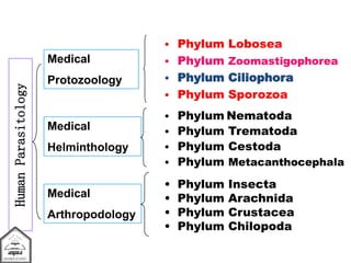
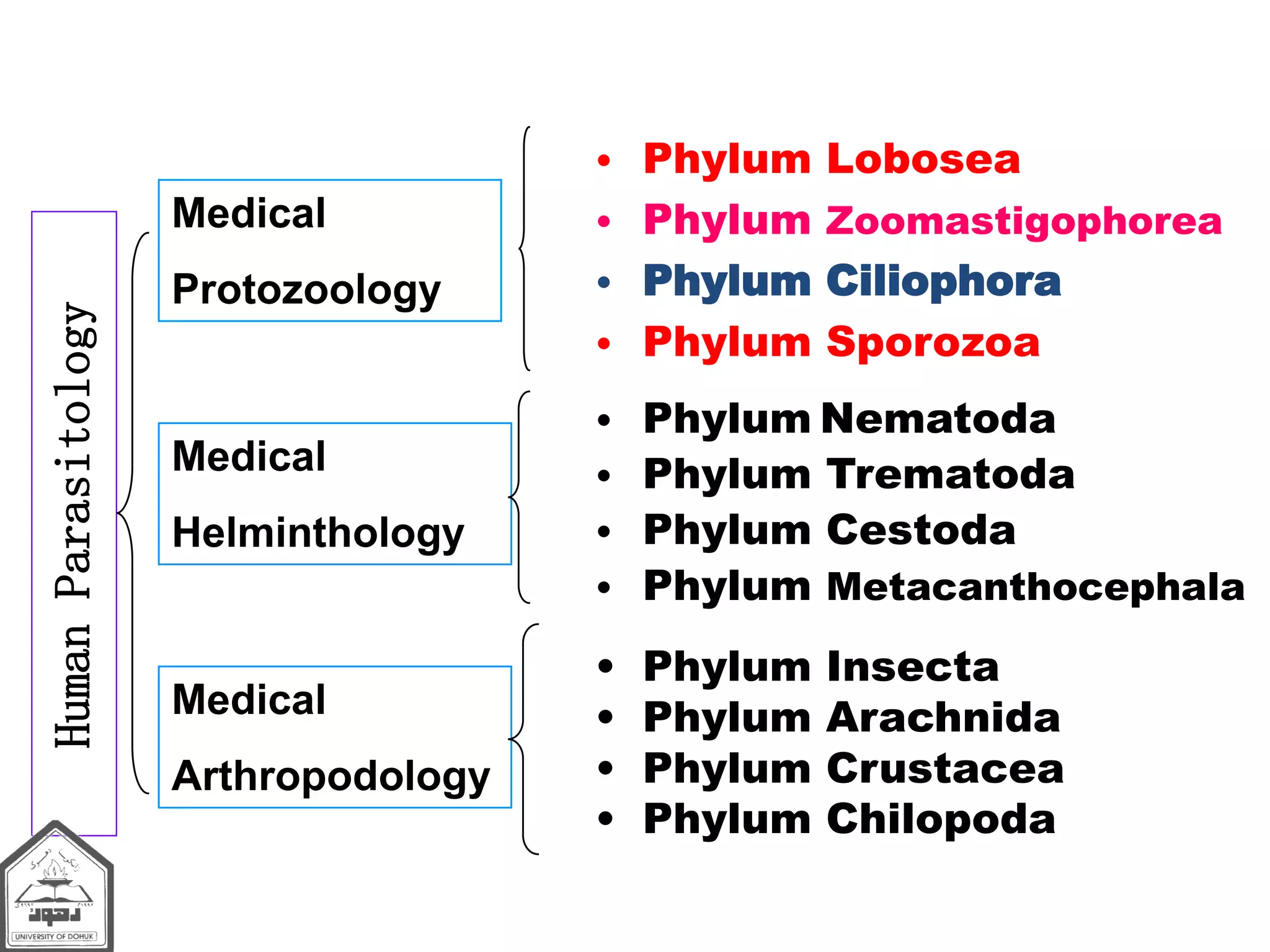
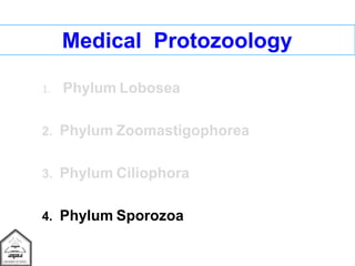
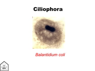
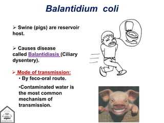
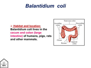
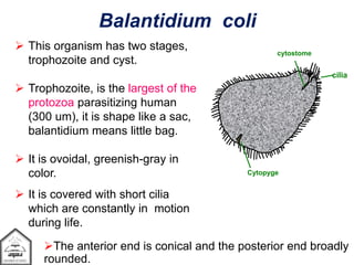
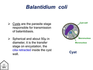
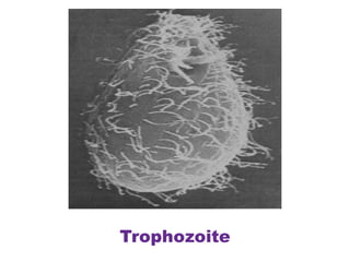
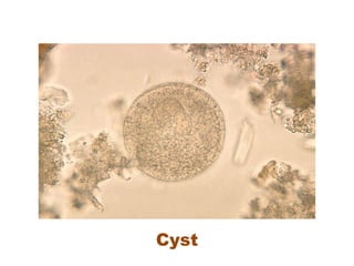
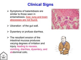
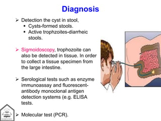
HumanParasitology
Medical
Helminthology
Medical
Protozoology
Medical
Arthropodology
• Phylum Nematoda
• Phylum Trematoda
• Phylum Cestoda
• Phylum Metacanthocephala
• Phylum Lobosea
• Phylum Zoomastigophorea
• Phylum Ciliophora
• Phylum Sporozoa
• Phylum Insecta
• Phylum Arachnida
• Phylum Crustacea
• Phylum Chilopoda
PHYLUM CILIOPHORA
Balantidium coli
Dr. SouzanH. Eassa,
Department of Medical Microbiology, College of Medicine,
University Of Duhok, Kurdistan Region, Iraq.
Medical Protozoology
1. Phylum Lobosea
2. Phylum Zoomastigophorea
3. Phylum Ciliophora
4. Phylum Sporozoa
Ciliophora
Balantidium coli
 Most ciliates are free-living; however, a few groups are
commensal or parasitic.
 These organisms characterized by having numerous cilia, which
are present both in the trophozoite and encysted stage.
 Multiplication is by transverse binary fission,
mean division starts from the central region of the cell.
 Most species have 2 kinds of nuclei: macronuclei and
micronuclei.
 Several species are pathogenic to animals, but only one,
Balantidium coli, occasionally establishes infection in humans.
Phylum Ciliophora
 Swine (pigs) are reservoir
host.
 Causes disease
called Balantidiasis (Ciliary
dysentery).
 Mode of transmission:
• By feco-oral route.
•Contaminated water is
the most common
mechanism of
transmission.
Balantidium coli
 Habitat and location:
Balantidium coli lives in the
cecum and colon (large
intestine) of humans, pigs, rats
and other mammals.
Balantidium coli
Balantidium coli
 This organism has two stages,
trophozoite and cyst.
 Trophozoite, is the largest of the
protozoa parasitizing human
(300 um), it is shape like a sac,
balantidium means little bag.
 It is ovoidal, greenish-gray in
color.
 It is covered with short cilia
which are constantly in motion
during life.
Cytopyge
cytostome
cilia
The anterior end is conical and the posterior end broadly
rounded.
 To one side of the anterior tip there is
a funnel-shaped peristome, which
leads into the cytostome.
 A minute cytopyge is situated at the
opposite end.
 One or two contractile vacuoles are
found within the cytoplasm.
 There is an elongated kidney-shaped
macronucleus (larger and less dense)
and lying within the concave side of
the macronucleus a minute
micronucleus (small and dense ).
Balantidium coli
Cytopyge
Micronucleus
Contractile V
Macronucleus
cytostome
cilia
Cyst wall
Micronucleus
Macronucleus
Balantidium coli
Cyst
 Cysts are the parasite stage
responsible for transmission
of balantidiasis.
 Spherical and about 50µ in
diameter, it is the transfer
stage on encystation, the
cilia retracted inside the cyst
wall.
Trophozoite
Cyst
Reproduction
 Both asexual and sexual reproduction present.
 Asexual by binary fission.
 Sexual by conjugation.
Life cycle
 Symptoms of balantidiasis are
similar to those seen in
entamebiasis. liver, lung and brain
abscesses are not found.
 Ulceration of the gut wall.
 Dysentery or profuse diarrhea.
 The resultant erosion of the
intestinal mucosa produces
varying degrees of irritation and
injury, leading to nausea,
vomiting, diarrhea, dysentery, and
abdominal colic.
Clinical Signs
 Detection the cyst in stool,
 Cysts-formed stools.
 Active trophzoites-diarrheic
stools.
 Sigmoidoscopy, trophozoite can
also be detected in tissue. In order
to collect a tissue specimen from
the large intestine.
 Serological tests such as enzyme
immunoassay and fluorescent-
antibody monoclonal antigen
detection systems (e.g. ELISA
tests.
 Molecular test (PCR).
Diagnosis
Treatment and Control
 Balantidium coli infection can be
treated effectively with antibiotics.
Three drugs are commonly used
and administered orally. They are
listed below in order of
recommendation.
1. Tetracyclines.
2. Metronidazole.
3. Iodoquinol.
Treatment and Control
 Eradication of fecal contamination of food and
water.
 Cysts killed by boiling, they are resistant to low
doses chlorine (halide tablets)
 Improved sanitation in institutions.
PHYLUM CILIOPHORA Balantidium coli

PHYLUM CILIOPHORA Balantidium coli

  • 1.
    HumanParasitology Medical Helminthology Medical Protozoology Medical Arthropodology • Phylum Nematoda •Phylum Trematoda • Phylum Cestoda • Phylum Metacanthocephala • Phylum Lobosea • Phylum Zoomastigophorea • Phylum Ciliophora • Phylum Sporozoa • Phylum Insecta • Phylum Arachnida • Phylum Crustacea • Phylum Chilopoda
  • 2.
    PHYLUM CILIOPHORA Balantidium coli Dr.SouzanH. Eassa, Department of Medical Microbiology, College of Medicine, University Of Duhok, Kurdistan Region, Iraq.
  • 3.
    Medical Protozoology 1. PhylumLobosea 2. Phylum Zoomastigophorea 3. Phylum Ciliophora 4. Phylum Sporozoa
  • 4.
  • 5.
     Most ciliatesare free-living; however, a few groups are commensal or parasitic.  These organisms characterized by having numerous cilia, which are present both in the trophozoite and encysted stage.  Multiplication is by transverse binary fission, mean division starts from the central region of the cell.  Most species have 2 kinds of nuclei: macronuclei and micronuclei.  Several species are pathogenic to animals, but only one, Balantidium coli, occasionally establishes infection in humans. Phylum Ciliophora
  • 6.
     Swine (pigs)are reservoir host.  Causes disease called Balantidiasis (Ciliary dysentery).  Mode of transmission: • By feco-oral route. •Contaminated water is the most common mechanism of transmission. Balantidium coli
  • 7.
     Habitat andlocation: Balantidium coli lives in the cecum and colon (large intestine) of humans, pigs, rats and other mammals. Balantidium coli
  • 8.
    Balantidium coli  Thisorganism has two stages, trophozoite and cyst.  Trophozoite, is the largest of the protozoa parasitizing human (300 um), it is shape like a sac, balantidium means little bag.  It is ovoidal, greenish-gray in color.  It is covered with short cilia which are constantly in motion during life. Cytopyge cytostome cilia The anterior end is conical and the posterior end broadly rounded.
  • 9.
     To oneside of the anterior tip there is a funnel-shaped peristome, which leads into the cytostome.  A minute cytopyge is situated at the opposite end.  One or two contractile vacuoles are found within the cytoplasm.  There is an elongated kidney-shaped macronucleus (larger and less dense) and lying within the concave side of the macronucleus a minute micronucleus (small and dense ). Balantidium coli Cytopyge Micronucleus Contractile V Macronucleus cytostome cilia
  • 10.
    Cyst wall Micronucleus Macronucleus Balantidium coli Cyst Cysts are the parasite stage responsible for transmission of balantidiasis.  Spherical and about 50µ in diameter, it is the transfer stage on encystation, the cilia retracted inside the cyst wall.
  • 11.
  • 13.
  • 14.
    Reproduction  Both asexualand sexual reproduction present.  Asexual by binary fission.  Sexual by conjugation.
  • 15.
  • 16.
     Symptoms ofbalantidiasis are similar to those seen in entamebiasis. liver, lung and brain abscesses are not found.  Ulceration of the gut wall.  Dysentery or profuse diarrhea.  The resultant erosion of the intestinal mucosa produces varying degrees of irritation and injury, leading to nausea, vomiting, diarrhea, dysentery, and abdominal colic. Clinical Signs
  • 17.
     Detection thecyst in stool,  Cysts-formed stools.  Active trophzoites-diarrheic stools.  Sigmoidoscopy, trophozoite can also be detected in tissue. In order to collect a tissue specimen from the large intestine.  Serological tests such as enzyme immunoassay and fluorescent- antibody monoclonal antigen detection systems (e.g. ELISA tests.  Molecular test (PCR). Diagnosis
  • 18.
    Treatment and Control Balantidium coli infection can be treated effectively with antibiotics. Three drugs are commonly used and administered orally. They are listed below in order of recommendation. 1. Tetracyclines. 2. Metronidazole. 3. Iodoquinol.
  • 19.
    Treatment and Control Eradication of fecal contamination of food and water.  Cysts killed by boiling, they are resistant to low doses chlorine (halide tablets)  Improved sanitation in institutions.