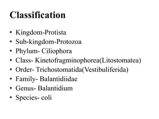
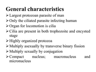
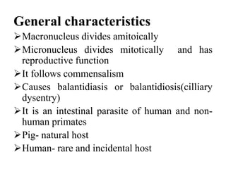
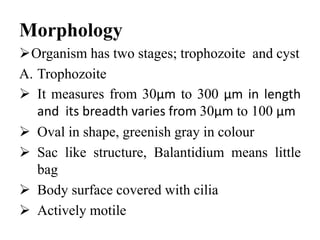
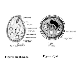
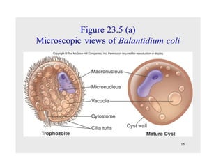
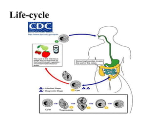
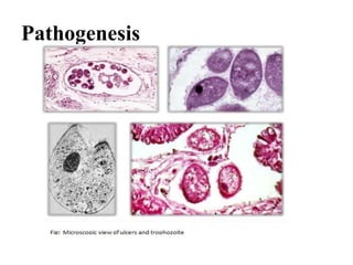
Balantidium coli
Classification
• Kingdom-Protista
• Sub-kingdom-Protozoa
• Phylum- Ciliophora
• Class- Kinetofragminophorea(Litostomatea)
• Order- Trichostomatida(Vestibuliferida)
• Family- Balantidiidae
• Genus- Balantidium
• Species- coli
General characteristics
Largest protozoan parasite of man
Only the ciliated parasite infecting human
Organ for locomotion is cilia
Cilia are present in both trophozoite and encysted
stage
Highly organized protozoa
Multiply asexually by transverse binary fission
Multiply sexually by conjugation
Compact nucleus; macronucleus and
micronucleus
General characteristics
Macronucleus divides amitoically
Micronucleus divides mitotically and has
reproductive function
It follows commensalism
Causes balantidiasis or balantidiosis(cilliary
dysentry)
It is an intestinal parasite of human and non-
human primates
Pig- natural host
Human- rare and incidental host
History
Malmsten reported the first case at Stockholm
in 1857
He was first to discovered it and named as
Paramacium coli
It was renamed as Balantidium coli by Stein in
1863
Morphologically different specimens gathered
from different host species, in 1992 by
McDonard and in1934 by Hagner
History
Different morphological species have different
nutritional requirement in vitro culture,
Barbosa et al., 2015; Levine, 1940
Levine in 1961 considered all these species to
be synonyms of Balantidium coli
Genetic analysis from different species
In 2011, Ponce-Gordo et al., concluded only
one specie infects warm blooded animals,
Balantidium coli
Geographical distribution
Cosmopolitan in distribution
Mainly found in pig raising areas
More common in temperate and tropical
regions
The Philippines, Papua New Guinea, some
Pacific Islands, some regions of Central and
South America and central Asia are considered
endemic areas
Geographical distribution
But also reported in Norway, Sweden, Finland,
and northern Russia
Epidemiology
The overall prevalence is estimated to be 0.02
to 1%
The number may be more as disease is
asymptomatic
In endemic areas,
o In South America the prevalence rate was 1 to
12% in late nineties
o Nearly, 30% cases in Oceania in 2013
o West Irian district of Indonesia, prevalence
rates has been reported upto 20%
Epidemiology
o It frequency of occurrance is less reported
from Indin
o In some pig-raising areas of New Guinea,
human infection rates are as high as 28%
An outbreak of balantidiasis on the Pacific
island of Truk in 1971 led to 110 human
infections
Infection tends to be more common among
humans who handles pigs
Habitat
Lives in lumen of large intestine of human,
pigs, rats and other mammals
Reservoir
Swine(pigs) are reservoir host
Source
Contaminated food and water with cysts
Infected hands with faeces
Morphology
Organism has two stages; trophozoite and cyst
A. Trophozoite
 It measures from 30µm to 300 µm in length
and its breadth varies from 30µm to 100 µm
 Oval in shape, greenish gray in colour
 Sac like structure, Balantidium means little
bag
 Body surface covered with cilia
 Actively motile
Morphology
 It has pointed/conical anterior end and rounded
posterior end
 At one side of anterior end, located a
conspicuous V- shaped cytostome
 Cytostome leads to funnel-shaped cytopharynx
 No intestine
 Posterior end contains a minute cytopyge
 Contains two contractile vacuoles in cytoplasm
 Many food vacuoles
Morphology
 Two nuclei: Macronucleus:
I. A large kidney shaped
II. Situated in the middle of the body
Micronucleus:
I. Lies in the concavity of macronucleus
 Invasive form
 Found in lumen of large intestine
 Found in dysenteric stool
 It can ingest Rbc, fat droplets and bacteria
Morphology
A. Cyst:
 Round and smaller than trophozoit
 Contains single cell inside cyst
 Measures 40µm to 60µm in diameter
 Yellowish or greenish with hyaline cytoplasm
 Cyst wall has two membrane
 A thick-transparent double-layered wall
surrounds the cyst
 It has granular cytoplasm
 Contains both micro and macro nucleus and
refractile body
Morphology
 Younger cyst may have cilia but mature cysts
do not
 Cilia retracted inside cyst wall
 Sometimes contractile vacuole may remains
active during encystment
 Encystment in host, under condition like
constipation and bowel dehydration
 Encystment may undergo outside of host
body
 Non- replicating encysted stage, develops in
lower colon
Morphology
 Excreted in feces
 This stage is responsible for transmission of
Balantidiasis
 It is resistive form and infective stage
 Found in chronic case and carriers
 In stained, macronucleus and some vacuoles
are easily seen
 Cilia can be seen
Figure: Trophozoite Figure: Cyst
Life-cycle
Life-cycle
Balantidium coli passes its life cycle in two
stages, but in single host
Pig- The natural host
Man- Rare and incidental host
Cyst-The infective stage of the parasite
Portal of entry-Mouth, by ingestion
Once the cyst is ingested via feces-
contaminated food or water
It passes through the host digestive system
Life-cycle
The tough cyst wall resist degradation in the
acidic environment of the stomach
It also resist basic environment of the small
intestine until it reaches the large intestine
Excystation takes place
Excystation produces a trophozoite from the
cyst stage.
Single cyst give rise single trophozoite
Trophozoite may remain in lumen or enter sub-
mucosa of large intestine
Life-cycle
Grows and multiplies asexually by transverse
binary fission
Each division produces two daughter
trophozoite
Binary fission occurs by division of
micronucleus followed by division of
macronucleus and the cytoplasm
Successive division produces a large number
of trophozoite
Life-cycle
Sexually divides by conjugation
Conjugation takes place when two trophozoite
enclosed in a cyst
An exchange of nuclear material and individual
separates
After certain period, trophozoite encyst into cyst
in lower part of intestine
Excreted in feces
Trophozoite in feces do not encyst and
disintegrate in the environment
But cyst are infective to man
When man ingest cyst , life cycle repeats again
Life-cycle
Transmission
Transmitted by fecal-oral route, with cyst and
less probably with trophozoite
Contaminated water and food is the main
source of transmission
Transmission by coprophagia could occur in
animals
Pathogenesis
Found both as a trophozoite and a cyst
Found in lumen of large intestine but invasion
of intestinal mucosa is its key feature of
pathology
Invasion is mediated by
I. Mechanical action by ciliary movement of
the trophozoite
II. Production of hyaluronidase by parasite
 Invades mucosa and sub-mucosa causing
ulcers
Pathogenesis
Ulcers:
I. Ulcers are similar to that of intestinal
amoebiasis
II. Found in caecum, ascending colon, sigmoid
colon and rectum
III. Oval or round in shape
IV. Are multiple in numbers having undermined
edges
V. Floor of ulcers contains pus and necrotic
materials
Pathogenesis
VI. Trophozoites do found in pus and at the
edges of ulcers
VII. Ulcers invades polymorphonuclear cells,
eosinophils and lymphocytes
VII. Ulcers do not invade muscular layer like
that of amoebic ulcers
VIII. Presence of trophozoite confirms the
Balantidium ulcers and diagnosis proceed
IX. Major complication is perforation
X. Liver , lung and brain abscesses are not found
Pathogenesis
Clinical manifestation
Generally, it is a self-limiting and asymptomatic
infection
Causes Balantidiasis or ciliate dysentery
A. Diarrhoea alternating with constipation is typical
symptoms in mild infection
B. Stool contains blood and mucus in acute
infection
Symptoms are similar to amoebic dysentery
headache, fever, nausea, vomiting, severe
abdominal pain and intestinal colic
Liver , lung and brain abscesses are not found
Clinical manifestation
Ulceration of gut wall
Perforation of intestine
Haemorrhage, shock and even death may
occur
C. In chronic cases, diarrhoea alternating with
constipation occurs
Diarrhoeal stool contains rare trace of blood
and lots of mucus
Presence of parasite in stool is less common
Intestinal perforation and extra-intestinal
infection are rarely seen
Laboratory Diagnosis
Microscopic examination of stool
 Trophozoites can be easily detected in saline
smears by their size and their slow motion
 Morphology of the macronucleus can be
easily recognized in both trophozoites and
cysts in temporary smears stained with iodine
 Other staining methods such as hematoxylin-
eosin or trichrome are also useful
 Concentration method by centrifugation
Laboratory Diagnosis
Molecular technique: Genetic analysis by
sequencing, PCR
Sigmoidoscopy
Serological test: Immuno-assay, Immuno-
fluorescent assay and ELISA
Treatment
Tetracycline is drugs of choice,500mg for 10
days, four times daily
Metronidazole,750 mg, 3 times daily for 5
days
Iodoquinol, 650mg, 3 times daily for 20 days
Metronidazole and Iodoquinol are alternative
drugs
Prognosis
Recover occurs without treatment
Severe and fatal in malnourished and immuno-
compromised patients
Prophylaxis
Sanitary disposal of human and pig feces
Eradication of contaminated food and water
either from pig or human
boiling kills cyst
Resistant to low dose of chlorine
Stool examination of food handlers of endemic
area
Avoid eating uncooked vegetables and fruits
that cannot be peeled off of sewage disposal
area
Notes
There are no data on prepatent and incubation
periods of B. coli in humans
 In piglets and non-human primates, the
incubation period ranges from 3 to 6 days
The infectious dose or dose-response for
trophozoites or cysts is not known.
The disease is communicable (infectious) for as
long as organisms are excreted
Cyst can survive for several weeks and
trophozoite die at faster rate (hours) outside host
Thank you

Balantidium coli

  • 1.
  • 2.
    Classification • Kingdom-Protista • Sub-kingdom-Protozoa •Phylum- Ciliophora • Class- Kinetofragminophorea(Litostomatea) • Order- Trichostomatida(Vestibuliferida) • Family- Balantidiidae • Genus- Balantidium • Species- coli
  • 3.
    General characteristics Largest protozoanparasite of man Only the ciliated parasite infecting human Organ for locomotion is cilia Cilia are present in both trophozoite and encysted stage Highly organized protozoa Multiply asexually by transverse binary fission Multiply sexually by conjugation Compact nucleus; macronucleus and micronucleus
  • 4.
    General characteristics Macronucleus dividesamitoically Micronucleus divides mitotically and has reproductive function It follows commensalism Causes balantidiasis or balantidiosis(cilliary dysentry) It is an intestinal parasite of human and non- human primates Pig- natural host Human- rare and incidental host
  • 5.
    History Malmsten reported thefirst case at Stockholm in 1857 He was first to discovered it and named as Paramacium coli It was renamed as Balantidium coli by Stein in 1863 Morphologically different specimens gathered from different host species, in 1992 by McDonard and in1934 by Hagner
  • 6.
    History Different morphological specieshave different nutritional requirement in vitro culture, Barbosa et al., 2015; Levine, 1940 Levine in 1961 considered all these species to be synonyms of Balantidium coli Genetic analysis from different species In 2011, Ponce-Gordo et al., concluded only one specie infects warm blooded animals, Balantidium coli
  • 7.
    Geographical distribution Cosmopolitan indistribution Mainly found in pig raising areas More common in temperate and tropical regions The Philippines, Papua New Guinea, some Pacific Islands, some regions of Central and South America and central Asia are considered endemic areas
  • 8.
    Geographical distribution But alsoreported in Norway, Sweden, Finland, and northern Russia
  • 9.
    Epidemiology The overall prevalenceis estimated to be 0.02 to 1% The number may be more as disease is asymptomatic In endemic areas, o In South America the prevalence rate was 1 to 12% in late nineties o Nearly, 30% cases in Oceania in 2013 o West Irian district of Indonesia, prevalence rates has been reported upto 20%
  • 10.
    Epidemiology o It frequencyof occurrance is less reported from Indin o In some pig-raising areas of New Guinea, human infection rates are as high as 28% An outbreak of balantidiasis on the Pacific island of Truk in 1971 led to 110 human infections Infection tends to be more common among humans who handles pigs
  • 11.
    Habitat Lives in lumenof large intestine of human, pigs, rats and other mammals Reservoir Swine(pigs) are reservoir host Source Contaminated food and water with cysts Infected hands with faeces
  • 12.
    Morphology Organism has twostages; trophozoite and cyst A. Trophozoite  It measures from 30µm to 300 µm in length and its breadth varies from 30µm to 100 µm  Oval in shape, greenish gray in colour  Sac like structure, Balantidium means little bag  Body surface covered with cilia  Actively motile
  • 13.
    Morphology  It haspointed/conical anterior end and rounded posterior end  At one side of anterior end, located a conspicuous V- shaped cytostome  Cytostome leads to funnel-shaped cytopharynx  No intestine  Posterior end contains a minute cytopyge  Contains two contractile vacuoles in cytoplasm  Many food vacuoles
  • 14.
    Morphology  Two nuclei:Macronucleus: I. A large kidney shaped II. Situated in the middle of the body Micronucleus: I. Lies in the concavity of macronucleus  Invasive form  Found in lumen of large intestine  Found in dysenteric stool  It can ingest Rbc, fat droplets and bacteria
  • 15.
    Morphology A. Cyst:  Roundand smaller than trophozoit  Contains single cell inside cyst  Measures 40µm to 60µm in diameter  Yellowish or greenish with hyaline cytoplasm  Cyst wall has two membrane  A thick-transparent double-layered wall surrounds the cyst  It has granular cytoplasm  Contains both micro and macro nucleus and refractile body
  • 16.
    Morphology  Younger cystmay have cilia but mature cysts do not  Cilia retracted inside cyst wall  Sometimes contractile vacuole may remains active during encystment  Encystment in host, under condition like constipation and bowel dehydration  Encystment may undergo outside of host body  Non- replicating encysted stage, develops in lower colon
  • 17.
    Morphology  Excreted infeces  This stage is responsible for transmission of Balantidiasis  It is resistive form and infective stage  Found in chronic case and carriers  In stained, macronucleus and some vacuoles are easily seen  Cilia can be seen
  • 19.
  • 21.
  • 22.
    Life-cycle Balantidium coli passesits life cycle in two stages, but in single host Pig- The natural host Man- Rare and incidental host Cyst-The infective stage of the parasite Portal of entry-Mouth, by ingestion Once the cyst is ingested via feces- contaminated food or water It passes through the host digestive system
  • 23.
    Life-cycle The tough cystwall resist degradation in the acidic environment of the stomach It also resist basic environment of the small intestine until it reaches the large intestine Excystation takes place Excystation produces a trophozoite from the cyst stage. Single cyst give rise single trophozoite Trophozoite may remain in lumen or enter sub- mucosa of large intestine
  • 24.
    Life-cycle Grows and multipliesasexually by transverse binary fission Each division produces two daughter trophozoite Binary fission occurs by division of micronucleus followed by division of macronucleus and the cytoplasm Successive division produces a large number of trophozoite
  • 25.
    Life-cycle Sexually divides byconjugation Conjugation takes place when two trophozoite enclosed in a cyst An exchange of nuclear material and individual separates After certain period, trophozoite encyst into cyst in lower part of intestine Excreted in feces Trophozoite in feces do not encyst and disintegrate in the environment But cyst are infective to man When man ingest cyst , life cycle repeats again
  • 26.
  • 27.
    Transmission Transmitted by fecal-oralroute, with cyst and less probably with trophozoite Contaminated water and food is the main source of transmission Transmission by coprophagia could occur in animals
  • 28.
    Pathogenesis Found both asa trophozoite and a cyst Found in lumen of large intestine but invasion of intestinal mucosa is its key feature of pathology Invasion is mediated by I. Mechanical action by ciliary movement of the trophozoite II. Production of hyaluronidase by parasite  Invades mucosa and sub-mucosa causing ulcers
  • 29.
    Pathogenesis Ulcers: I. Ulcers aresimilar to that of intestinal amoebiasis II. Found in caecum, ascending colon, sigmoid colon and rectum III. Oval or round in shape IV. Are multiple in numbers having undermined edges V. Floor of ulcers contains pus and necrotic materials
  • 30.
    Pathogenesis VI. Trophozoites dofound in pus and at the edges of ulcers VII. Ulcers invades polymorphonuclear cells, eosinophils and lymphocytes VII. Ulcers do not invade muscular layer like that of amoebic ulcers VIII. Presence of trophozoite confirms the Balantidium ulcers and diagnosis proceed IX. Major complication is perforation X. Liver , lung and brain abscesses are not found
  • 31.
  • 32.
    Clinical manifestation Generally, itis a self-limiting and asymptomatic infection Causes Balantidiasis or ciliate dysentery A. Diarrhoea alternating with constipation is typical symptoms in mild infection B. Stool contains blood and mucus in acute infection Symptoms are similar to amoebic dysentery headache, fever, nausea, vomiting, severe abdominal pain and intestinal colic Liver , lung and brain abscesses are not found
  • 33.
    Clinical manifestation Ulceration ofgut wall Perforation of intestine Haemorrhage, shock and even death may occur C. In chronic cases, diarrhoea alternating with constipation occurs Diarrhoeal stool contains rare trace of blood and lots of mucus Presence of parasite in stool is less common
  • 34.
    Intestinal perforation andextra-intestinal infection are rarely seen
  • 35.
    Laboratory Diagnosis Microscopic examinationof stool  Trophozoites can be easily detected in saline smears by their size and their slow motion  Morphology of the macronucleus can be easily recognized in both trophozoites and cysts in temporary smears stained with iodine  Other staining methods such as hematoxylin- eosin or trichrome are also useful  Concentration method by centrifugation
  • 36.
    Laboratory Diagnosis Molecular technique:Genetic analysis by sequencing, PCR Sigmoidoscopy Serological test: Immuno-assay, Immuno- fluorescent assay and ELISA
  • 37.
    Treatment Tetracycline is drugsof choice,500mg for 10 days, four times daily Metronidazole,750 mg, 3 times daily for 5 days Iodoquinol, 650mg, 3 times daily for 20 days Metronidazole and Iodoquinol are alternative drugs
  • 38.
    Prognosis Recover occurs withouttreatment Severe and fatal in malnourished and immuno- compromised patients
  • 39.
    Prophylaxis Sanitary disposal ofhuman and pig feces Eradication of contaminated food and water either from pig or human boiling kills cyst Resistant to low dose of chlorine Stool examination of food handlers of endemic area Avoid eating uncooked vegetables and fruits that cannot be peeled off of sewage disposal area
  • 40.
    Notes There are nodata on prepatent and incubation periods of B. coli in humans  In piglets and non-human primates, the incubation period ranges from 3 to 6 days The infectious dose or dose-response for trophozoites or cysts is not known. The disease is communicable (infectious) for as long as organisms are excreted Cyst can survive for several weeks and trophozoite die at faster rate (hours) outside host
  • 41.