Download as PDF, PPTX


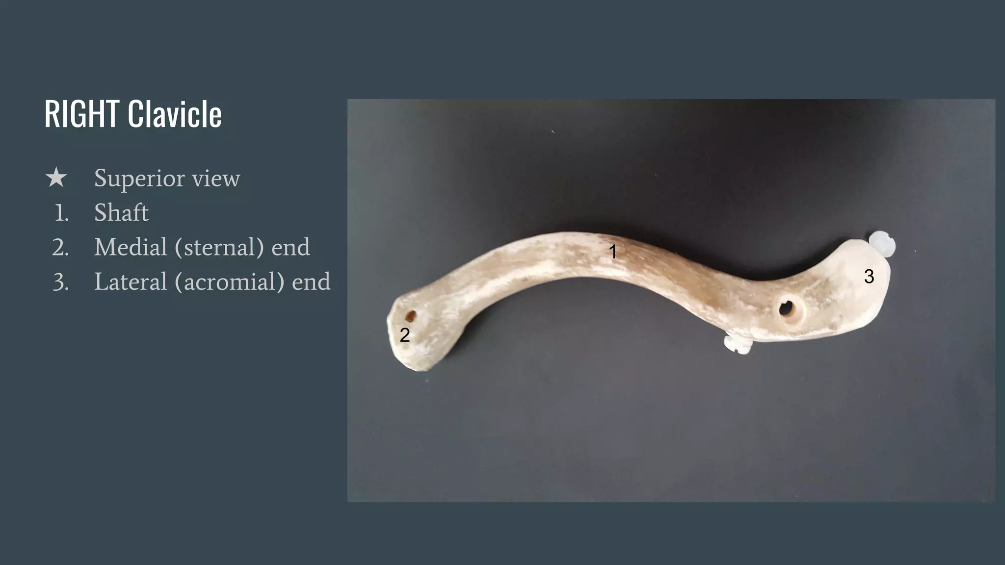
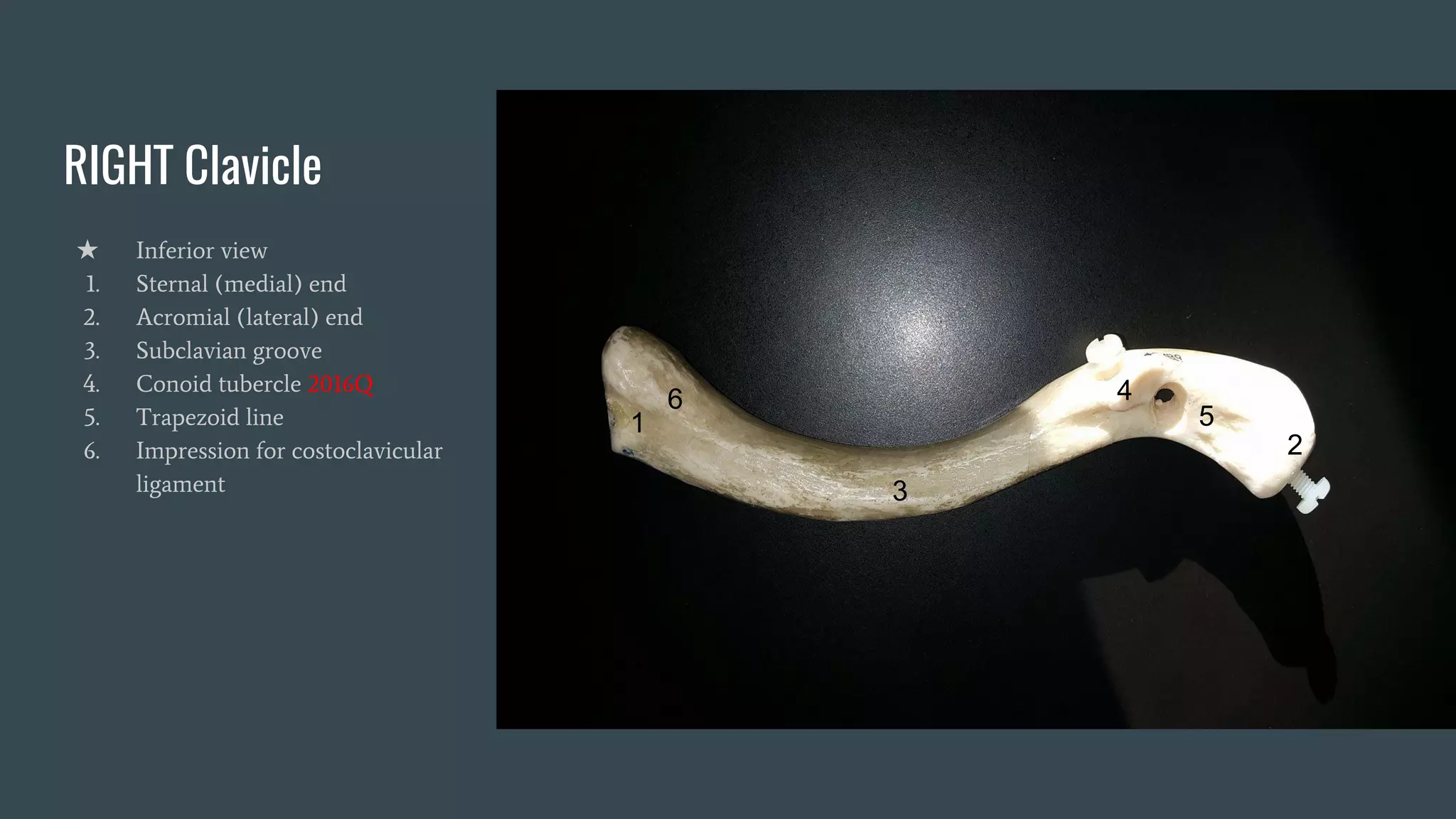
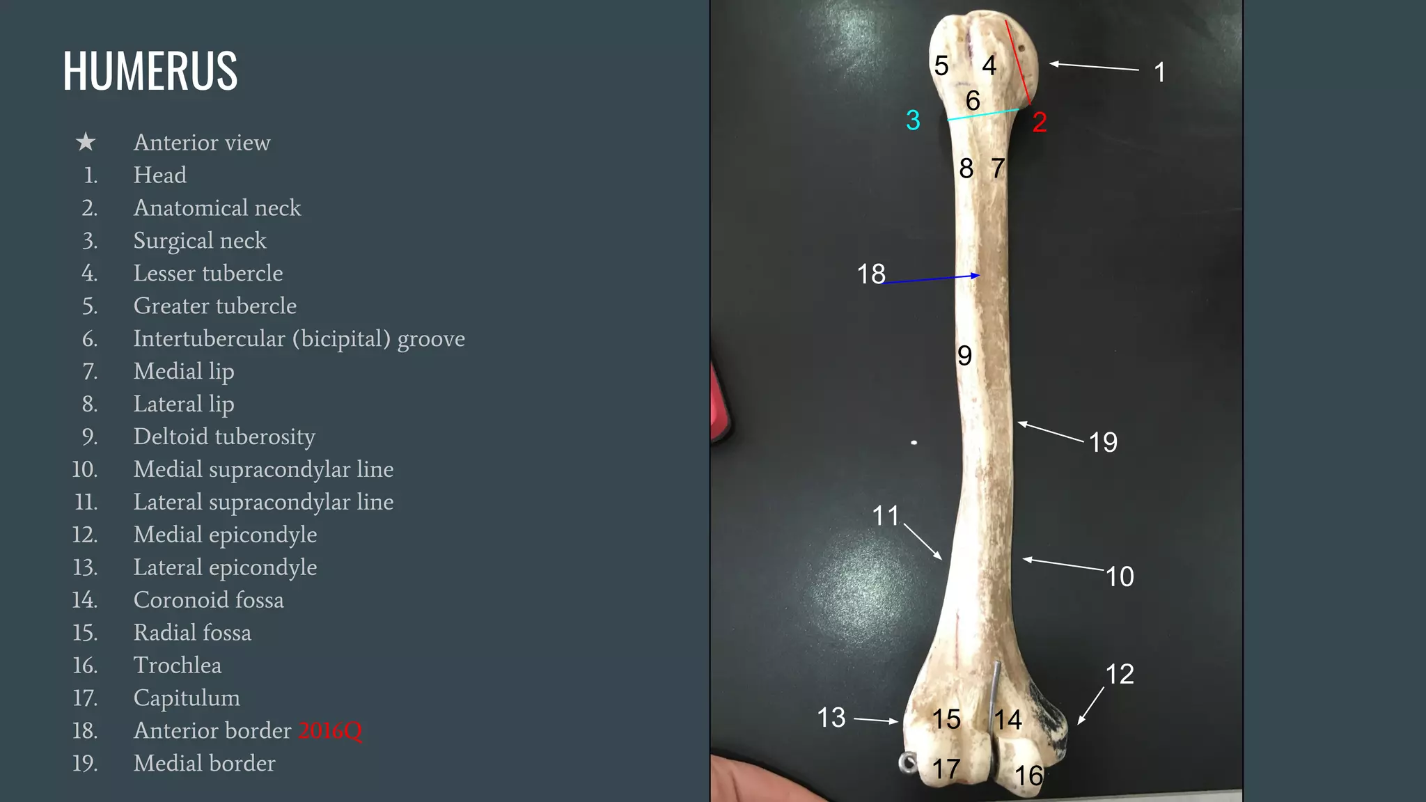

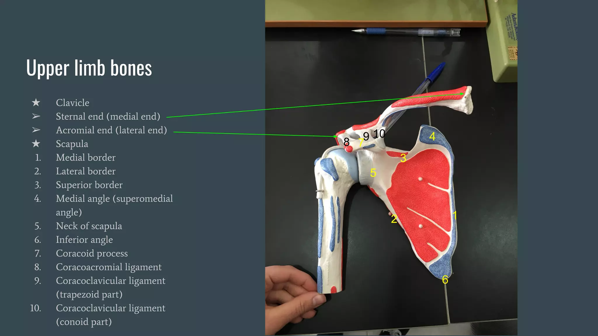
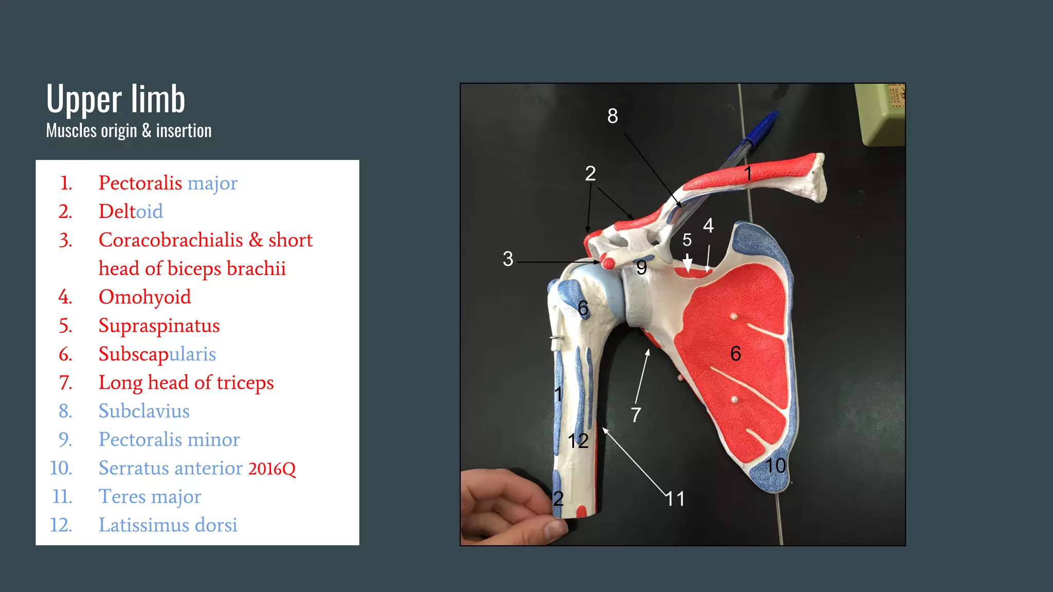
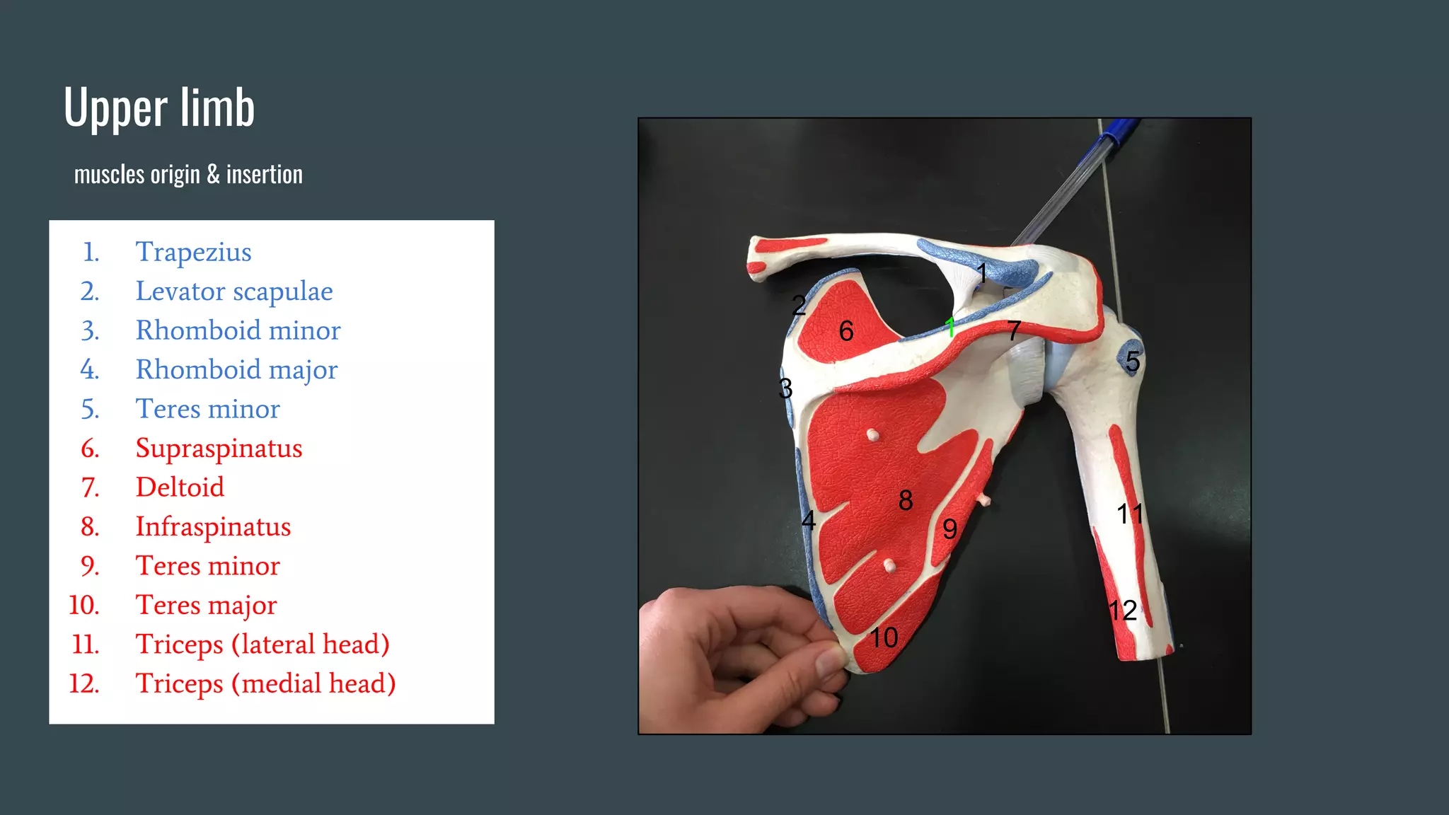
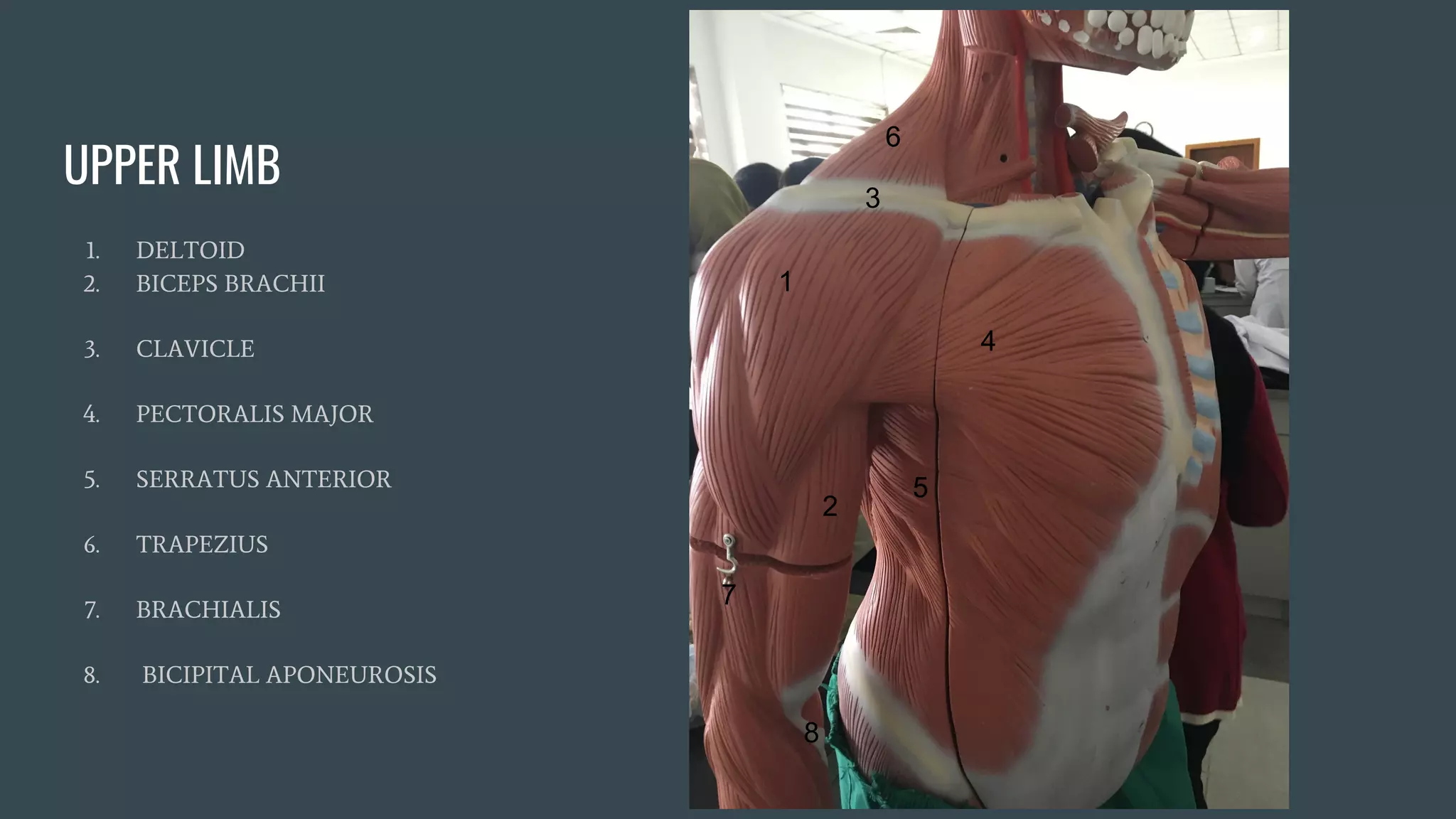
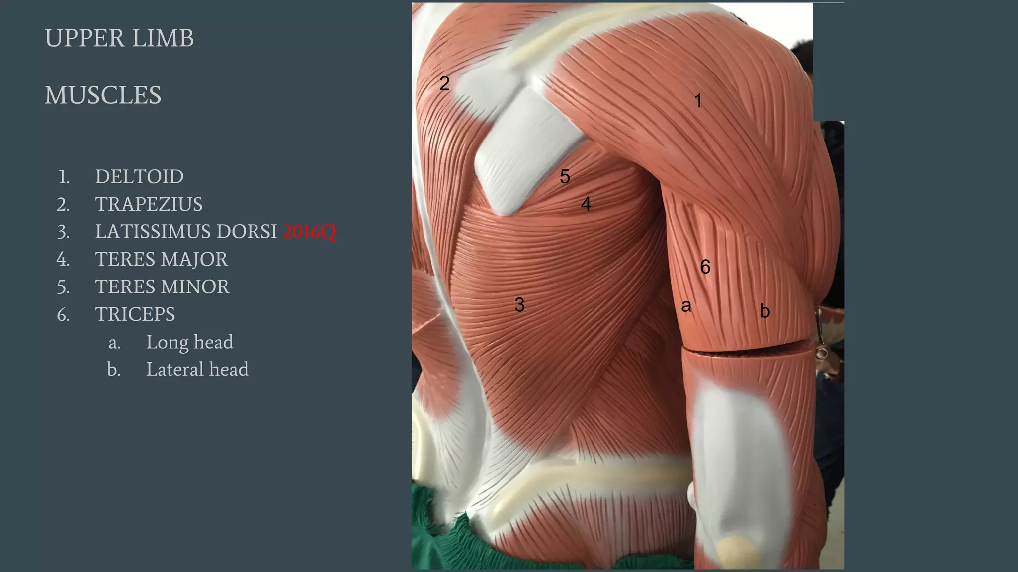
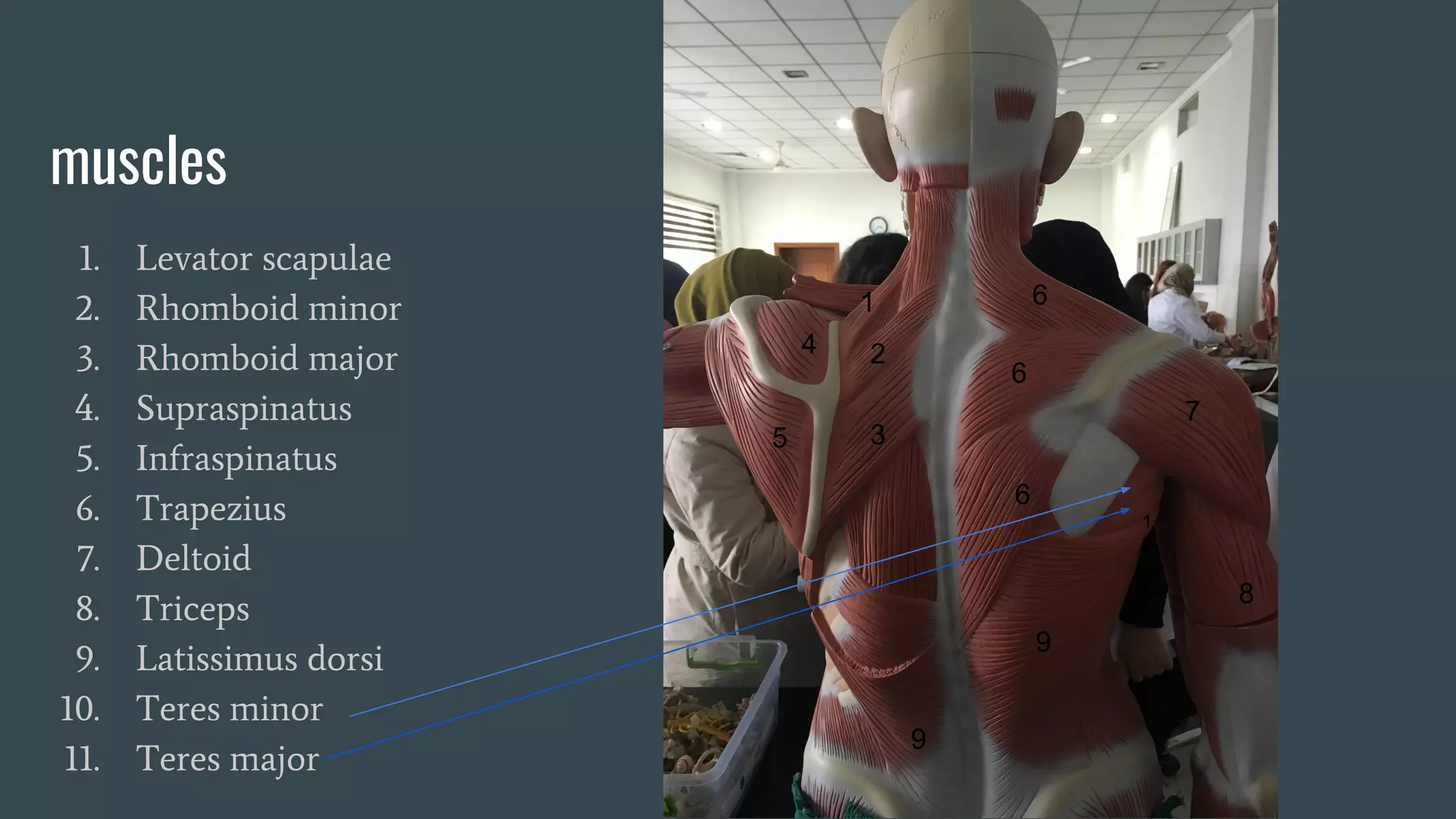

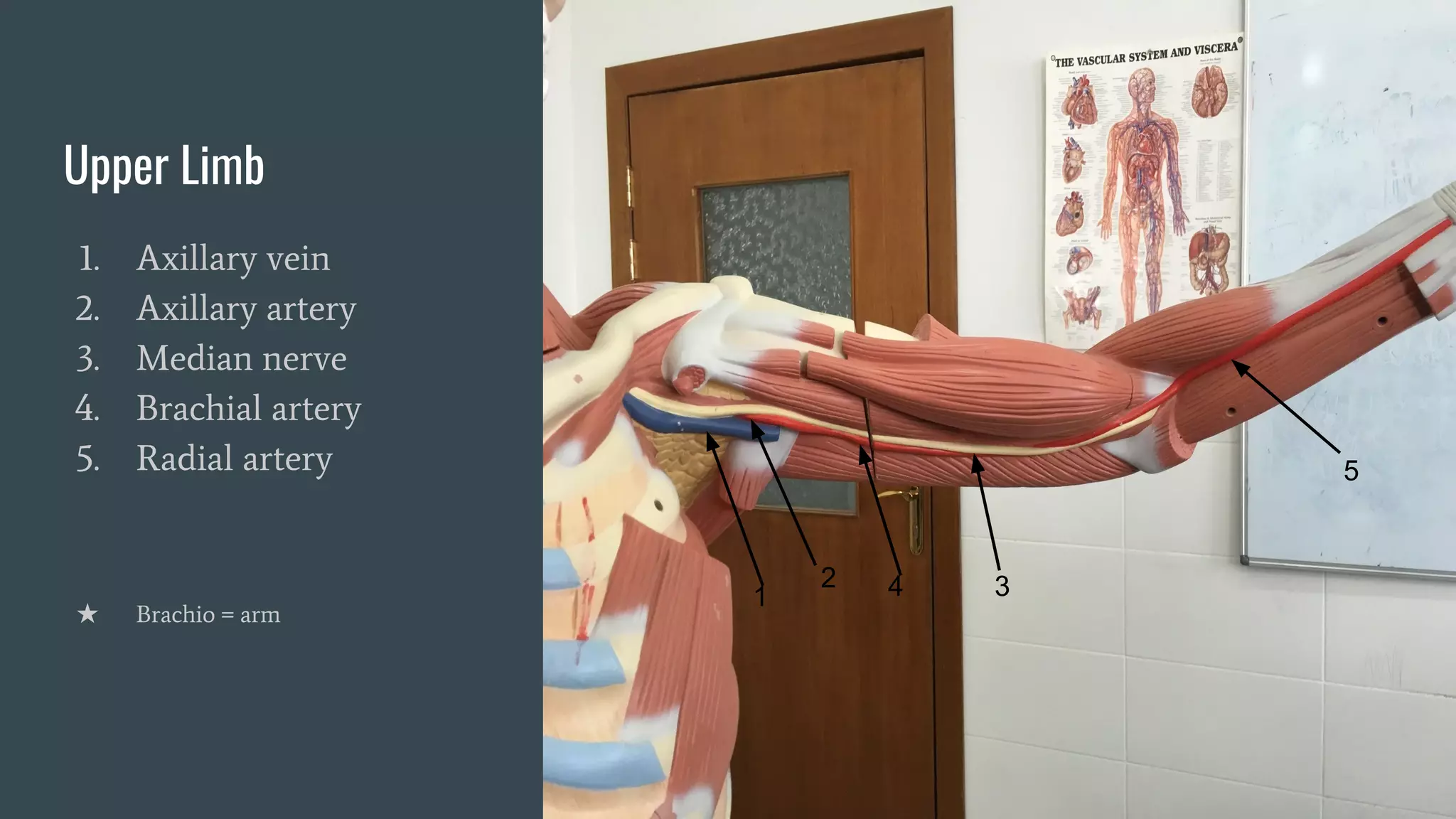
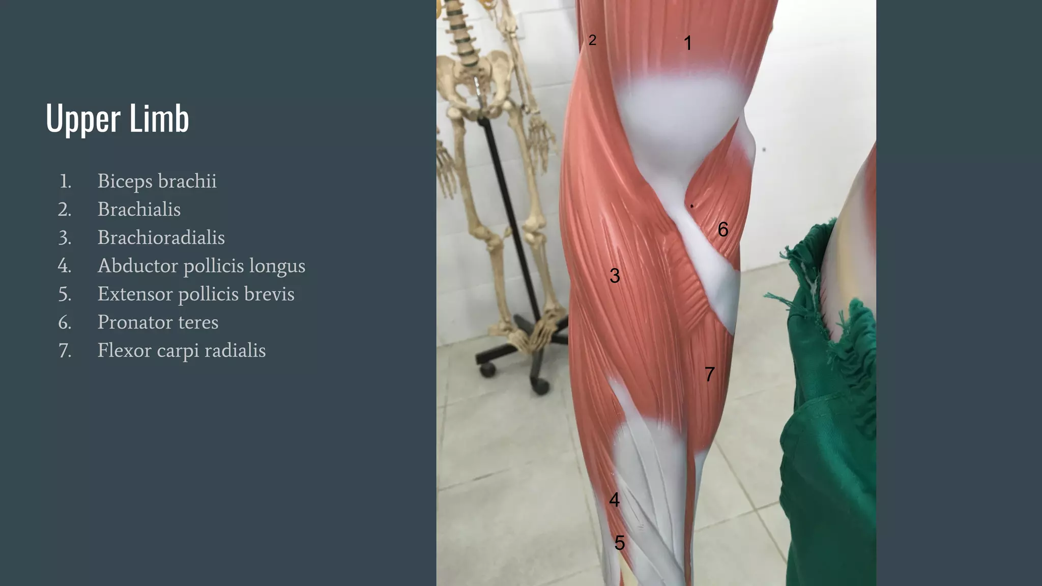

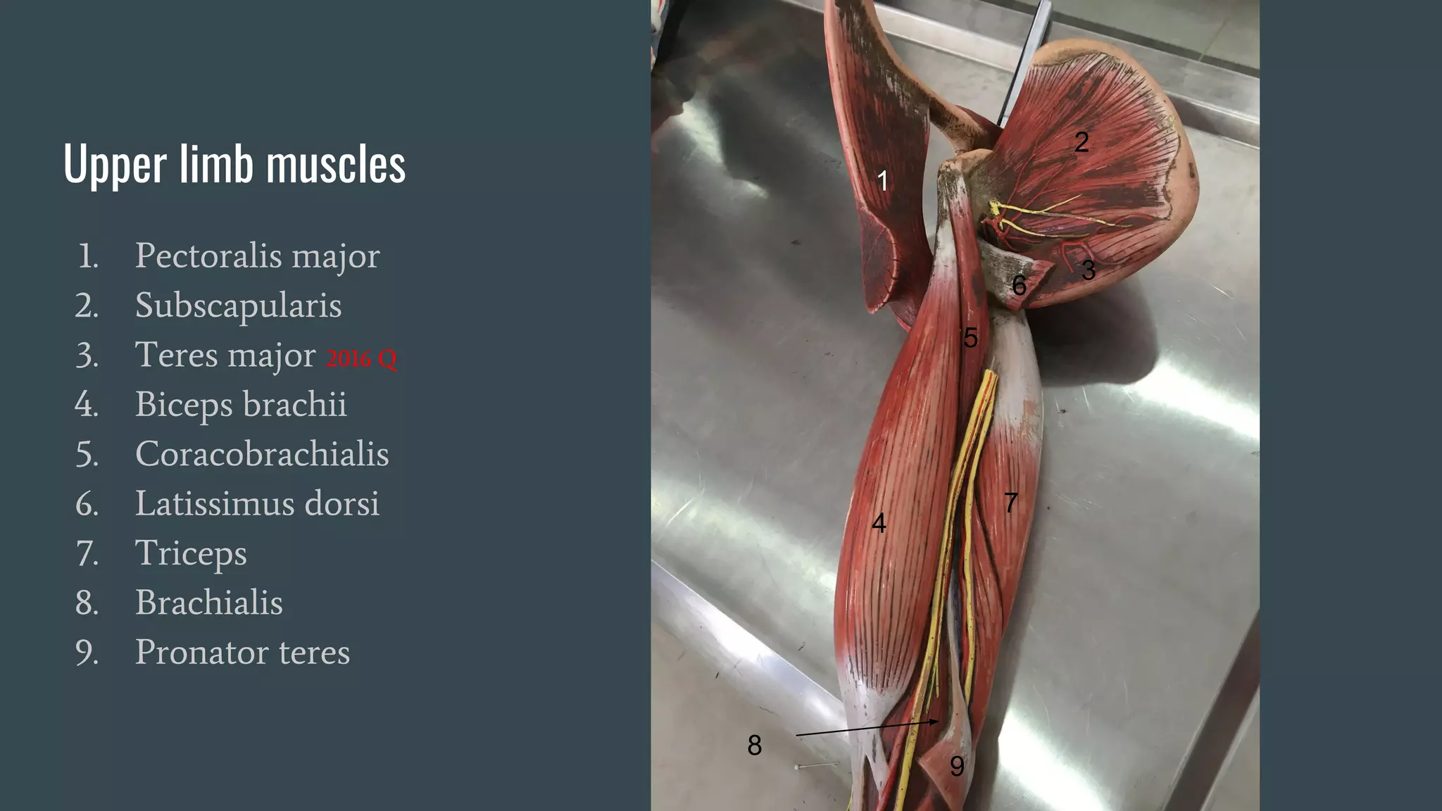
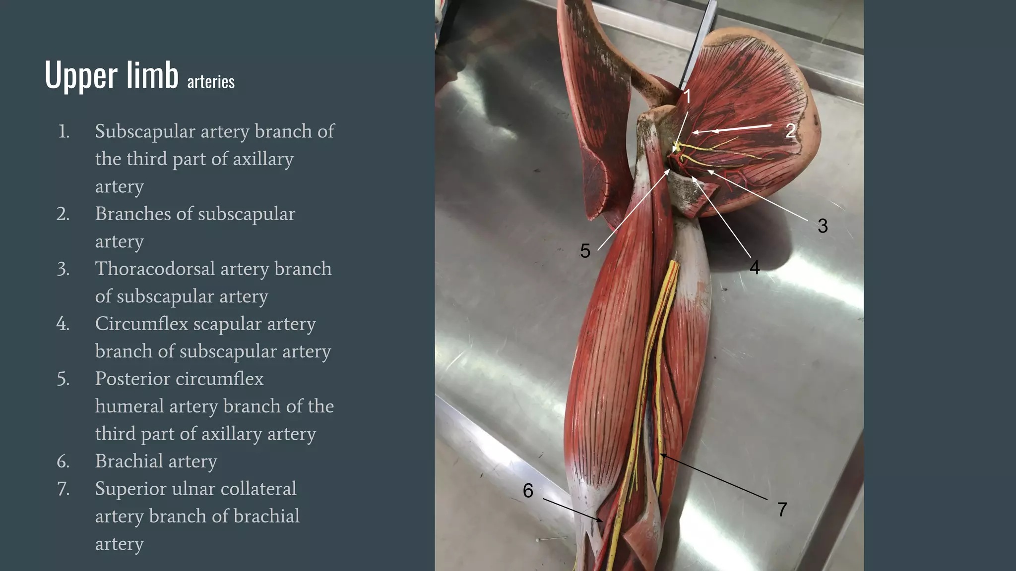
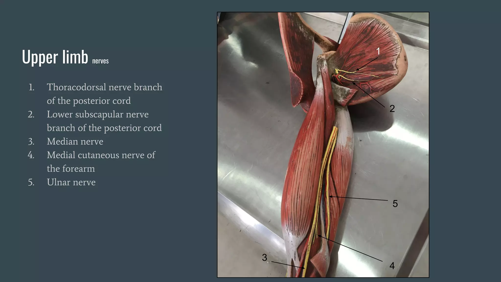
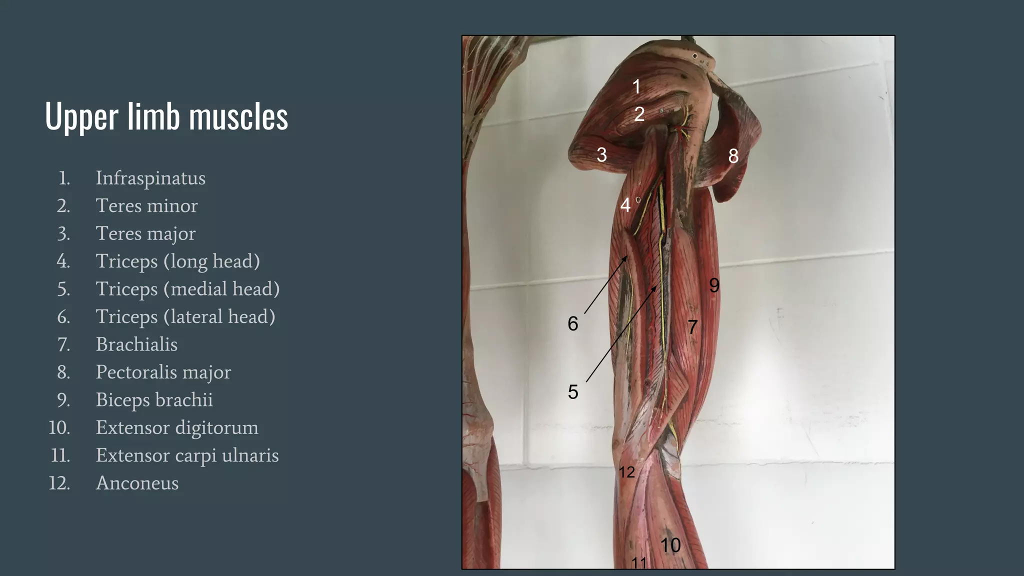
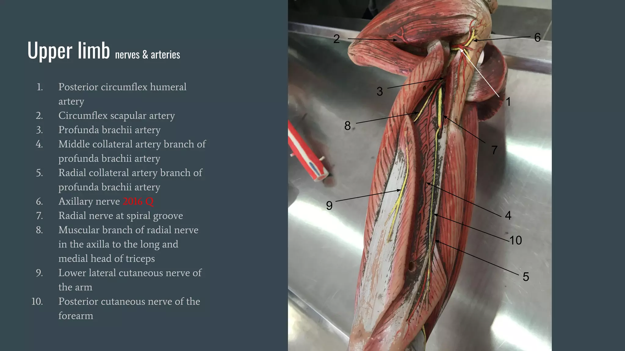
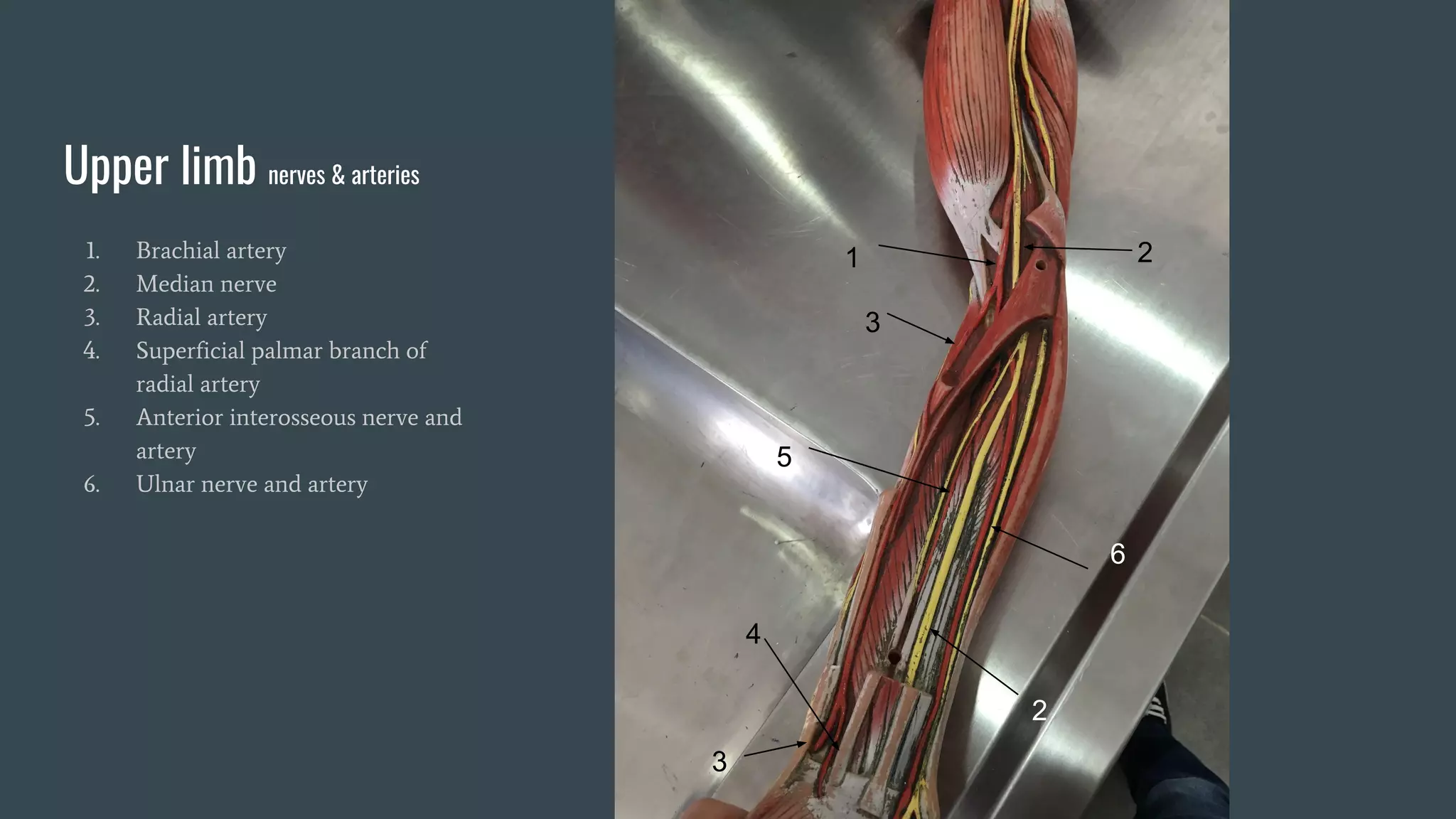
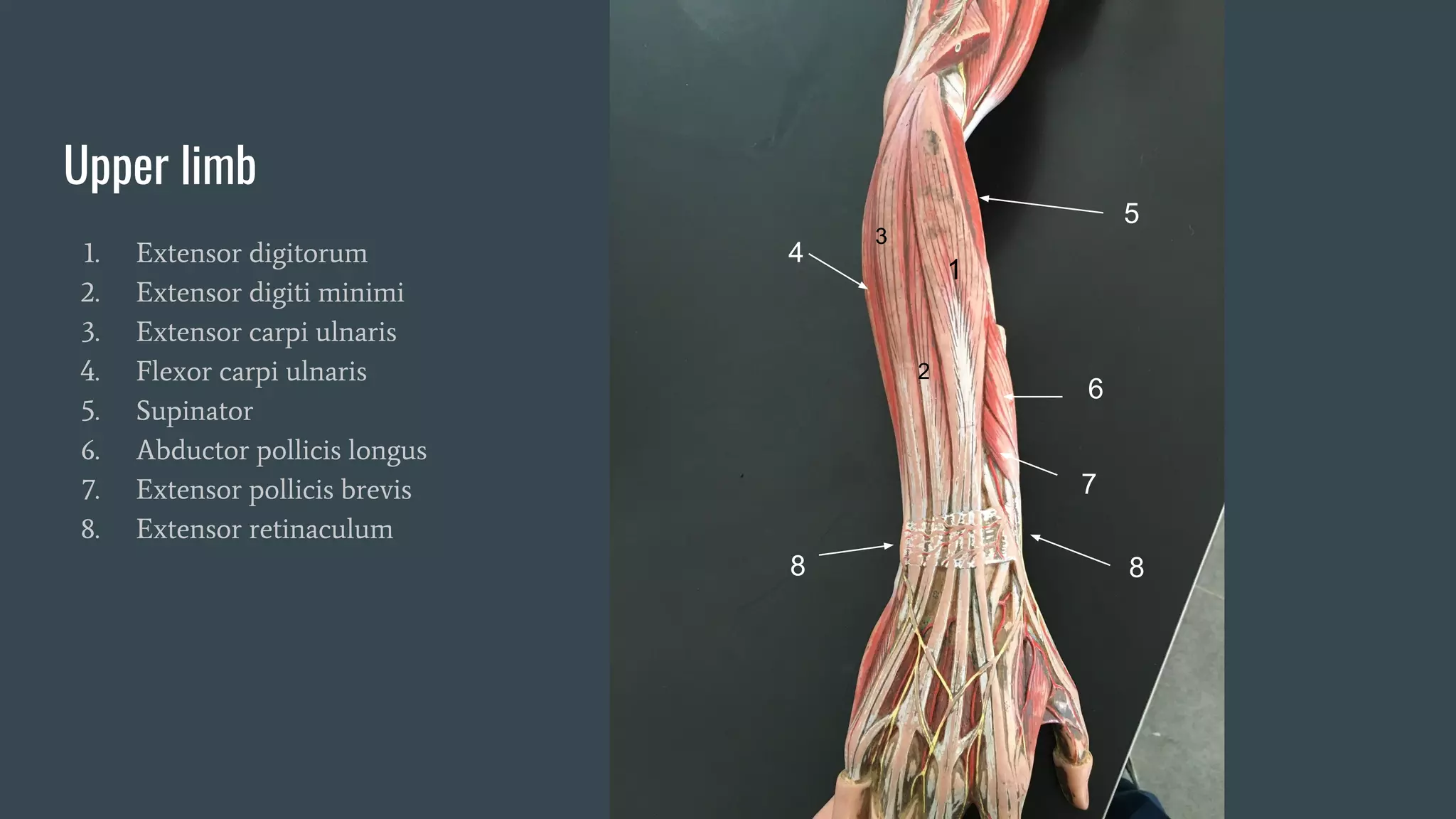
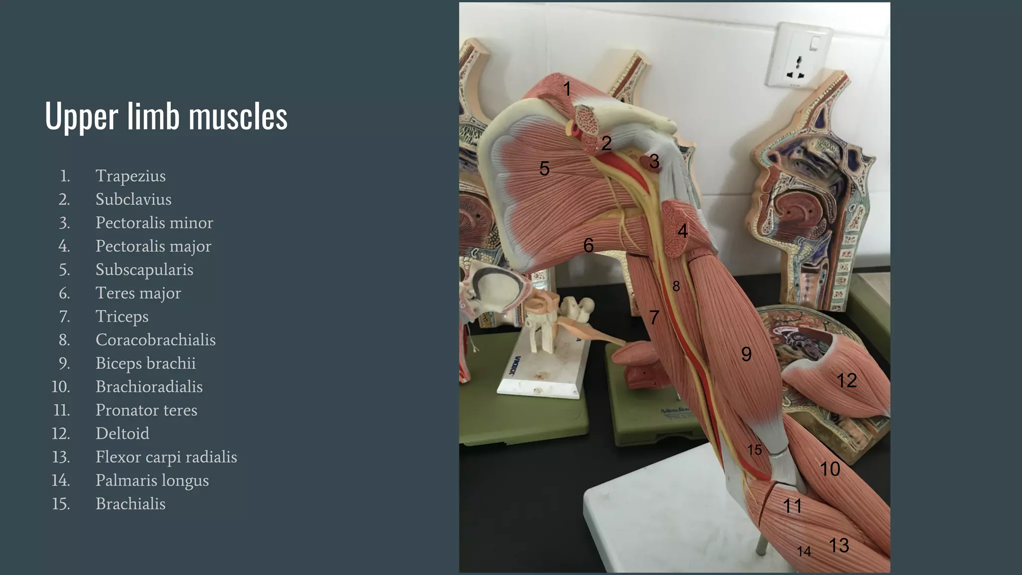


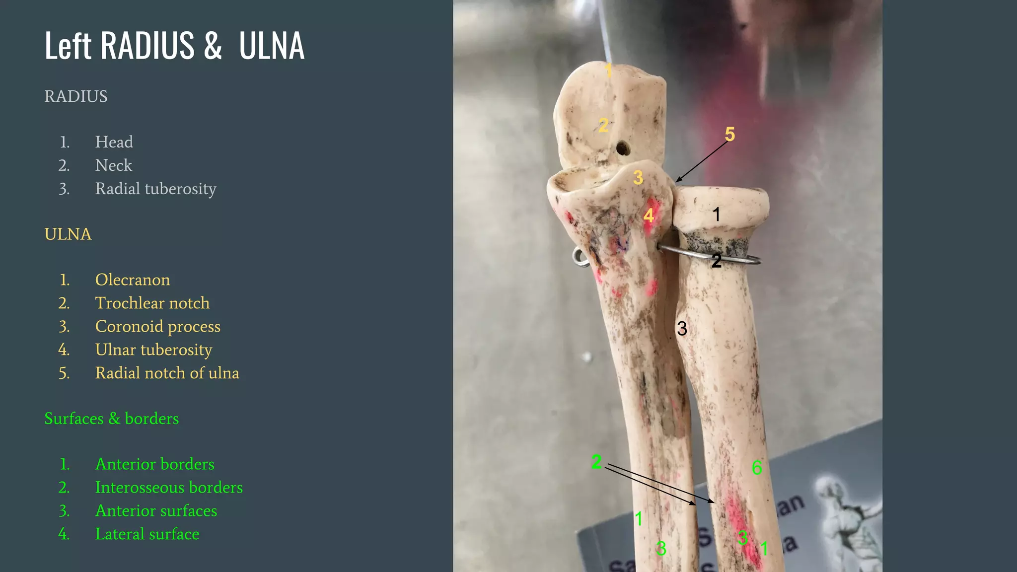
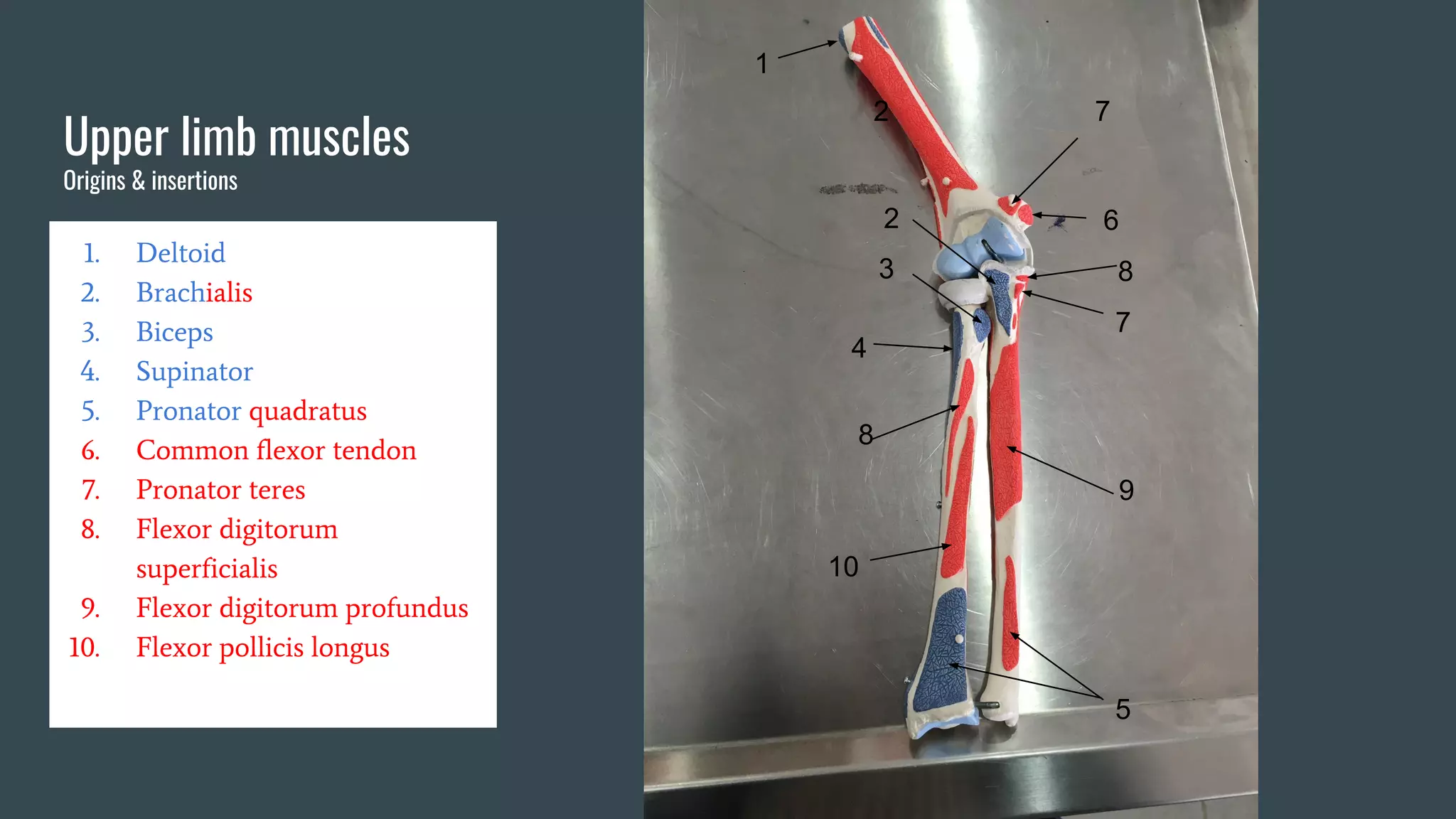
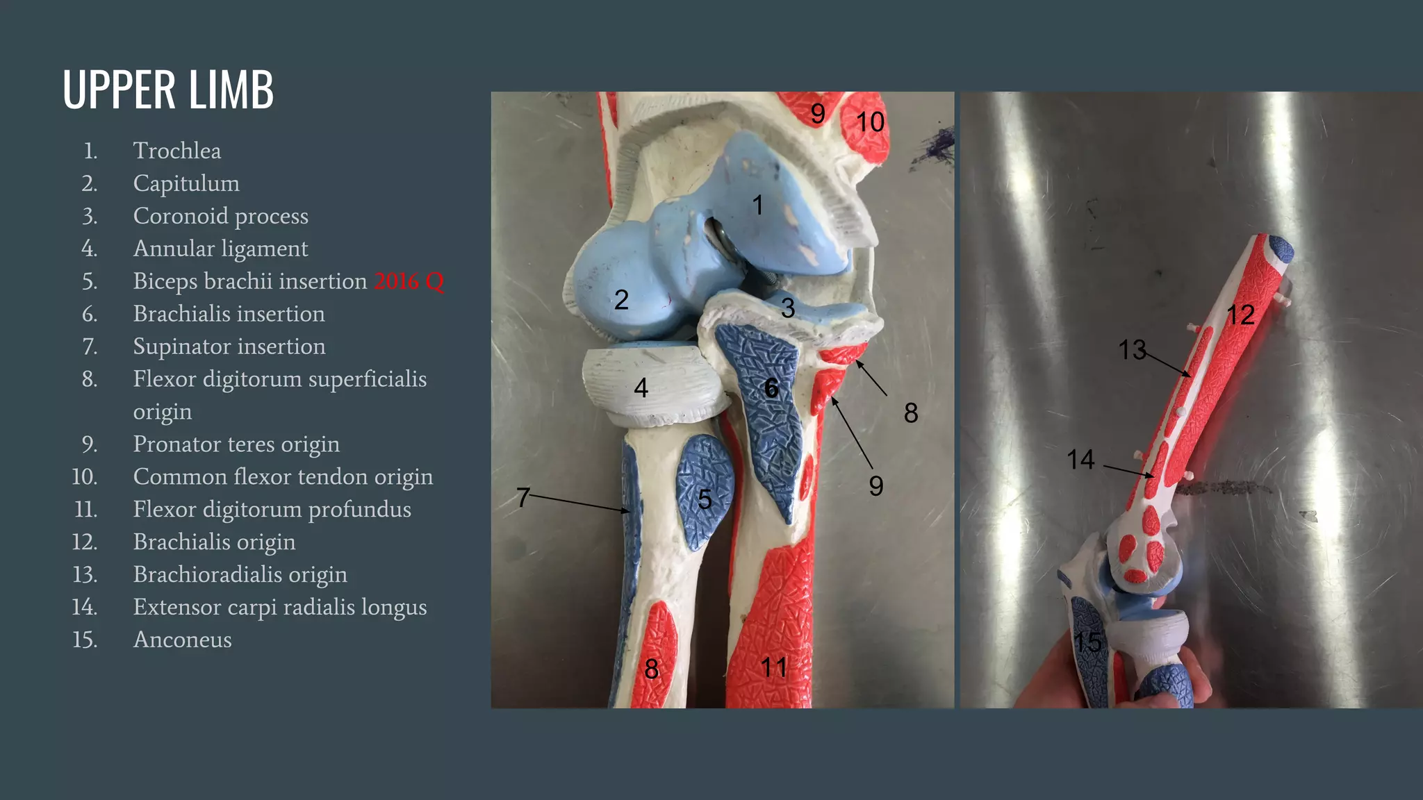
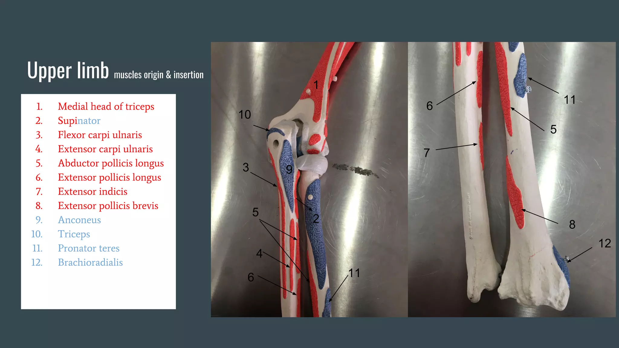
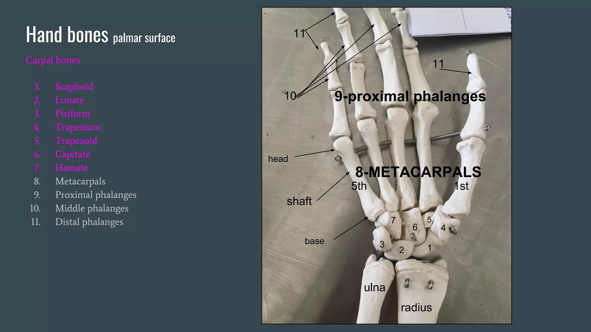

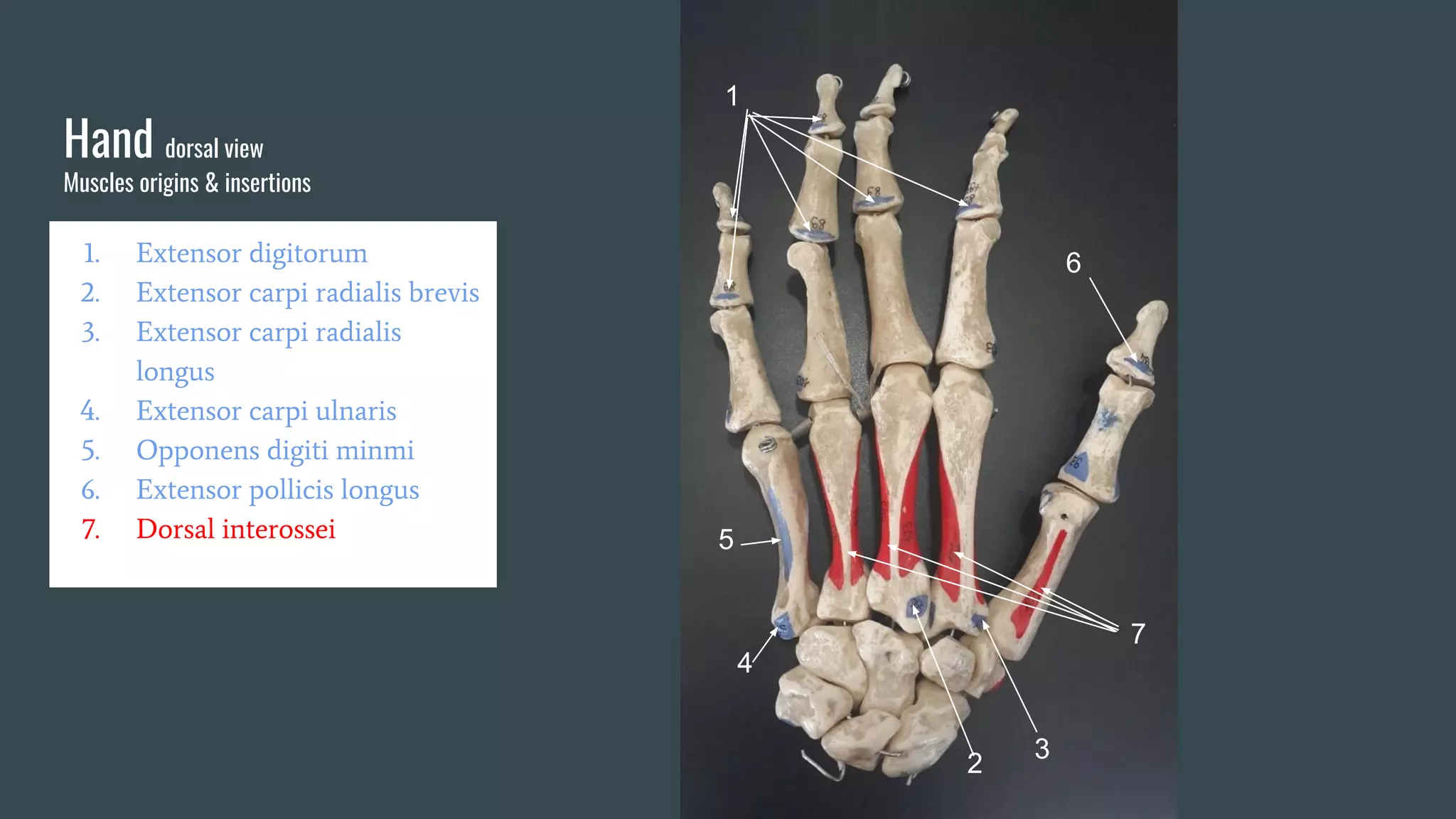

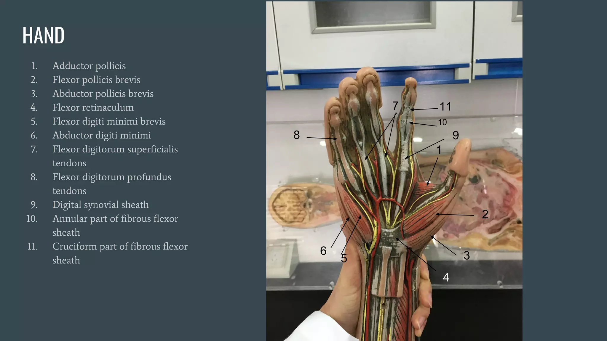
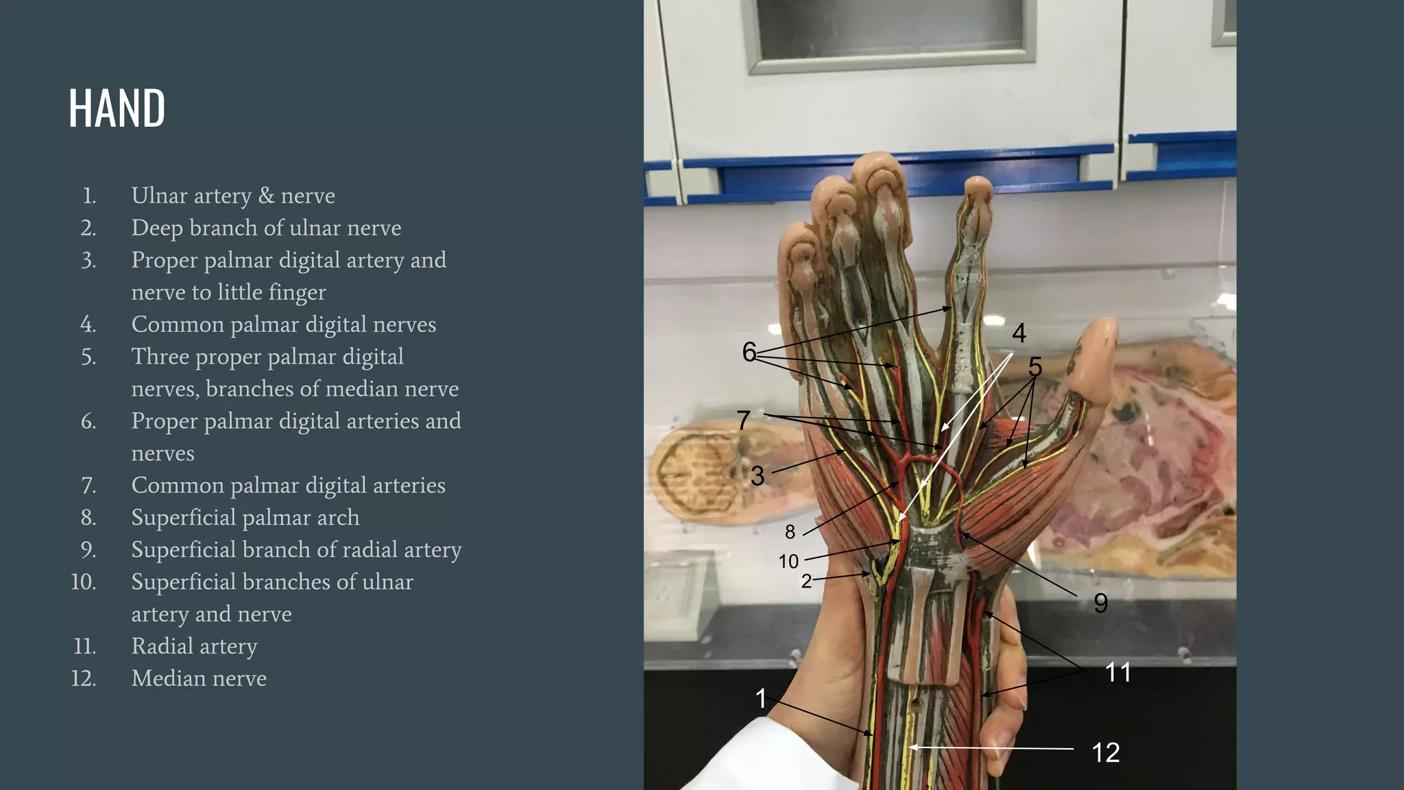
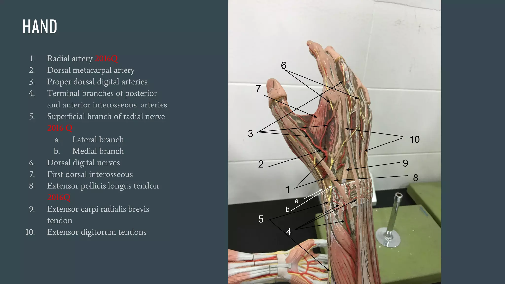
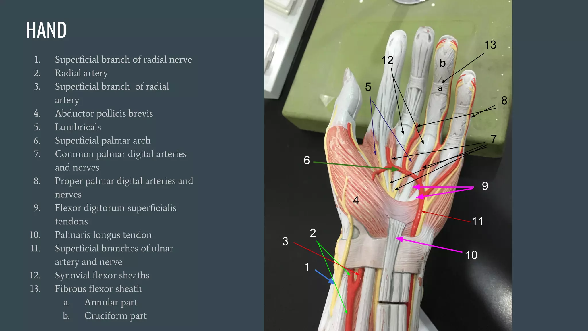
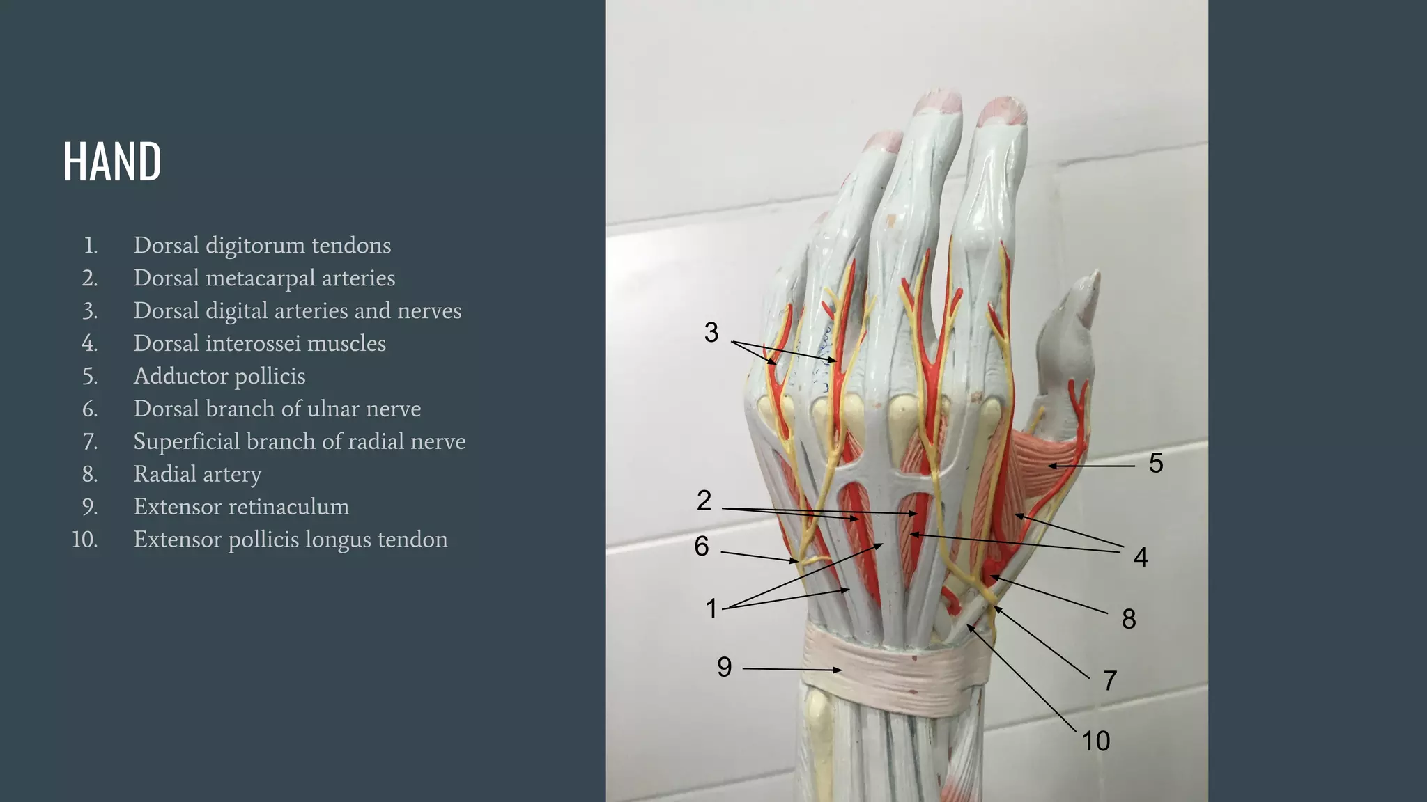
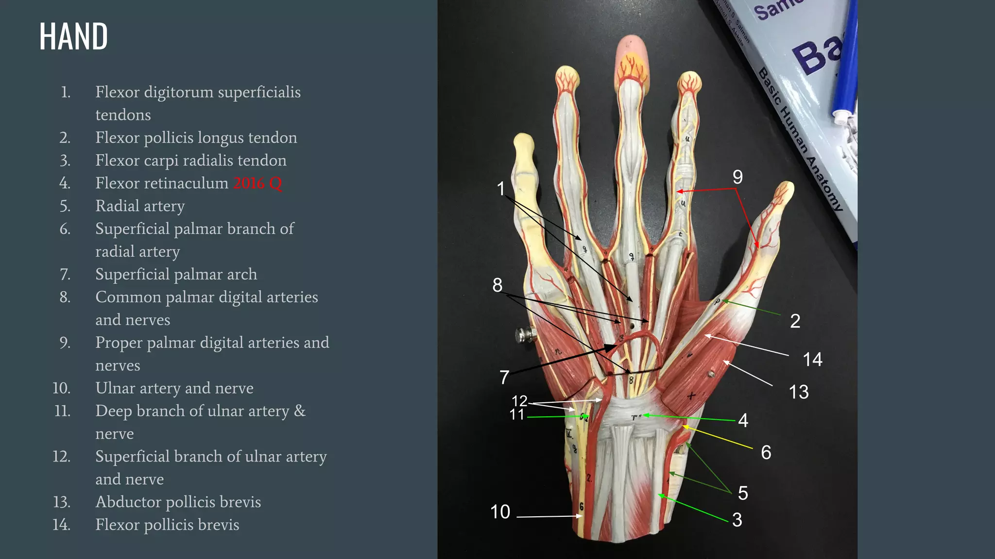
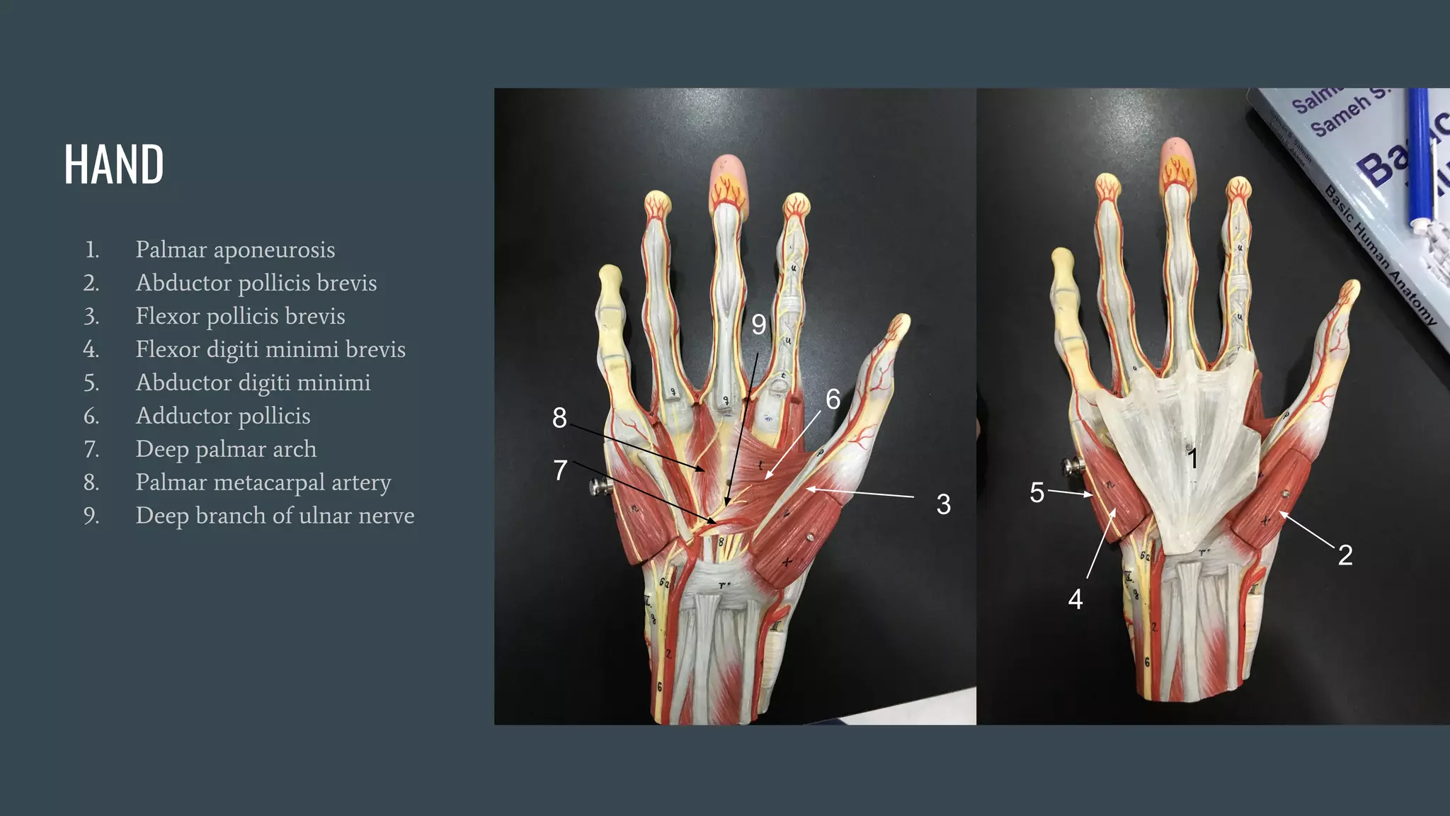
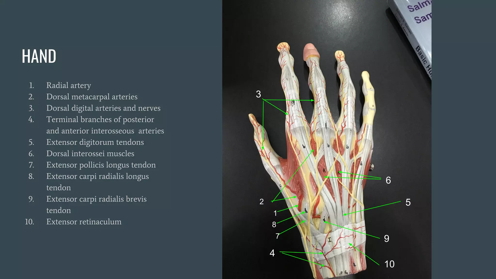
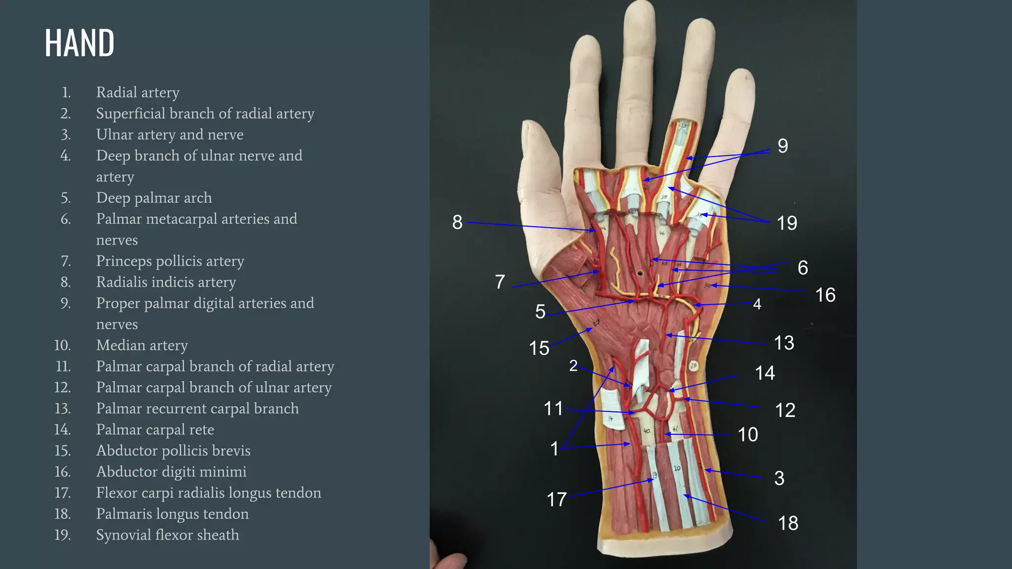


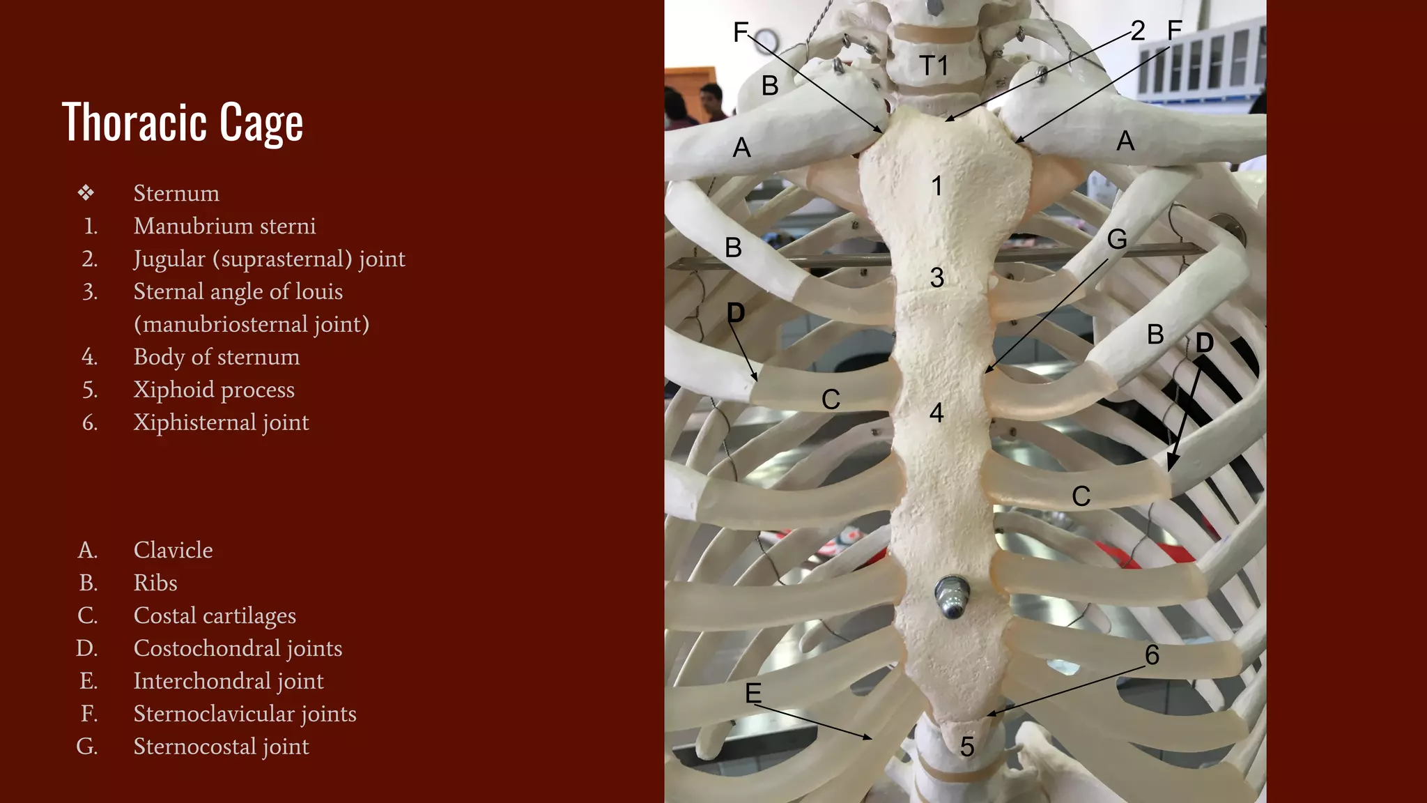


This document provides labelled diagrams of anatomical structures from the practical anatomy lab. It contains over 50 labelled diagrams of the skin, spinal cord, upper limb bones, muscles, arteries, and nerves. For each diagram, all major relevant structures are labelled with numbering. Brief explanatory text provides the names for each numbered structure. The purpose is to help students learn anatomy through these labelled diagrams from the author's anatomy lab.


