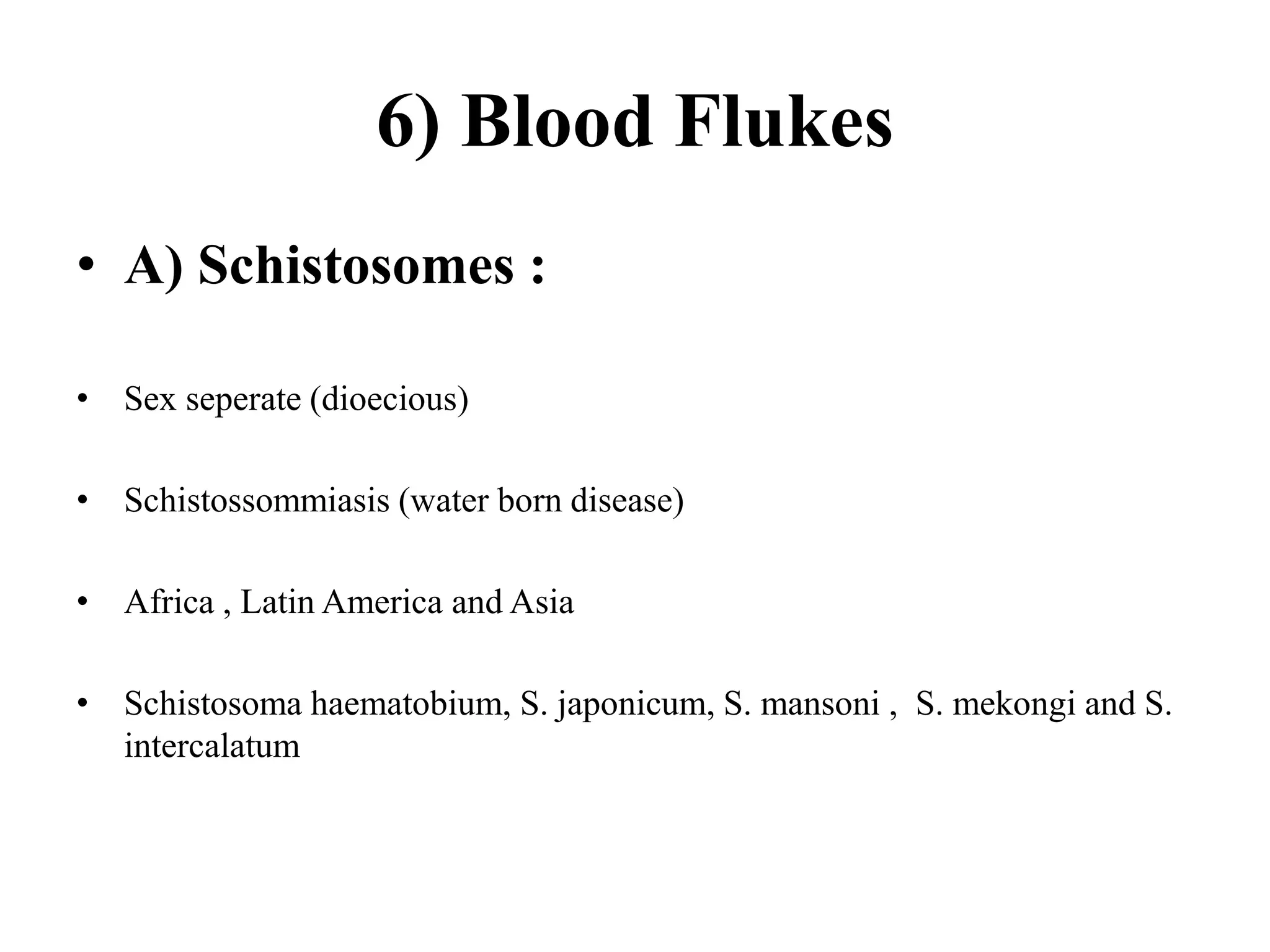
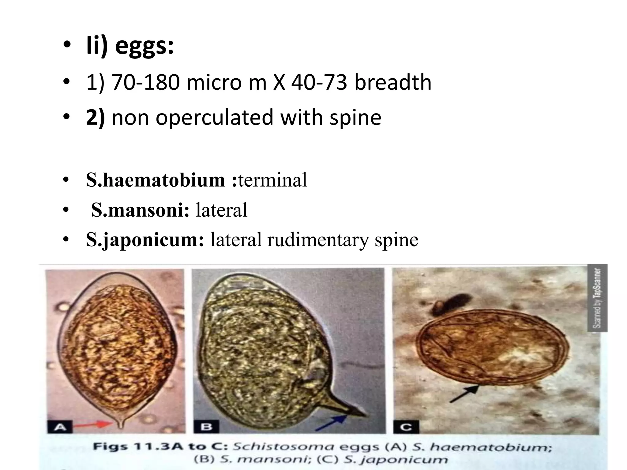
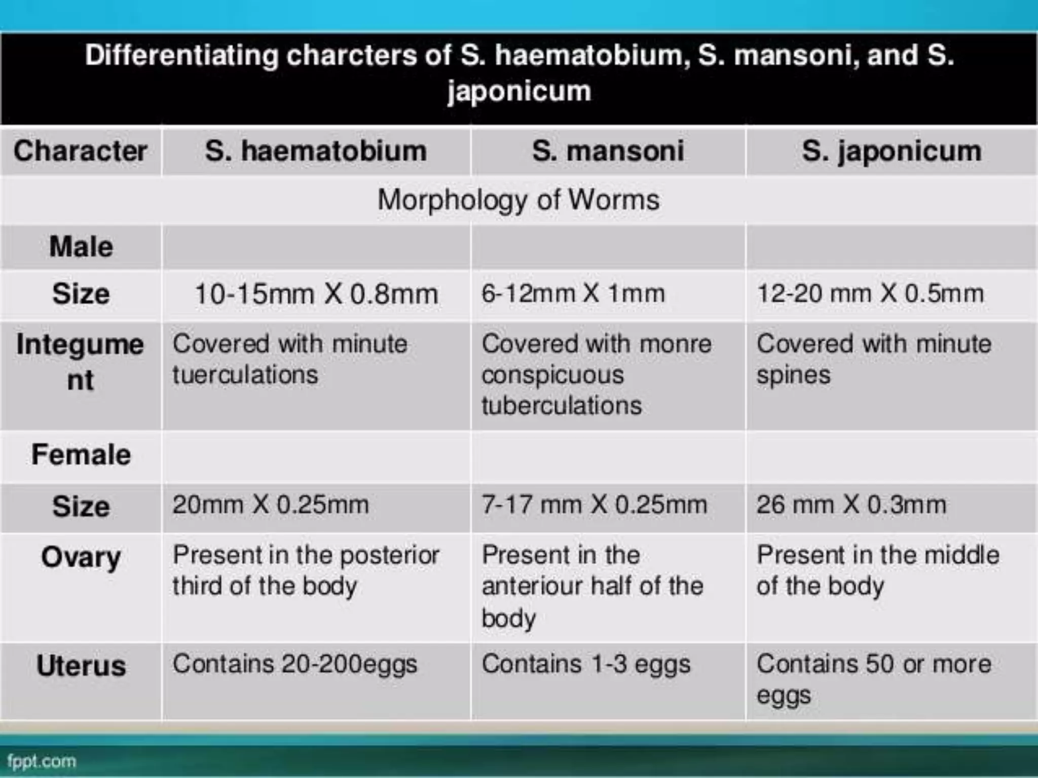
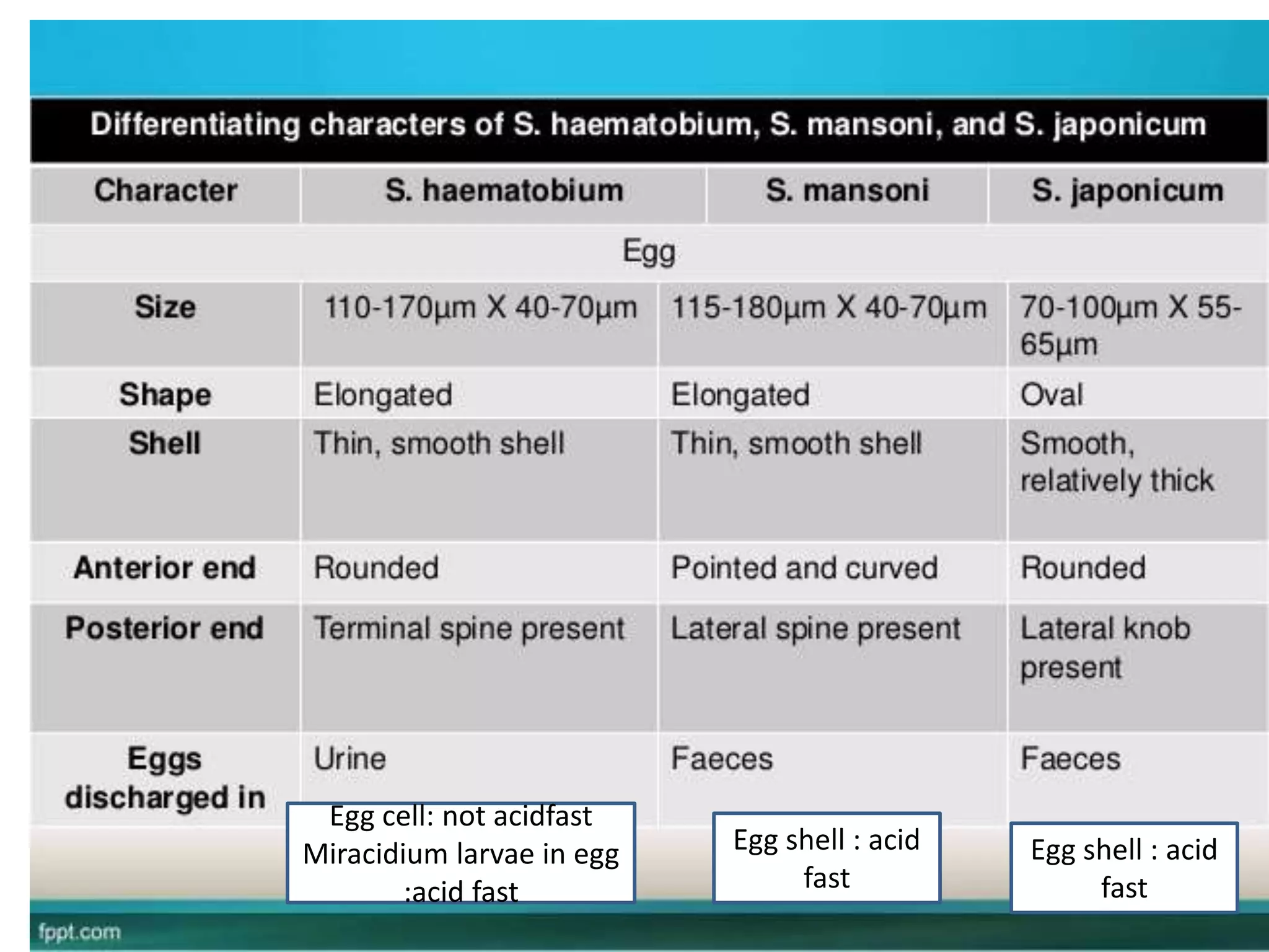
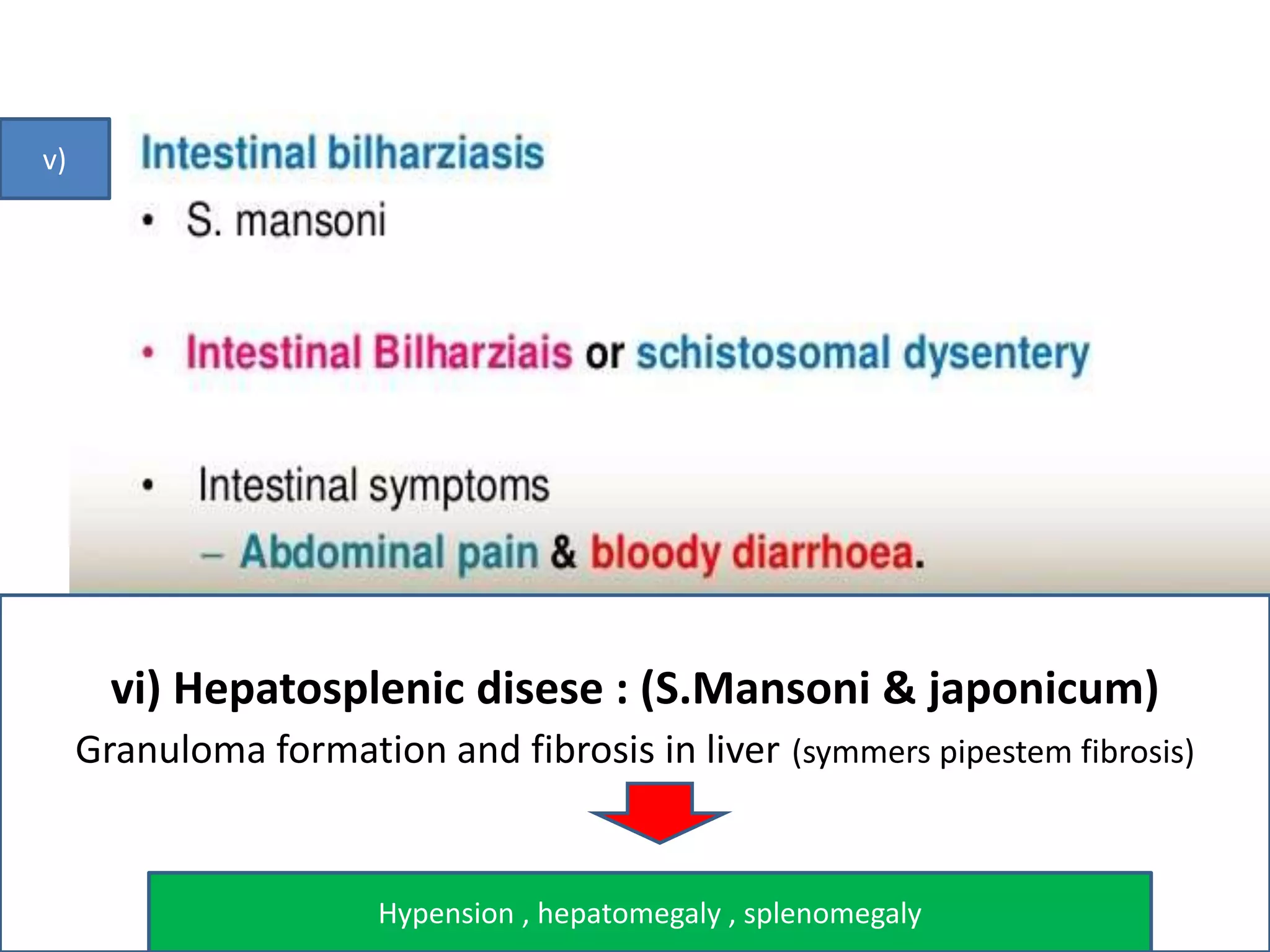
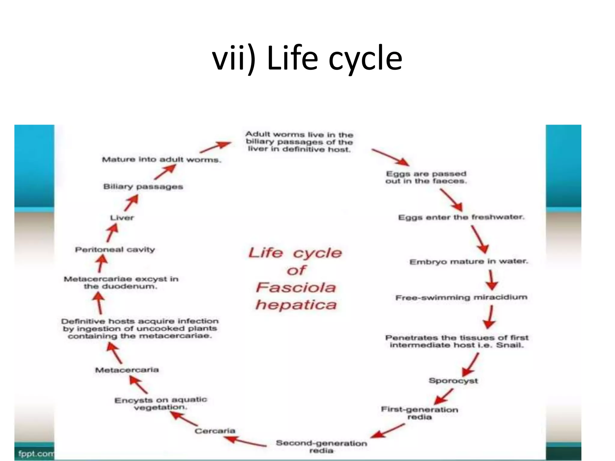
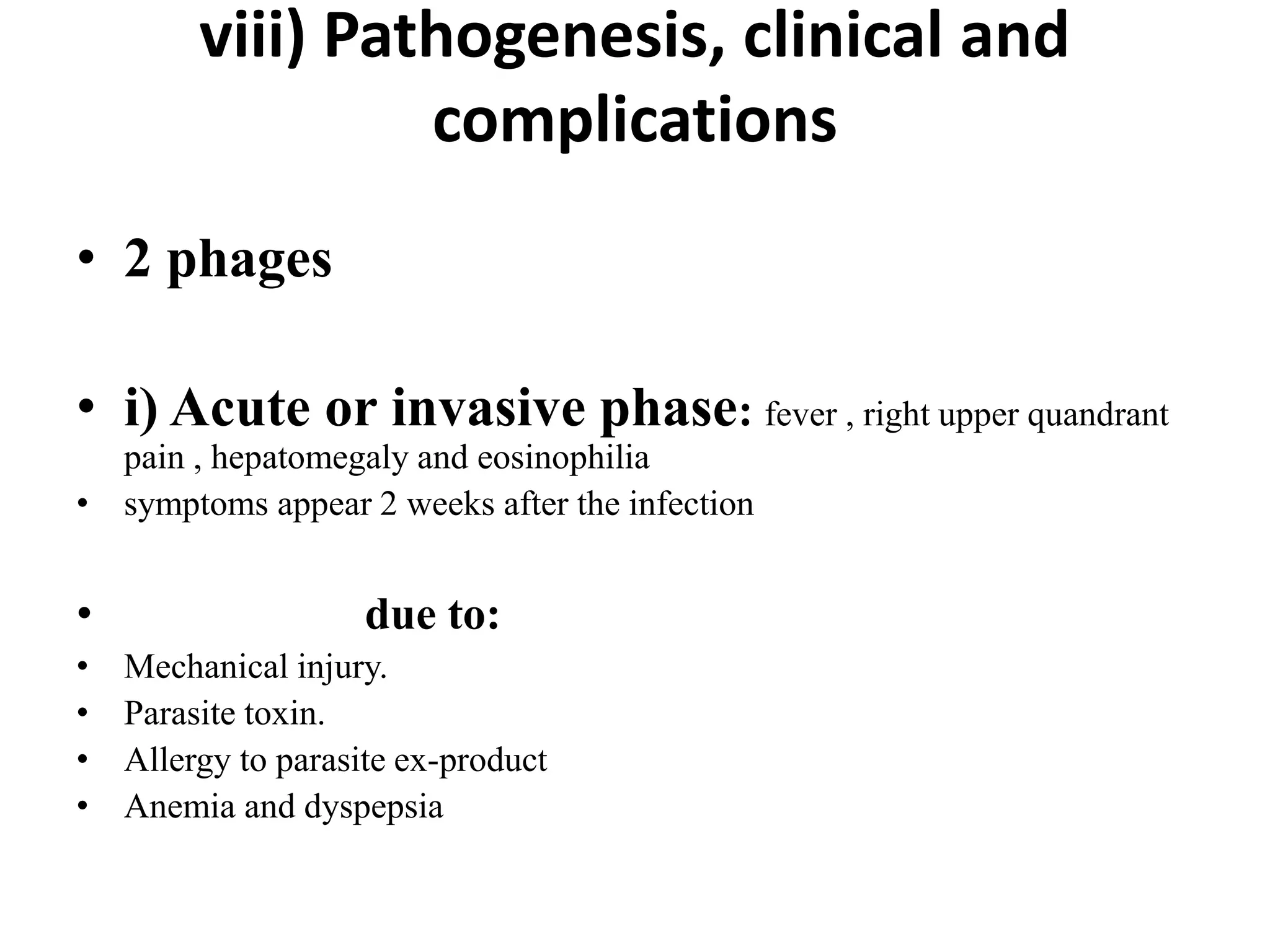
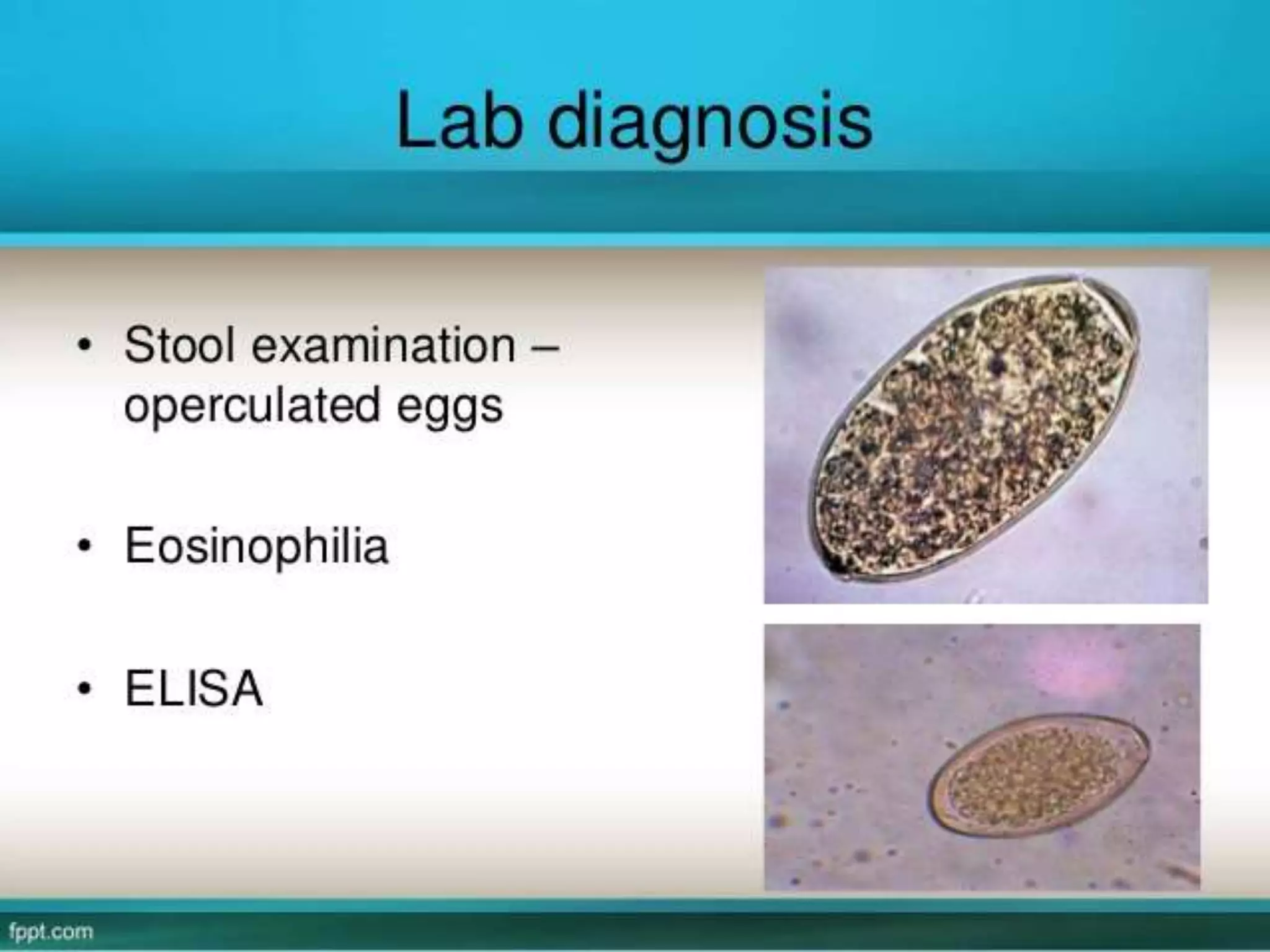
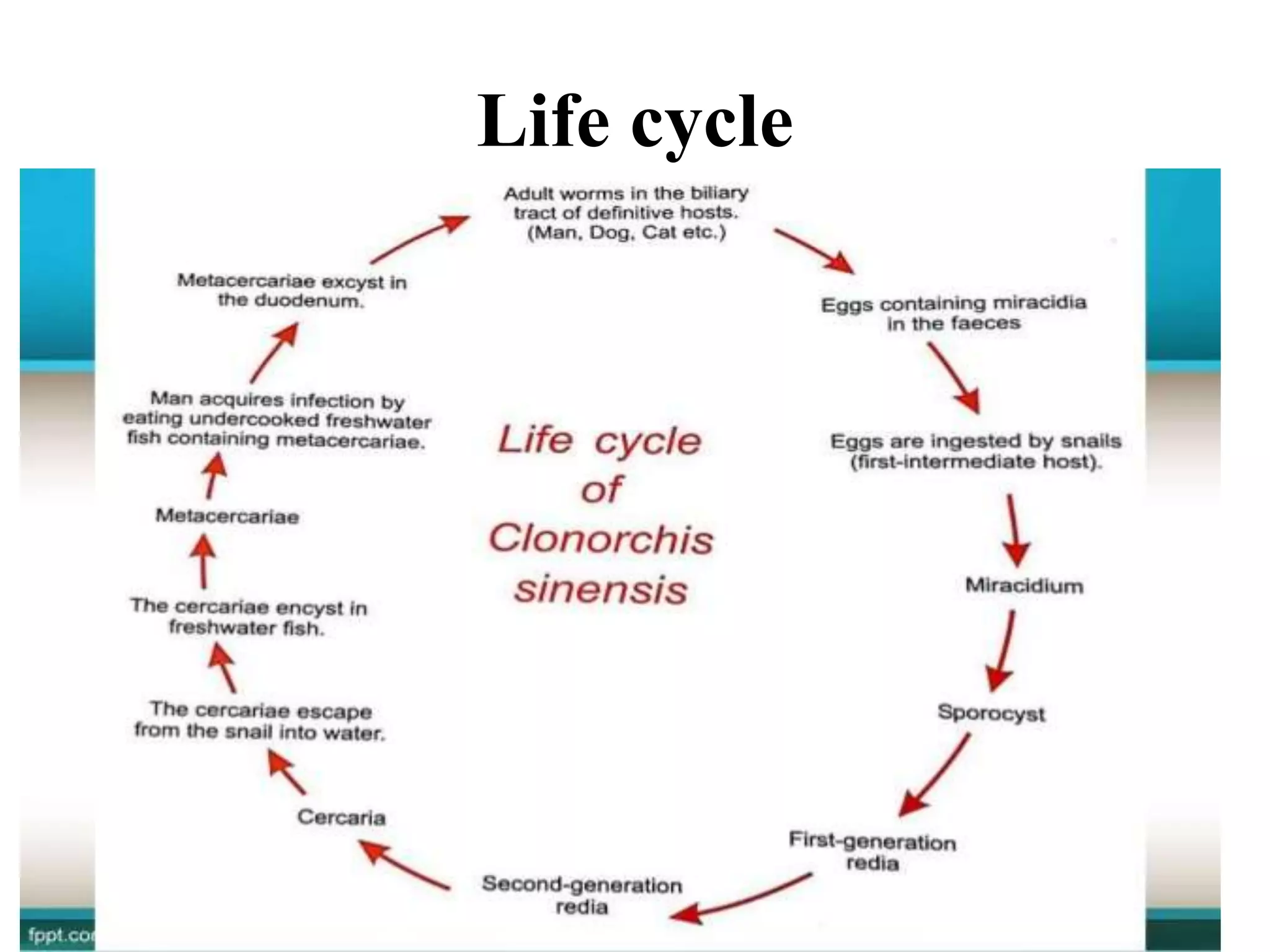
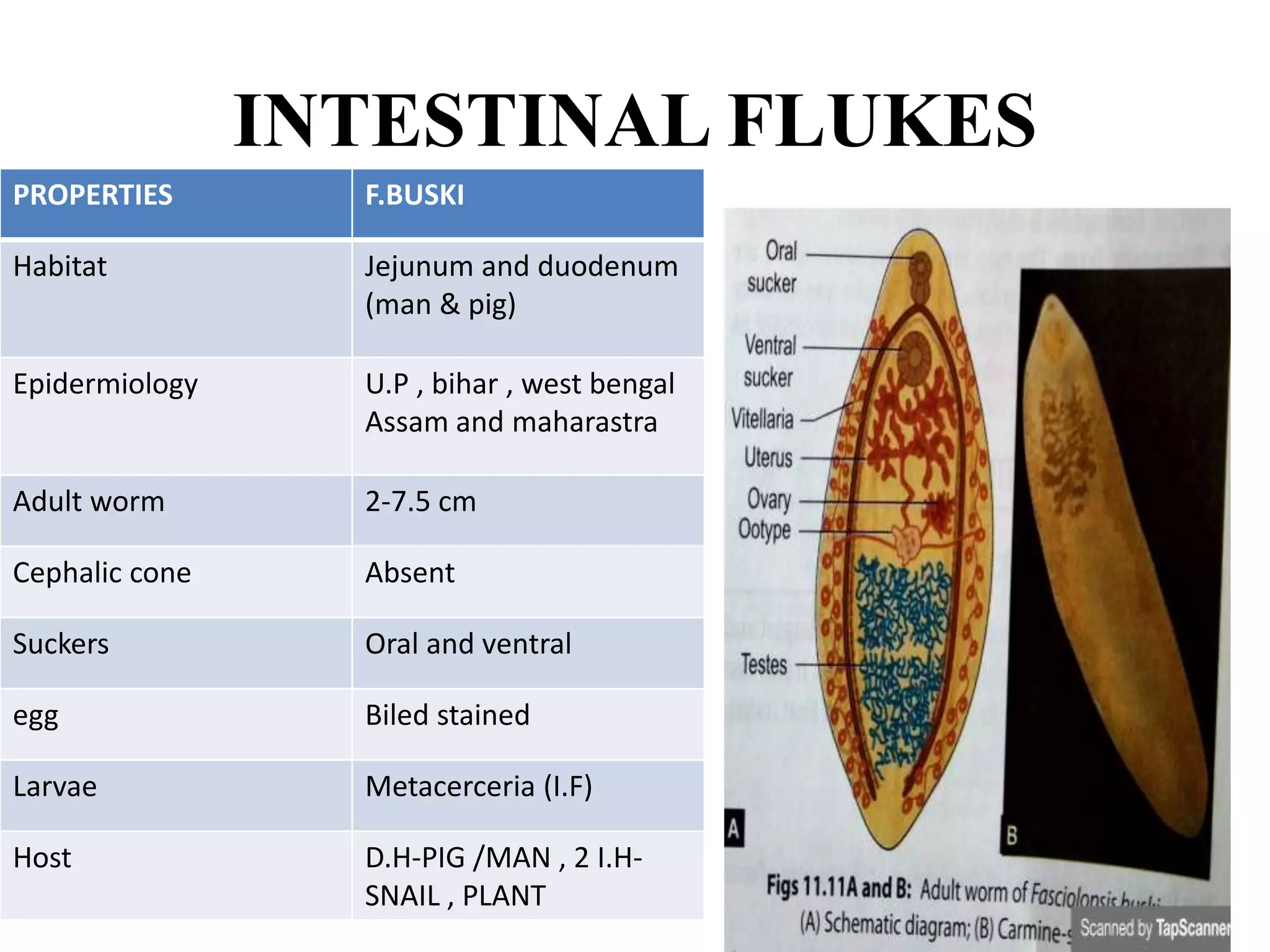
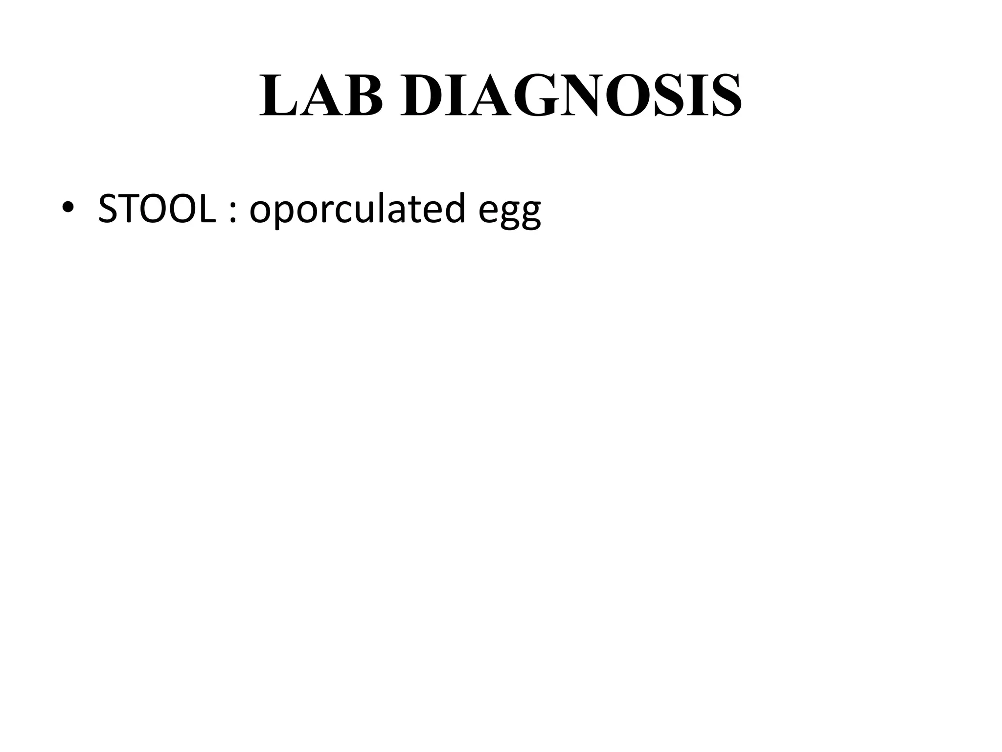
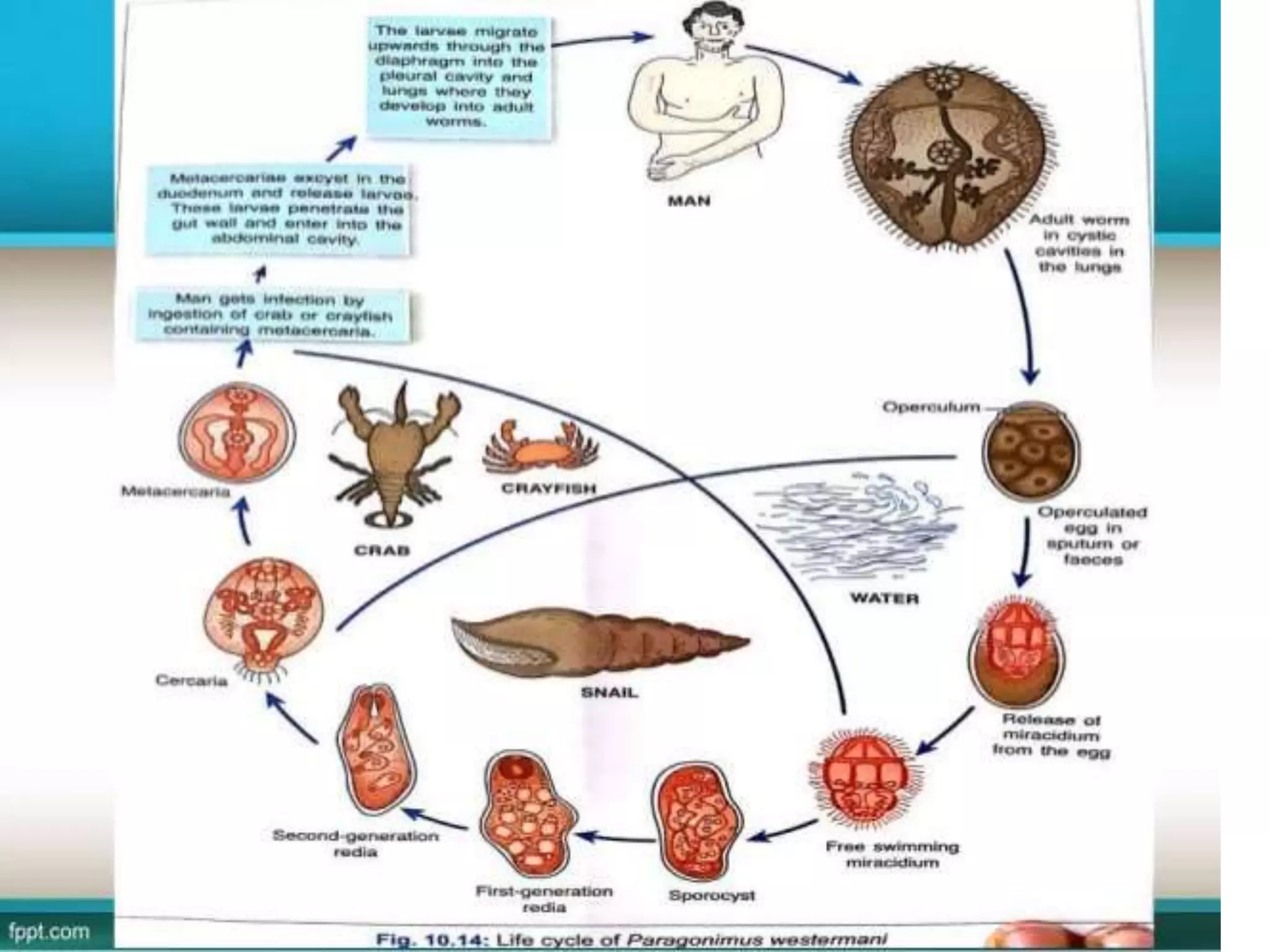
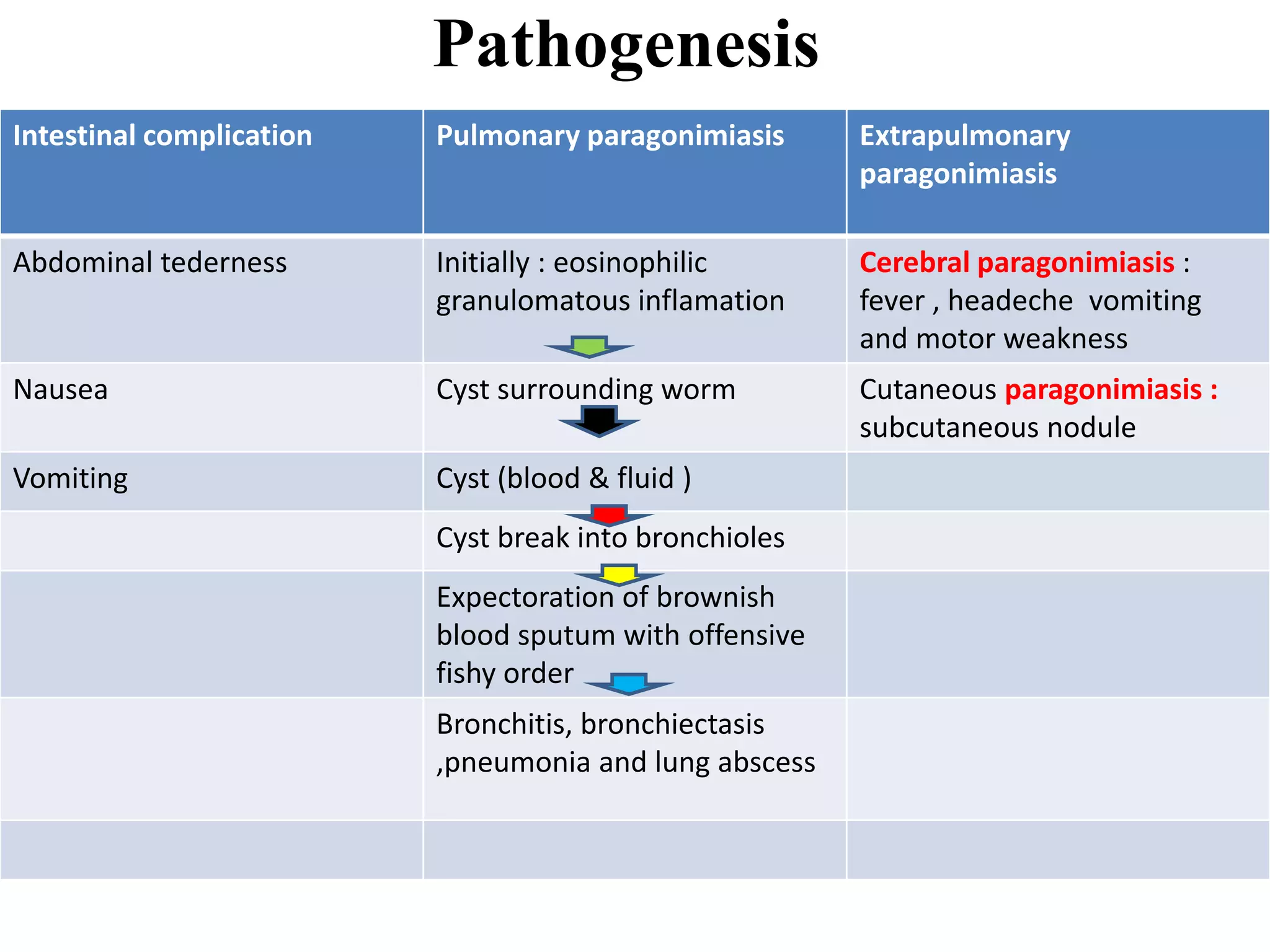
Trematodes : Flukes
• 1) INTRODUCTION
• Unsegemented helminthes
• Leaf of tree or flatfish (fluke)
• Trematode (suckers with hole in middle)
• Trematode : (trema : hole and eidos : appearance)
• I) Habitat classification :
3) General characters
• 1)Morphology : adult worm , egg and larva
• i) Adult worm: unsegmented , Flattened dorsoventrally.
• A) size : 1mm to 6mm
• B) suckers: oral (mouth) and ventral suckers
• SEX NOT SEPERATE (EXCEPT SCHISTOSOMES)
AC , NO
ANUS
EC
MRC
MRC
FRC
FRC
SCHISTOSOMES MALE AND FEMALE
• Ii)Egg: oviparous(egg larva)
• All operculated except schistosome (nonoperculated –urine (spine)
•
enviroment
• III) Larvae : 5-larval (miracidium , sporocyst , redia, cercaria and
metacerceria)
4)Lifecycle
• i) Definitive host :Mammals , humans or animals
• Ii) Intermediate host : freshwater molluscs or snail
• Second intermediate host : Fish or Crab (encystment)
• Schistosomes not need 2nd intermediate host
Rediae : 2nd generation
No rediae in schistosomes
Difference in life cycle of other trematodes and schistosomes
Metacerceria larvae (I.F in s.i.h -fish)
human by ingestion
lung, liver, intestine, bile duct
Adult worm
self fertilized (egg)
feses and sputum
miracidium larvae
Ingested by snail (1st i.h)
sporocyte
redia(1st & 2nd )
infective to man cercaria
metacercaria S.i.h (fish)
cerceria larvae (water)
Human Skin penetration
Enter venus circulation intestine &kidney
Adult worm
Cross fertilized
Egg in urine and stool
Miracisium in water
Snail (1st i.h)
Sporocyte (1st and 2nd )
Cercaria in water
Clonorchis
&
opisthorchis
5)MODE OF INFECTION
• i) Ingestion (cercaeria):
• A) Vegetables: F.Hepatica , F.buski , W.watsoni
• B) fish: C.cinensis , H.heterophyes , M.yokogawai
• C) fish of crab or crayfish: P.westermani
• Ii) skin penetration: S.haematobium , S.mansoni , S.japonicum
6) Blood Flukes
• A) Schistosomes :
• Sex seperate (dioecious)
• Schistossommiasis (water born disease)
• Africa , Latin America and Asia
• Schistosoma haematobium, S. japonicum, S. mansoni , S. mekongi and S.
intercalatum
• B) Order :strigeida
• C) Superfamily : schistosomatoidea
• D) Family : schistosomatidae
• E) Morphology: : adult worm , egg and larva
•
• Ii) eggs:
• 1) 70-180 micro m X 40-73 breadth
• 2) non operculated with spine
• S.haematobium :terminal
• S.mansoni: lateral
• S.japonicum: lateral rudimentary spine
• Iii) larva:miracidium , sporocyts (1st & 2nd ) &
cercaria (infective form )
Egg cell: not acidfast
Miracidium larvae in egg
:acid fast
Egg shell : acid
fast
Egg shell : acid
fast
f) Mechanism of Egg Expulsion
LAID
• EGG LAID enter into vesicle wall by spine
Small venus and pelvic
pelvic plexus ,
(mescentric portal
system ,pulmonary
arteries )
Release :
lytic
substance
passes
Lumen of urinary
bladder
Discharged :
urine
Skin –local
tissue
irritation
Egg –in feces
g) Life cycle of schistosomiasis
i)
Antigen from egg stimulate HMI Immune complex and serum sikness (c/d katyama
fever)
h) Pathogenesis and clinical features
Cercariae (skin penetration )
dermatitis , pruritis
Lung :cough with
fever
• a) urogenital disease : (s.hematobium)
• Symptomatic after 3 months
• EGG: pathogenic
• Egg urinary bladder (dysuria and hematuria)
D.Hypersensitivity
Urinary
bladder
Granuloma of
macrophage ,
lymphocyte
etc
Granuloma
join together
Granular
metaplasia
Heavy
infection
Elephanthasis
of scrotum
and penis
Important
pathogen in HIV
ii) Chronic schistosomiasis
• iii) Obstructive utropathies : (s.hematobium)
• Fibrosis
• iv) Bladder carcinoma: (s.hematobium) due to
metaplastic change
• i) predisposing factor :
• Diet containing nitroso compound (cheese , beans etc)
• Secondary bacterial infection
Obstruction
in lower
ureters
Hydroureter and
hydronephrosis
v)
vi) Hepatosplenic disese : (S.Mansoni & japonicum)
Granuloma formation and fibrosis in liver (symmers pipestem fibrosis)
Hypension , hepatomegaly , splenomegaly
• vii) cerebral schistosomiasis : s.japonicum
• 2-4% of cases
• Symptoms : jacksonian convulsion and grand male
fever
j) LABORATORY DIAGNOSIS
Urine microscopy
: urine and stool
(rarely) for
nonoporculated
egg
Collection time :
12pm to 3pm
Histopathology :
egg in bladder
mucosa biopsy
Ab detection in
serum
IgE & igG4 :
I) HAMA – FALCON
ASSAY SCREENING
TEST –ELISA
II) HAMA –EITB
(ENZYME LINKED
IMMUNOTRANSFER
BLOT
1) ANTIGEN
DETECTION : A)
Circulating
cathodic antige
(CCA) in serum
and urine by
ELISA
B) ELISA based
specific
monocolonal AB
against (M Ab –
SEA)
h) TREATMENT
• A) Praziquantel : 20 mg /kg/dose , two dose in
single day
• B) Metrifonate : inhibits acetylcholine
receptor on adult male worm . Multiple dose
for a week
7)Liver flukes
• 1)Fasciola hepatica and F.gigantica
• 2)Colonorchis sinensis
• 3)Opisthorichis species
• 4)Dicrocoelium dendriticum
1)Fasciola hepatica
• i) largest and most common in human(Greater liver fluke)
• Ii) Sheep (sheep liver fluke)
• Iii) G.D : Egypt
• Iv) host: Herbivorous animals and accidently man
• V)Habitat :liver and bile duct
• Vi) Disease: Fascioliasis(Liver Rot)
• Vii) Morphology :
• A) Adult:
• Leaf
• length 30mm long and 15mm
broad.
• Anteriorly (conical) and
posteriorly (round)
• Testes: Two
• Ovary: One
• B) Eggs:
• 140umx80um
• Oval
• Operculated
• immature embryo
• Bile stained
• C) larva:
• metacercaria (infective form)
• Miracidia
• Rediae
• Sporocyst
vii) Life cycle
viii) Pathogenesis, clinical and
complications
• 2 phages
• i) Acute or invasive phase: fever , right upper quandrant
pain , hepatomegaly and eosinophilia
• symptoms appear 2 weeks after the infection
• due to:
• Mechanical injury.
• Parasite toxin.
• Allergy to parasite ex-product
• Anemia and dyspepsia
• Ii) Chronic stage:
• maturing worm in the bile duct
• A) Ectopic fascioliasis
• In lung
• Subcutaneous tissue
• Rare central nervous system(rare)
Cholecystitis Cholelithiasis
,Jaundice , Liver fibrosis
• B) Halzoun (marrare sybdrome)
• Uncooked meat of sheep goat
• form of acute laryngopharyngitis.
2)
• Eggs :
• flask shaped
• Operculum (prominent shoulder and
common shaped knob)
• Larvae :
• metacercaria (infective form)
• Miracidia
• Rediae
• Sporocyst
• Mode of transmission
• Eating of frozen, dried or pickled fish (metacercariae).
Life cycle
Pathogenesis
• cholangitis.
• Cholangiocarcinoma.
• Calculus formation.
• Few cases :
• Biliary cirrhosis.
• Portalhypertension.
• Clinical Picture :
• Fever
• epigastric pain
• diarrhea
• tender hepatomegaly
• biliary colic
• obstructive jaundice.
Laboratory diagnosis
INTESTINAL FLUKES
PROPERTIES F.BUSKI
Habitat Jejunum and duodenum
(man & pig)
Epidermiology U.P , bihar , west bengal
Assam and maharastra
Adult worm 2-7.5 cm
Cephalic cone Absent
Suckers Oral and ventral
egg Biled stained
Larvae Metacerceria (I.F)
Host D.H-PIG /MAN , 2 I.H-
SNAIL , PLANT
PATHOGRNESIS
MALABSORPTION WITH YELLOWISH GREEN COLOUR STOOL
LAB DIAGNOSIS
• STOOL : oporculated egg
Lung fluke (paragonium
westermani/oriental lung fluke)
Properties paragonium westermani
Epidermiology Manipur , north east states of india
Habitat Paranchyma of lung
Adult worm Reddish brown , broad anterior end ,
Excretory bladder : divided body into 2
part
egg Oval , golden brown, 80-120 micro m X 45-
65 micro m
larvae Metacerceria (I.F) ,
Host D.H- (Man , dog , cat) , 2 I.H – (Snail , crab)
Pathogenesis
Intestinal complication Pulmonary paragonimiasis Extrapulmonary
paragonimiasis
Abdominal tederness Initially : eosinophilic
granulomatous inflamation
Cerebral paragonimiasis :
fever , headeche vomiting
and motor weakness
Nausea Cyst surrounding worm Cutaneous paragonimiasis :
subcutaneous nodule
Vomiting Cyst (blood & fluid )
Cyst break into bronchioles
Expectoration of brownish
blood sputum with offensive
fishy order
Bronchitis, bronchiectasis
,pneumonia and lung abscess
Laboratory diagnosis
1) Sputum
microscopy : early
morning sample mount
with saline (operculated
egg)
2) Formal ether
method
3) Stool
microscope in
children (sputum difficult
to collect)
Serology :
1) Ab detection :
a) Rapid test : dot immunogold
filtration assay , ELISA (IgG
or IgE)
2) Antigen detection : dot
ELISA (ACTIVE INFECTION)
MRI , CT scan (cyst
in CNS)
Trematodes

Trematodes

  • 2.
    Trematodes : Flukes •1) INTRODUCTION • Unsegemented helminthes • Leaf of tree or flatfish (fluke) • Trematode (suckers with hole in middle) • Trematode : (trema : hole and eidos : appearance)
  • 3.
    • I) Habitatclassification :
  • 4.
    3) General characters •1)Morphology : adult worm , egg and larva • i) Adult worm: unsegmented , Flattened dorsoventrally. • A) size : 1mm to 6mm • B) suckers: oral (mouth) and ventral suckers • SEX NOT SEPERATE (EXCEPT SCHISTOSOMES)
  • 5.
  • 6.
  • 7.
    • Ii)Egg: oviparous(egglarva) • All operculated except schistosome (nonoperculated –urine (spine) • enviroment
  • 8.
    • III) Larvae: 5-larval (miracidium , sporocyst , redia, cercaria and metacerceria)
  • 9.
    4)Lifecycle • i) Definitivehost :Mammals , humans or animals • Ii) Intermediate host : freshwater molluscs or snail • Second intermediate host : Fish or Crab (encystment) • Schistosomes not need 2nd intermediate host
  • 10.
    Rediae : 2ndgeneration No rediae in schistosomes
  • 11.
    Difference in lifecycle of other trematodes and schistosomes Metacerceria larvae (I.F in s.i.h -fish) human by ingestion lung, liver, intestine, bile duct Adult worm self fertilized (egg) feses and sputum miracidium larvae Ingested by snail (1st i.h) sporocyte redia(1st & 2nd ) infective to man cercaria metacercaria S.i.h (fish) cerceria larvae (water) Human Skin penetration Enter venus circulation intestine &kidney Adult worm Cross fertilized Egg in urine and stool Miracisium in water Snail (1st i.h) Sporocyte (1st and 2nd ) Cercaria in water Clonorchis & opisthorchis
  • 12.
    5)MODE OF INFECTION •i) Ingestion (cercaeria): • A) Vegetables: F.Hepatica , F.buski , W.watsoni • B) fish: C.cinensis , H.heterophyes , M.yokogawai • C) fish of crab or crayfish: P.westermani • Ii) skin penetration: S.haematobium , S.mansoni , S.japonicum
  • 13.
    6) Blood Flukes •A) Schistosomes : • Sex seperate (dioecious) • Schistossommiasis (water born disease) • Africa , Latin America and Asia • Schistosoma haematobium, S. japonicum, S. mansoni , S. mekongi and S. intercalatum
  • 14.
    • B) Order:strigeida • C) Superfamily : schistosomatoidea • D) Family : schistosomatidae • E) Morphology: : adult worm , egg and larva •
  • 15.
    • Ii) eggs: •1) 70-180 micro m X 40-73 breadth • 2) non operculated with spine • S.haematobium :terminal • S.mansoni: lateral • S.japonicum: lateral rudimentary spine
  • 16.
    • Iii) larva:miracidium, sporocyts (1st & 2nd ) & cercaria (infective form )
  • 19.
    Egg cell: notacidfast Miracidium larvae in egg :acid fast Egg shell : acid fast Egg shell : acid fast
  • 20.
    f) Mechanism ofEgg Expulsion LAID • EGG LAID enter into vesicle wall by spine Small venus and pelvic pelvic plexus , (mescentric portal system ,pulmonary arteries ) Release : lytic substance passes Lumen of urinary bladder Discharged : urine Skin –local tissue irritation Egg –in feces
  • 21.
    g) Life cycleof schistosomiasis
  • 22.
    i) Antigen from eggstimulate HMI Immune complex and serum sikness (c/d katyama fever) h) Pathogenesis and clinical features Cercariae (skin penetration ) dermatitis , pruritis Lung :cough with fever
  • 23.
    • a) urogenitaldisease : (s.hematobium) • Symptomatic after 3 months • EGG: pathogenic • Egg urinary bladder (dysuria and hematuria) D.Hypersensitivity Urinary bladder Granuloma of macrophage , lymphocyte etc Granuloma join together Granular metaplasia Heavy infection Elephanthasis of scrotum and penis Important pathogen in HIV ii) Chronic schistosomiasis
  • 24.
    • iii) Obstructiveutropathies : (s.hematobium) • Fibrosis • iv) Bladder carcinoma: (s.hematobium) due to metaplastic change • i) predisposing factor : • Diet containing nitroso compound (cheese , beans etc) • Secondary bacterial infection Obstruction in lower ureters Hydroureter and hydronephrosis
  • 25.
    v) vi) Hepatosplenic disese: (S.Mansoni & japonicum) Granuloma formation and fibrosis in liver (symmers pipestem fibrosis) Hypension , hepatomegaly , splenomegaly
  • 26.
    • vii) cerebralschistosomiasis : s.japonicum • 2-4% of cases • Symptoms : jacksonian convulsion and grand male fever
  • 27.
    j) LABORATORY DIAGNOSIS Urinemicroscopy : urine and stool (rarely) for nonoporculated egg Collection time : 12pm to 3pm Histopathology : egg in bladder mucosa biopsy Ab detection in serum IgE & igG4 : I) HAMA – FALCON ASSAY SCREENING TEST –ELISA II) HAMA –EITB (ENZYME LINKED IMMUNOTRANSFER BLOT 1) ANTIGEN DETECTION : A) Circulating cathodic antige (CCA) in serum and urine by ELISA B) ELISA based specific monocolonal AB against (M Ab – SEA)
  • 28.
    h) TREATMENT • A)Praziquantel : 20 mg /kg/dose , two dose in single day • B) Metrifonate : inhibits acetylcholine receptor on adult male worm . Multiple dose for a week
  • 29.
    7)Liver flukes • 1)Fasciolahepatica and F.gigantica • 2)Colonorchis sinensis • 3)Opisthorichis species • 4)Dicrocoelium dendriticum
  • 30.
    1)Fasciola hepatica • i)largest and most common in human(Greater liver fluke) • Ii) Sheep (sheep liver fluke) • Iii) G.D : Egypt • Iv) host: Herbivorous animals and accidently man • V)Habitat :liver and bile duct • Vi) Disease: Fascioliasis(Liver Rot)
  • 31.
    • Vii) Morphology: • A) Adult: • Leaf • length 30mm long and 15mm broad. • Anteriorly (conical) and posteriorly (round) • Testes: Two • Ovary: One
  • 32.
    • B) Eggs: •140umx80um • Oval • Operculated • immature embryo • Bile stained
  • 33.
    • C) larva: •metacercaria (infective form) • Miracidia • Rediae • Sporocyst
  • 34.
  • 36.
    viii) Pathogenesis, clinicaland complications • 2 phages • i) Acute or invasive phase: fever , right upper quandrant pain , hepatomegaly and eosinophilia • symptoms appear 2 weeks after the infection • due to: • Mechanical injury. • Parasite toxin. • Allergy to parasite ex-product • Anemia and dyspepsia
  • 37.
    • Ii) Chronicstage: • maturing worm in the bile duct • A) Ectopic fascioliasis • In lung • Subcutaneous tissue • Rare central nervous system(rare) Cholecystitis Cholelithiasis ,Jaundice , Liver fibrosis
  • 38.
    • B) Halzoun(marrare sybdrome) • Uncooked meat of sheep goat • form of acute laryngopharyngitis.
  • 40.
  • 41.
    • Eggs : •flask shaped • Operculum (prominent shoulder and common shaped knob)
  • 42.
    • Larvae : •metacercaria (infective form) • Miracidia • Rediae • Sporocyst
  • 43.
    • Mode oftransmission • Eating of frozen, dried or pickled fish (metacercariae).
  • 44.
  • 46.
    Pathogenesis • cholangitis. • Cholangiocarcinoma. •Calculus formation. • Few cases : • Biliary cirrhosis. • Portalhypertension.
  • 47.
    • Clinical Picture: • Fever • epigastric pain • diarrhea • tender hepatomegaly • biliary colic • obstructive jaundice.
  • 48.
  • 49.
    INTESTINAL FLUKES PROPERTIES F.BUSKI HabitatJejunum and duodenum (man & pig) Epidermiology U.P , bihar , west bengal Assam and maharastra Adult worm 2-7.5 cm Cephalic cone Absent Suckers Oral and ventral egg Biled stained Larvae Metacerceria (I.F) Host D.H-PIG /MAN , 2 I.H- SNAIL , PLANT
  • 50.
  • 51.
    LAB DIAGNOSIS • STOOL: oporculated egg
  • 52.
    Lung fluke (paragonium westermani/orientallung fluke) Properties paragonium westermani Epidermiology Manipur , north east states of india Habitat Paranchyma of lung Adult worm Reddish brown , broad anterior end , Excretory bladder : divided body into 2 part egg Oval , golden brown, 80-120 micro m X 45- 65 micro m larvae Metacerceria (I.F) , Host D.H- (Man , dog , cat) , 2 I.H – (Snail , crab)
  • 55.
    Pathogenesis Intestinal complication Pulmonaryparagonimiasis Extrapulmonary paragonimiasis Abdominal tederness Initially : eosinophilic granulomatous inflamation Cerebral paragonimiasis : fever , headeche vomiting and motor weakness Nausea Cyst surrounding worm Cutaneous paragonimiasis : subcutaneous nodule Vomiting Cyst (blood & fluid ) Cyst break into bronchioles Expectoration of brownish blood sputum with offensive fishy order Bronchitis, bronchiectasis ,pneumonia and lung abscess
  • 56.
    Laboratory diagnosis 1) Sputum microscopy: early morning sample mount with saline (operculated egg) 2) Formal ether method 3) Stool microscope in children (sputum difficult to collect) Serology : 1) Ab detection : a) Rapid test : dot immunogold filtration assay , ELISA (IgG or IgE) 2) Antigen detection : dot ELISA (ACTIVE INFECTION) MRI , CT scan (cyst in CNS)