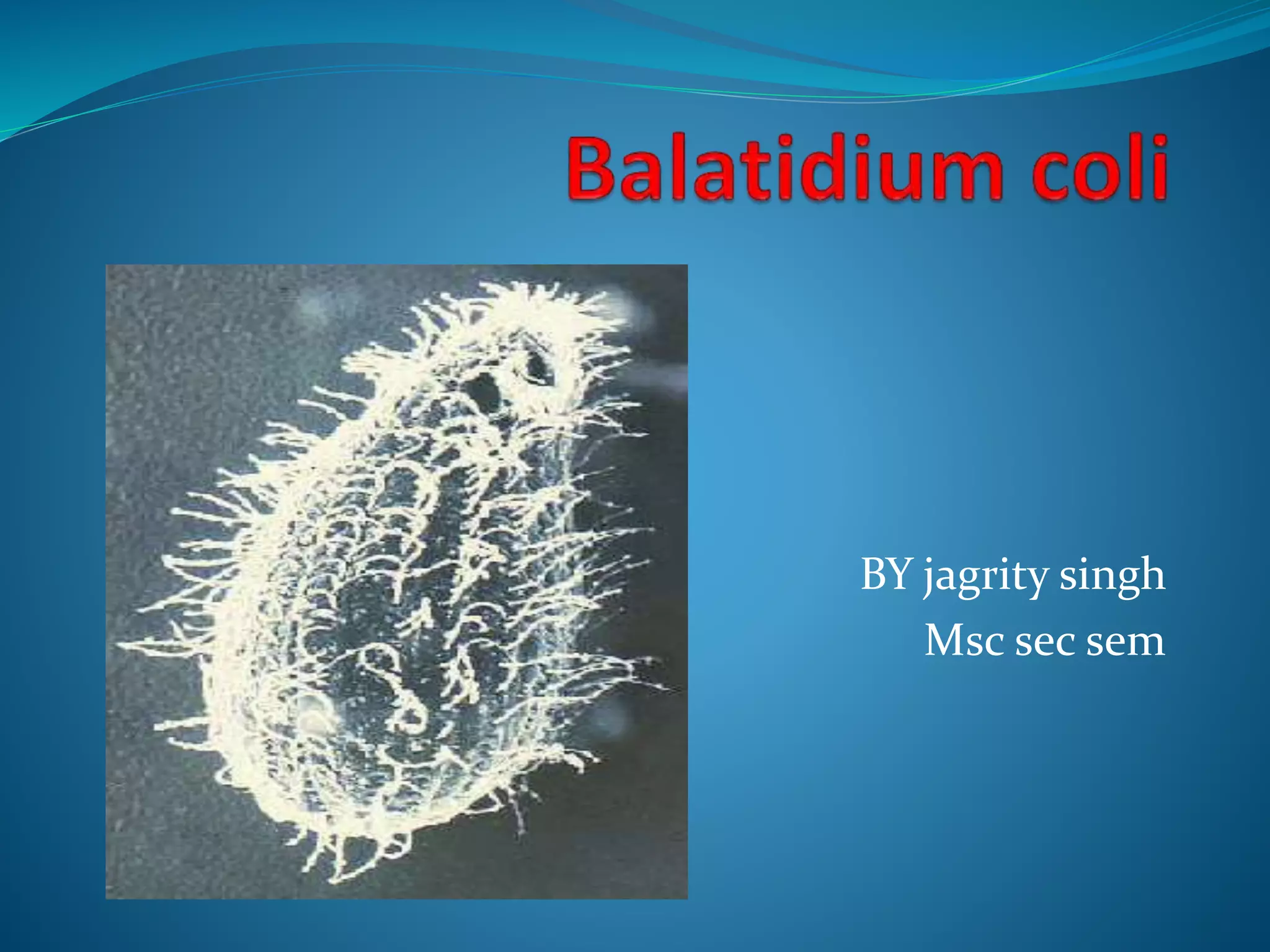
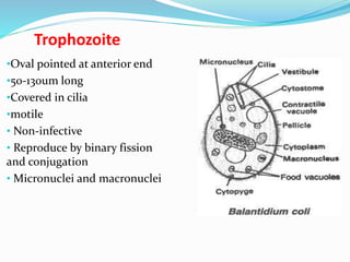
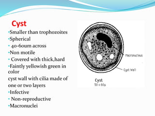
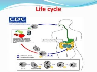
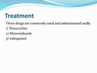
BY jagrity singh
Msc sec sem
Scientific classification
 Domain: Eukaryota
 Phylum: Ciliophora (Doflein, 1901)
 Class: Kinetofragminophorea (De Puytorac et al., 1974)
 Order: Vestibuliferida
 Family: Balantiididae
 Genus: Balantidium
 Species: B. coli (Malmsten, 1857)
 A parasitic species of ciliate protozoan that causes the
disease Balantidiasis.
 Balantidium coli is the largest protozoan and the only
ciliate known to parasitize humans.
Geographical distributation
 The protozoa are found worldwide
 Bolivia
 Papua New Guinea
 Philippines
Trophozoite
•Oval pointed at anterior end
•50-130um long
•Covered in cilia
•motile
• Non-infective
• Reproduce by binary fission
and conjugation
• Micronuclei and macronuclei
Cyst
•Smaller than trophozoites
•Spherical
• 40-60um across
•Non motile
• Covered with thick,hard
•Faintly yellowish green in
color
cyst wall with cilia made of
one or two layers
•Infective
• Non-reproductive
•Macronuclei
Transmission
 Fecal-oral route
 Eating meat, fruits, and vegetables that have been
 contaminated by an infected person or contaminated
by fecal matter from an infected animal
 Drinking and washing food with contaminated water
 Having poor hygiene habits
Life cycle
 Life Cycle Completed in a single host Natural hosts
 Pigs Accidental host
 Route : Ingestion
 Reproduction: asexual and sexual
Life cycle
Life cycle
 Balantidium coli has 2 developmental stages: a
trophozoite stage and a cyst stage.
 The cyst is the infective stage of Balantidium coli life
cycle.
 Once the cyst is ingested via feces-contaminated food
or water, it passes through the host digestive system.
 There, excystation takes place in small intestine.
 Excystation produces a trophozoite from the cyst
stage.
Life Cycle
 The motile trophozoite then resides in the lumen of the large
intestine, feeding on intestinal nutrients.
 Trophozoites multiply by asexual binary fission or sexual
conjugation.
 The trophozoite may become invasive and penetrate the
mucosa of the large intestine.
 Trophozoites are released with the feces, and encyst to form
new cyst.
 Encystation takes place in the rectum of the host as
 feces are dehydrated or soon after the feces have been excreted.
 Cysts in the environment are then ready to infect another host.
Clinical Presentation of Balantidiasis
 Trophozoites can invade the mucosa of the large
intestine (cecum and colon) and cause ulcerations.
 The parasite secretes a substance called hyaluronidase
enzyme, which helps degrade intestinal tissue and
facilitates penetration of the mucosa.
 Other bacteria in the intestine may enter the ulcer
along with Balantidium coli, leading to secondary
infections. Ulcerations of the large intestine can be
viewed using sigmoidoscopy
symptoms
 Acute ,even hemorrhagic
 Diarrhea
 Ulceration to gut wall
 Dysentery
 Colitis
 Abdominal pain
Epidemiology
 Balantidiosis is most often found in tropical regions
throughout the world ,It is not a common human
disease;
 The infection rate is less than 1% ,The parasite is
nonpathogenic in pigs and is much more prevalent
(20-100%) among these hosts.
 Pigs are a good source of infection for humans in areas
where they share habitation.
Diagnosis
 Examination of patient`s stool
 A stool sample is collected and a wet mount is
prepared
 Biopsy
 sigmoid scope is used to visually inspect the last
sections of the large intestine
unstained trichrome eosin
Treatment
Three drugs are commonly used and administered orally
1) Tetracycline
2) Metronidazole
3) Iodoquinol

Balatidium coli

  • 1.
  • 2.
    Scientific classification  Domain:Eukaryota  Phylum: Ciliophora (Doflein, 1901)  Class: Kinetofragminophorea (De Puytorac et al., 1974)  Order: Vestibuliferida  Family: Balantiididae  Genus: Balantidium  Species: B. coli (Malmsten, 1857)
  • 3.
     A parasiticspecies of ciliate protozoan that causes the disease Balantidiasis.  Balantidium coli is the largest protozoan and the only ciliate known to parasitize humans.
  • 4.
    Geographical distributation  Theprotozoa are found worldwide  Bolivia  Papua New Guinea  Philippines
  • 5.
    Trophozoite •Oval pointed atanterior end •50-130um long •Covered in cilia •motile • Non-infective • Reproduce by binary fission and conjugation • Micronuclei and macronuclei
  • 6.
    Cyst •Smaller than trophozoites •Spherical •40-60um across •Non motile • Covered with thick,hard •Faintly yellowish green in color cyst wall with cilia made of one or two layers •Infective • Non-reproductive •Macronuclei
  • 7.
    Transmission  Fecal-oral route Eating meat, fruits, and vegetables that have been  contaminated by an infected person or contaminated by fecal matter from an infected animal  Drinking and washing food with contaminated water  Having poor hygiene habits
  • 8.
    Life cycle  LifeCycle Completed in a single host Natural hosts  Pigs Accidental host  Route : Ingestion  Reproduction: asexual and sexual
  • 9.
  • 10.
    Life cycle  Balantidiumcoli has 2 developmental stages: a trophozoite stage and a cyst stage.  The cyst is the infective stage of Balantidium coli life cycle.  Once the cyst is ingested via feces-contaminated food or water, it passes through the host digestive system.  There, excystation takes place in small intestine.  Excystation produces a trophozoite from the cyst stage.
  • 11.
    Life Cycle  Themotile trophozoite then resides in the lumen of the large intestine, feeding on intestinal nutrients.  Trophozoites multiply by asexual binary fission or sexual conjugation.  The trophozoite may become invasive and penetrate the mucosa of the large intestine.  Trophozoites are released with the feces, and encyst to form new cyst.  Encystation takes place in the rectum of the host as  feces are dehydrated or soon after the feces have been excreted.  Cysts in the environment are then ready to infect another host.
  • 12.
    Clinical Presentation ofBalantidiasis  Trophozoites can invade the mucosa of the large intestine (cecum and colon) and cause ulcerations.  The parasite secretes a substance called hyaluronidase enzyme, which helps degrade intestinal tissue and facilitates penetration of the mucosa.  Other bacteria in the intestine may enter the ulcer along with Balantidium coli, leading to secondary infections. Ulcerations of the large intestine can be viewed using sigmoidoscopy
  • 13.
    symptoms  Acute ,evenhemorrhagic  Diarrhea  Ulceration to gut wall  Dysentery  Colitis  Abdominal pain
  • 14.
    Epidemiology  Balantidiosis ismost often found in tropical regions throughout the world ,It is not a common human disease;  The infection rate is less than 1% ,The parasite is nonpathogenic in pigs and is much more prevalent (20-100%) among these hosts.  Pigs are a good source of infection for humans in areas where they share habitation.
  • 15.
    Diagnosis  Examination ofpatient`s stool  A stool sample is collected and a wet mount is prepared  Biopsy  sigmoid scope is used to visually inspect the last sections of the large intestine
  • 16.
  • 17.
    Treatment Three drugs arecommonly used and administered orally 1) Tetracycline 2) Metronidazole 3) Iodoquinol