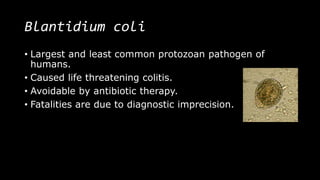
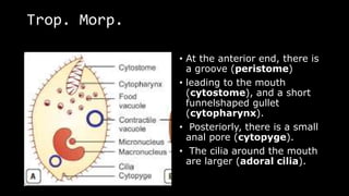
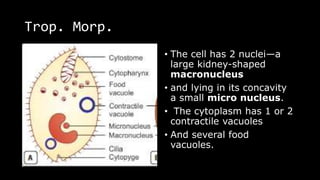
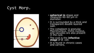
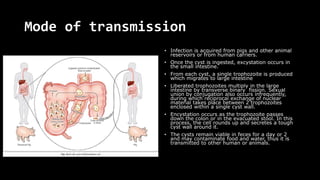
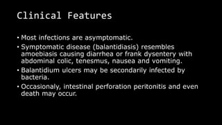
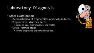
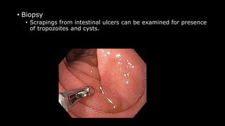
BALANTIDIUM COLI
By, Leyon Selvin Raj,
Tutor,
BN Patel institute of paramedical & Science, (Paramedical Division).
Blantidium coli
• Largest and least common protozoan pathogen of
humans.
• Caused life threatening colitis.
• Avoidable by antibiotic therapy.
• Fatalities are due to diagnostic imprecision.
Epidemiology
• Most commonly reported in
• Tropical and sub tropical
• Particularly central and south America
• Iran, Papu New Guinea and Philippines
• Prevalence is less than 1%
• Found in many mammals other than humans
• Pigs and monkeys- especially
• Swine- imp. Animal reservoir
*The largest reported balantidiasis, involves 110
persons, resulted from gross contamination of ground
and surface water supplies by pig feces after a severe
typhoon.
Morphology
• B. coli occurs in two stages
• Trophozoite
• Cyst
Trophozoite
• Lives in large intestine.
• Feeding on cell debris, bacteria, starch grains, and
other particles.
• Motile – found in dysenteric stool.
• Large ovoid cell. 60-70µm in length and 40-50µm in
breath. (200µm-large one)
Trop. Morp.
• At the anterior end, there is
a groove (peristome)
• leading to the mouth
(cytostome), and a short
funnelshaped gullet
(cytopharynx).
• Posteriorly, there is a small
anal pore (cytopyge).
• The cilia around the mouth
are larger (adoral cilia).
Trop. Morp.
• The cell has 2 nuclei—a
large kidney-shaped
macronucleus
• and lying in its concavity
a small micro nucleus.
• The cytoplasm has 1 or 2
contractile vacuoles
• And several food
vacuoles.
Cyst Morp.
• spherical in shape and
measures 40–60 μm in
diameter.
• It is surrounded by a thick and
transparent double layered
wall.
• The cytoplasm is granular.
Macronucleus, micro nucleus,
and vacuoles are also present
in the cyst.
• The cyst is the infective
stage of B. coli.
• It is found in chronic cases
and carriers.
Life cycle
• B. coli passes its life cycle in one host only
(monoxenous).
• Natural host: Pig
• Accidental host: Man.
• Reservoirs: Pig, monkey, and rat.
• Infective form: Cyst.
Mode of transmission
• Balantidiasis is a zoonosis. Human beings acquire infection by ingestion of
food and water contaminated with feces containing the cysts of B. coli.
Mode of transmission
• Infection is acquired from pigs and other animal
reservoirs or from human carriers.
• Once the cyst is ingested, excystation occurs in
the small intestine.
• From each cyst, a single trophozoite is produced
which migrates to large intestine
• Liberated trophozoites multiply in the large
intestine by transverse binary fission. Sexual
union by conjugation also occurs infrequently,
during which reciprocal exchange of nuclear
material takes place between 2 trophozoites
enclosed within a single cyst wall.
• Encystation occurs as the trophozoite passes
down the colon or in the evacuated stool. In this
process, the cell rounds up and secretes a tough
cyst wall around it.
• The cysts remain viable in feces for a day or 2
and may contaminate food and water, thus it is
transmitted to other human or animals.
Pathogenesis
• In Healthy individual, B. coli lives as lumen commensal
and is asymptomatic.
• Clinical disease occurs only when the resistance of host
is lowered.
• Trophozoites burrow into the intestinal mucosa, set up
colonies, and initiate inflammatory reaction.
• Leads to mucosal ulcers and submucosal abcesses,
resembling lesions in amoebiasis.
• They don’t invade liver, like E. histolytica,
Clinical Features
• Most infections are asymptomatic.
• Symptomatic disease (balantidiasis) resembles
amoebiasis causing diarrhea or frank dysentery with
abdominal colic, tenesmus, nausea and vomiting.
• Balantidium ulcers may be secondarily infected by
bacteria.
• Occasionaly, intestinal perforation peritonitis and even
death may occur.
Laboratory Diagnosis
• Stool Examination
• Demonstration of trophozoites and cysts in feces.
• Trophozoites- diarrheic feces
• Large in size, macronucleus, and motile
• Cysts- formed stool
• Round shape and large macronucleus.
• Biopsy
• Scrapings from intestinal ulcers can be examined for presence
of tropozoites and cysts.
• Culture
• B. coli – Locke’s egg albumin medium or NIH polyxenic
mrdium
• *rarely needed
Treatment
• Tetracycline- 500mg 4times/day for 10 days
• Alternative
• Doxycycline
Prevention (prophylaxis)
• Avoidance- Contamination of food and water with
animal and human feces
• Prevention of human-pig contact
• Treatment of infected pigs
Reference
• Farrar, J., Hotez, P., Junghanss, T., Kang, G., Lalloo, D.
and White, N.J., 2013. Manson's Tropical Diseases E-
Book. Elsevier Health Sciences.
• Paniker, C.J., 2007. Textbook of medical
parasitology (No. Ed. 6). Jaypee Brothers Medical
Publishers (P) Ltd.

Balantidium coli

  • 1.
    BALANTIDIUM COLI By, LeyonSelvin Raj, Tutor, BN Patel institute of paramedical & Science, (Paramedical Division).
  • 2.
    Blantidium coli • Largestand least common protozoan pathogen of humans. • Caused life threatening colitis. • Avoidable by antibiotic therapy. • Fatalities are due to diagnostic imprecision.
  • 3.
    Epidemiology • Most commonlyreported in • Tropical and sub tropical • Particularly central and south America • Iran, Papu New Guinea and Philippines
  • 4.
    • Prevalence isless than 1% • Found in many mammals other than humans • Pigs and monkeys- especially • Swine- imp. Animal reservoir *The largest reported balantidiasis, involves 110 persons, resulted from gross contamination of ground and surface water supplies by pig feces after a severe typhoon.
  • 5.
    Morphology • B. colioccurs in two stages • Trophozoite • Cyst
  • 6.
    Trophozoite • Lives inlarge intestine. • Feeding on cell debris, bacteria, starch grains, and other particles. • Motile – found in dysenteric stool. • Large ovoid cell. 60-70µm in length and 40-50µm in breath. (200µm-large one)
  • 8.
    Trop. Morp. • Atthe anterior end, there is a groove (peristome) • leading to the mouth (cytostome), and a short funnelshaped gullet (cytopharynx). • Posteriorly, there is a small anal pore (cytopyge). • The cilia around the mouth are larger (adoral cilia).
  • 9.
    Trop. Morp. • Thecell has 2 nuclei—a large kidney-shaped macronucleus • and lying in its concavity a small micro nucleus. • The cytoplasm has 1 or 2 contractile vacuoles • And several food vacuoles.
  • 10.
    Cyst Morp. • sphericalin shape and measures 40–60 μm in diameter. • It is surrounded by a thick and transparent double layered wall. • The cytoplasm is granular. Macronucleus, micro nucleus, and vacuoles are also present in the cyst. • The cyst is the infective stage of B. coli. • It is found in chronic cases and carriers.
  • 11.
    Life cycle • B.coli passes its life cycle in one host only (monoxenous). • Natural host: Pig • Accidental host: Man. • Reservoirs: Pig, monkey, and rat. • Infective form: Cyst.
  • 12.
    Mode of transmission •Balantidiasis is a zoonosis. Human beings acquire infection by ingestion of food and water contaminated with feces containing the cysts of B. coli.
  • 13.
    Mode of transmission •Infection is acquired from pigs and other animal reservoirs or from human carriers. • Once the cyst is ingested, excystation occurs in the small intestine. • From each cyst, a single trophozoite is produced which migrates to large intestine • Liberated trophozoites multiply in the large intestine by transverse binary fission. Sexual union by conjugation also occurs infrequently, during which reciprocal exchange of nuclear material takes place between 2 trophozoites enclosed within a single cyst wall. • Encystation occurs as the trophozoite passes down the colon or in the evacuated stool. In this process, the cell rounds up and secretes a tough cyst wall around it. • The cysts remain viable in feces for a day or 2 and may contaminate food and water, thus it is transmitted to other human or animals.
  • 14.
    Pathogenesis • In Healthyindividual, B. coli lives as lumen commensal and is asymptomatic. • Clinical disease occurs only when the resistance of host is lowered. • Trophozoites burrow into the intestinal mucosa, set up colonies, and initiate inflammatory reaction. • Leads to mucosal ulcers and submucosal abcesses, resembling lesions in amoebiasis. • They don’t invade liver, like E. histolytica,
  • 15.
    Clinical Features • Mostinfections are asymptomatic. • Symptomatic disease (balantidiasis) resembles amoebiasis causing diarrhea or frank dysentery with abdominal colic, tenesmus, nausea and vomiting. • Balantidium ulcers may be secondarily infected by bacteria. • Occasionaly, intestinal perforation peritonitis and even death may occur.
  • 16.
    Laboratory Diagnosis • StoolExamination • Demonstration of trophozoites and cysts in feces. • Trophozoites- diarrheic feces • Large in size, macronucleus, and motile • Cysts- formed stool • Round shape and large macronucleus.
  • 17.
    • Biopsy • Scrapingsfrom intestinal ulcers can be examined for presence of tropozoites and cysts.
  • 18.
    • Culture • B.coli – Locke’s egg albumin medium or NIH polyxenic mrdium • *rarely needed
  • 19.
    Treatment • Tetracycline- 500mg4times/day for 10 days • Alternative • Doxycycline
  • 20.
    Prevention (prophylaxis) • Avoidance-Contamination of food and water with animal and human feces • Prevention of human-pig contact • Treatment of infected pigs
  • 22.
    Reference • Farrar, J.,Hotez, P., Junghanss, T., Kang, G., Lalloo, D. and White, N.J., 2013. Manson's Tropical Diseases E- Book. Elsevier Health Sciences. • Paniker, C.J., 2007. Textbook of medical parasitology (No. Ed. 6). Jaypee Brothers Medical Publishers (P) Ltd.