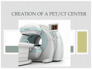
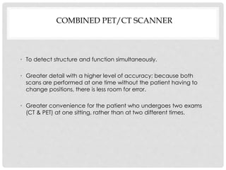
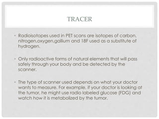
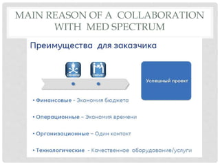
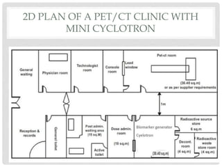
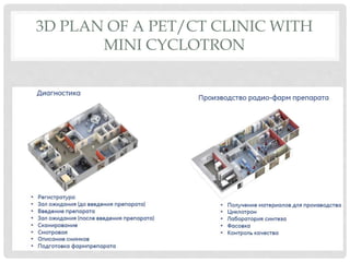
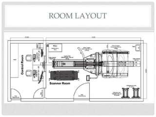
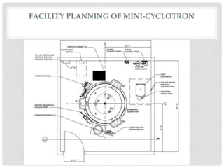
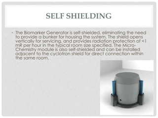
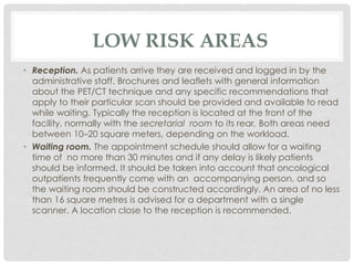
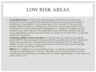
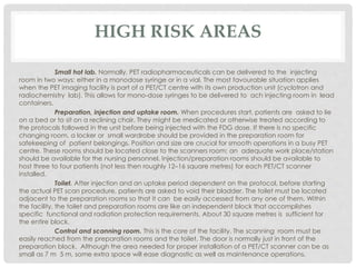
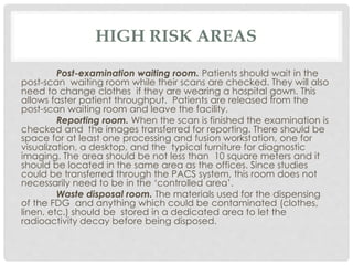
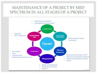
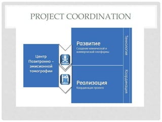
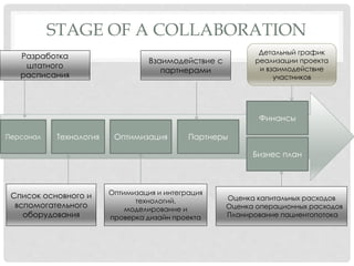
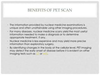
SlideShare a Scribd company logo
1 of 23
CREATION OF A PET/CT CENTER
HISTORY OF PET SCAN
• The concept of emission and transmission tomography was
introduced by David E. Kuhal and Roy Edwards in the late 1950s at
the university of Pennsylvania.
• In the 1970s, Tatsuo Ido at the Brookhaven National laboratory was
the first to describe the synthesis of 18-F FDG, the most commonly
used PET scanning isotope carrier.
• Now there is not one person who developed the PET scan but a
whole collection of people have made what it is today.
PET SCAN
COMBINED PET/CT SCANNER
• To detect structure and function simultaneously.
• Greater detail with a higher level of accuracy; because both
scans are performed at one time without the patient having to
change positions, there is less room for error.
• Greater convenience for the patient who undergoes two exams
(CT & PET) at one sitting, rather than at two different times.
PET/CT FUSION
TRACER
• Radioisotopes used in PET scans are isotopes of carbon,
nitrogen,oxygen,gallium and 18F used as a substitute of
hydrogen.
• Only radioactive forms of natural elements that will pass
safely through your body and be detected by the
scanner.
• The type of scanner used depends on what your doctor
wants to measure. For example, if your doctor is looking at
the tumor, he might use radio labeled glucose (FDG) and
watch how it is metabolized by the tumor.
MAIN REASON OF A COLLABORATION
WITH MED SPECTRUM
2D PLAN OF A PET/CT CLINIC WITH
MINI CYCLOTRON
3D PLAN OF A PET/CT CLINIC WITH
MINI CYCLOTRON
ROOM LAYOUT
FACILITY PLANNING OF MINI-CYCLOTRON
SELF SHIELDING
• The Biomarker Generator is self-shielded, eliminating the need
to provide a bunker for housing the system. The shield opens
vertically for servicing, and provides radiation protection of <1
mR per hour in the typical room size specified. The Micro-
Chemistry module is also self-shielded and can be installed
adjacent to the cyclotron shield for direct connection within
the same room.
LOW RISK AREAS
• Reception. As patients arrive they are received and logged in by the
administrative staff. Brochures and leaflets with general information
about the PET/CT technique and any specific recommendations that
apply to their particular scan should be provided and available to read
while waiting. Typically the reception is located at the front of the
facility, normally with the secretarial room to its rear. Both areas need
between 10–20 square meters, depending on the workload.
• Waiting room. The appointment schedule should allow for a waiting
time of no more than 30 minutes and if any delay is likely patients
should be informed. It should be taken into account that oncological
outpatients frequently come with an accompanying person, and so
the waiting room should be constructed accordingly. An area of no less
than 16 square metres is advised for a department with a single
scanner. A location close to the reception is recommended.
LOW RISK AREAS
• Consulting room. In this room the request and clinical records are
analysed and the patient is interviewed and physically examined, if
necessary. The patient is informed about the nature of the specific
examination he/she is undergoing. This room should be close to the
waiting room and adequately equipped. A supply of oxygen gas for
medical use and vacuum for aspiration and all other services as per
local regulations should be provided. An area of not less than 12 square
metres is necessary.
• Cleaning utilities room and store. A small room or cabinet should be
available for the storage of QC phantoms, supplies and other materials.
There should also be a dedicated space allocated to the cleaning
utilities. Those can be located at one end of the facility and 5 square
metres each would be sufficient.
• Offices. In addition to the reporting room, a certain number of rooms
should be available for clinical, scientific and technical staff, and for
meetings and teaching activities, the number depending on the size and
aims of the unit.
HIGH RISK AREAS
Small hot lab. Normally, PET radiopharmaceuticals can be delivered to the injecting
room in two ways: either in a monodose syringe or in a vial. The most favourable situation applies
when the PET imaging facility is part of a PET/CT centre with its own production unit (cyclotron and
radiochemistry lab). This allows for mono-dose syringes to be delivered to ach injecting room in lead
containers.
Preparation, injection and uptake room. When procedures start, patients are asked to lie
on a bed or to sit on a reclining chair. They might be medicated or otherwise treated according to
the protocols followed in the unit before being injected with the FDG dose. If there is no specific
changing room, a locker or small wardrobe should be provided in the preparation room for
safekeeping of patient belongings. Position and size are crucial for smooth operations in a busy PET
centre. These rooms should be located close to the scanners room; an adequate work place/station
should be available for the nursing personnel. Injection/preparation rooms should be available to
host three to four patients (not less then roughly 12–16 square metres) for each PET/CT scanner
installed.
Toilet. After injection and an uptake period dependent on the protocol, before starting
the actual PET scan procedure, patients are asked to void their bladder. The toilet must be located
adjacent to the preparation rooms so that it can be easily accessed from any one of them. Within
the facility, the toilet and preparation rooms are like an independent block that accomplishes
specific functional and radiation protection requirements. About 30 square metres is sufficient for
the entire block.
Control and scanning room. This is the core of the facility. The scanning room must be
easily reached from the preparation rooms and the toilet. The door is normally just in front of the
preparation block. Although the area needed for proper installation of a PET/CT scanner can be as
small as 7 m 5 m, some extra space will ease diagnostic as well as maintenance operations.
HIGH RISK AREAS
Post-examination waiting room. Patients should wait in the
post-scan waiting room while their scans are checked. They will also
need to change clothes if they are wearing a hospital gown. This
allows faster patient throughput. Patients are released from the
post-scan waiting room and leave the facility.
Reporting room. When the scan is finished the examination is
checked and the images transferred for reporting. There should be
space for at least one processing and fusion workstation, one for
visualization, a desktop, and the typical furniture for diagnostic
imaging. The area should be not less than 10 square meters and it
should be located in the same area as the offices. Since studies
could be transferred through the PACS system, this room does not
necessarily need to be in the ‘controlled area’.
Waste disposal room. The materials used for the dispensing
of the FDG and anything which could be contaminated (clothes,
linen, etc.) should be stored in a dedicated area to let the
radioactivity decay before being disposed.
NUMBER OF INSTALLED SCANNERS
Most PET/CT facilities have only one scanner. Creating a larger facility
with more scanners increases its efficiency significantly. Operating two scanners
requires less than twice the amount of radiopharmaceuticals and thereby reduces
production and transportation costs for each individual patient dose. Medical,
technical, nursing and secretarial personnel can also be used more efficiently. In
case of planned downtime or unexpected breakdowns of one scanner, the other
can be utilized for high priority procedures or operate for extended hours to
prevent cancellation of scheduled studies. This is particularly important for
patients already injected with radiotracer, thus avoiding unnecessary irradiation
resulting from administration without being scanned.
Planning for more than one
PET/CT scanner per facility is highly recommended.
STAGE OF A PROJECT
MAINTENANCE OF A PROJECT BY MED
SPECTRUM IN ALL STAGES OF A PROJECT
PROJECT COORDINATION
STAGE OF A COLLABORATION
Персонал Технология Оптимизация Партнеры
Финансы
Бизнес план
Список основного и
вспомогательного
оборудования
Оптимизация и интеграция
технологий,
моделирование и
проверка дизайн проекта
Оценка капитальных расходов
Оценка операционных расходов
Планирование пациентопотока
Детальный график
реализации проекта
и взаимодействие
участников
Разработка
штатного
расписания
Взаимодействие с
партнерами
MANNING TABLE
BENEFITS OF PET SCAN
• The information provided by nuclear medicine examinations is
unique and often unattainable using other imaging procedures.
• For many diseases, nuclear medicine scans yield the most useful
information needed to make a diagnosis or to determine
appropriate treatment, if any.
• Nuclear medicine is less expensive and may yield more precise
information than exploratory surgery.
• By identifying changes in the body at the cellular level, PET imaging
may detect the early onset of disease before it is evident on other
imaging tests such as CT or MRI.

More Related Content

What's hot

Setting up Radiology Diagnostic Centers
Setting up Radiology Diagnostic CentersSetting up Radiology Diagnostic Centers
Setting up Radiology Diagnostic Centers
metupalle
 
Ayush hospital standards.pptx
Ayush hospital standards.pptxAyush hospital standards.pptx
Ayush hospital standards.pptx
Dr Soumya Patil
 

What's hot (20)

Hospital architecture design planning
Hospital architecture design  planningHospital architecture design  planning
Hospital architecture design planning
 
Hospital planning
Hospital planningHospital planning
Hospital planning
 
CT SCAN equipment.pptx
CT SCAN equipment.pptxCT SCAN equipment.pptx
CT SCAN equipment.pptx
 
orthopedic requirements
orthopedic requirementsorthopedic requirements
orthopedic requirements
 
literature study for EMERGENCY DEPARTMENT in hospital
literature study for EMERGENCY DEPARTMENT in hospitalliterature study for EMERGENCY DEPARTMENT in hospital
literature study for EMERGENCY DEPARTMENT in hospital
 
Shielding Design
Shielding DesignShielding Design
Shielding Design
 
GYNECOLOGY AND OBSTETRICS DEPARTMENT
GYNECOLOGY AND OBSTETRICS DEPARTMENTGYNECOLOGY AND OBSTETRICS DEPARTMENT
GYNECOLOGY AND OBSTETRICS DEPARTMENT
 
gynecology , neurology , nephrology
gynecology , neurology , nephrologygynecology , neurology , nephrology
gynecology , neurology , nephrology
 
Hospital planning
Hospital planningHospital planning
Hospital planning
 
Operation theater DEMC
Operation theater DEMCOperation theater DEMC
Operation theater DEMC
 
Hospital design
Hospital designHospital design
Hospital design
 
Setting up Radiology Diagnostic Centers
Setting up Radiology Diagnostic CentersSetting up Radiology Diagnostic Centers
Setting up Radiology Diagnostic Centers
 
Department of cardiology
Department of cardiologyDepartment of cardiology
Department of cardiology
 
Advanced Diagnostic Center (radiology): An Introduction-DIAGNOSTIC RADIOLOGY ...
Advanced Diagnostic Center (radiology): An Introduction-DIAGNOSTIC RADIOLOGY ...Advanced Diagnostic Center (radiology): An Introduction-DIAGNOSTIC RADIOLOGY ...
Advanced Diagnostic Center (radiology): An Introduction-DIAGNOSTIC RADIOLOGY ...
 
Hospital 2
Hospital 2Hospital 2
Hospital 2
 
Healthcare facility design
Healthcare facility designHealthcare facility design
Healthcare facility design
 
Ayush hospital standards.pptx
Ayush hospital standards.pptxAyush hospital standards.pptx
Ayush hospital standards.pptx
 
FORTIS HOSPITAL NOIDA CASE STUDY WITH LIBRARY STUDY
FORTIS HOSPITAL NOIDA CASE STUDY WITH LIBRARY STUDY FORTIS HOSPITAL NOIDA CASE STUDY WITH LIBRARY STUDY
FORTIS HOSPITAL NOIDA CASE STUDY WITH LIBRARY STUDY
 
Cancer hospital litrature casestudy
Cancer hospital litrature casestudyCancer hospital litrature casestudy
Cancer hospital litrature casestudy
 
Report on hospital design
Report on hospital designReport on hospital design
Report on hospital design
 

Viewers also liked

COMPUTED TOMOGRAPHY SCAN
COMPUTED TOMOGRAPHY SCANCOMPUTED TOMOGRAPHY SCAN
COMPUTED TOMOGRAPHY SCAN
Shounak Nandi
 
Positron emission tomographic scan
Positron emission tomographic scanPositron emission tomographic scan
Positron emission tomographic scan
Prashant Makhija
 
Business plan for medical lab
Business plan for medical labBusiness plan for medical lab
Business plan for medical lab
Cochin University
 
Pet presentation, positron emission tomography
Pet presentation, positron emission tomography Pet presentation, positron emission tomography
Pet presentation, positron emission tomography
emicica
 

Viewers also liked (20)

COMPUTED TOMOGRAPHY SCAN
COMPUTED TOMOGRAPHY SCANCOMPUTED TOMOGRAPHY SCAN
COMPUTED TOMOGRAPHY SCAN
 
Computer tomography
Computer tomographyComputer tomography
Computer tomography
 
Carbohydrate
Carbohydrate Carbohydrate
Carbohydrate
 
PET
PETPET
PET
 
Pet scan short
Pet scan shortPet scan short
Pet scan short
 
Positron emission tomographic scan
Positron emission tomographic scanPositron emission tomographic scan
Positron emission tomographic scan
 
PET scan
PET scanPET scan
PET scan
 
Pet scan
Pet scanPet scan
Pet scan
 
Positron Emissions Tomography (PET SCAN)
Positron Emissions Tomography (PET SCAN)Positron Emissions Tomography (PET SCAN)
Positron Emissions Tomography (PET SCAN)
 
Mri 3
Mri 3Mri 3
Mri 3
 
Psma pet scan
Psma pet scanPsma pet scan
Psma pet scan
 
Magnetic Resonance Imaging and Its Benefits
Magnetic Resonance Imaging and Its BenefitsMagnetic Resonance Imaging and Its Benefits
Magnetic Resonance Imaging and Its Benefits
 
Marketing Plan for Multi Diagnostic Center
Marketing Plan for Multi Diagnostic CenterMarketing Plan for Multi Diagnostic Center
Marketing Plan for Multi Diagnostic Center
 
Business plan for medical lab
Business plan for medical labBusiness plan for medical lab
Business plan for medical lab
 
Positron Emission Tomography
Positron Emission TomographyPositron Emission Tomography
Positron Emission Tomography
 
What is a Positron Emission Tomography?
What is a Positron Emission Tomography?What is a Positron Emission Tomography?
What is a Positron Emission Tomography?
 
PET CAMBRIDGE ESOL
PET CAMBRIDGE ESOLPET CAMBRIDGE ESOL
PET CAMBRIDGE ESOL
 
Mri hardware
Mri hardwareMri hardware
Mri hardware
 
marketing of a diagnostic centre, medical lab, clinical lab
marketing of a diagnostic centre, medical lab, clinical labmarketing of a diagnostic centre, medical lab, clinical lab
marketing of a diagnostic centre, medical lab, clinical lab
 
Pet presentation, positron emission tomography
Pet presentation, positron emission tomography Pet presentation, positron emission tomography
Pet presentation, positron emission tomography
 

Similar to PET CENTER CREATION

Orag. of lab services
Orag. of lab servicesOrag. of lab services
Orag. of lab services
Nc Das
 
420620698-Administration-of-PICU-child-health-nursing.pptx
420620698-Administration-of-PICU-child-health-nursing.pptx420620698-Administration-of-PICU-child-health-nursing.pptx
420620698-Administration-of-PICU-child-health-nursing.pptx
DineshKumarSahu23
 
Pediatrics intensive care unit
Pediatrics intensive care unitPediatrics intensive care unit
Pediatrics intensive care unit
Sandy Kaur
 
Planning-Organizing-imaging-services-pdf.pdf
Planning-Organizing-imaging-services-pdf.pdfPlanning-Organizing-imaging-services-pdf.pdf
Planning-Organizing-imaging-services-pdf.pdf
yaskumaumaridriss
 

Similar to PET CENTER CREATION (20)

Radiology services.pptx
Radiology services.pptxRadiology services.pptx
Radiology services.pptx
 
HYBRID OPERATING THEATER ROOM
HYBRID OPERATING THEATER ROOM HYBRID OPERATING THEATER ROOM
HYBRID OPERATING THEATER ROOM
 
Radiologyservices.pptx
Radiologyservices.pptxRadiologyservices.pptx
Radiologyservices.pptx
 
Orag. of lab services
Orag. of lab servicesOrag. of lab services
Orag. of lab services
 
Tarun report ppt
Tarun report pptTarun report ppt
Tarun report ppt
 
Radiologydepartment_INTRODUCTION AND PLANING.pptx
Radiologydepartment_INTRODUCTION AND PLANING.pptxRadiologydepartment_INTRODUCTION AND PLANING.pptx
Radiologydepartment_INTRODUCTION AND PLANING.pptx
 
1425
14251425
1425
 
Icu
IcuIcu
Icu
 
Organisation of icu
Organisation of icuOrganisation of icu
Organisation of icu
 
OT & ICU LAYOUTS.pptx
OT & ICU LAYOUTS.pptxOT & ICU LAYOUTS.pptx
OT & ICU LAYOUTS.pptx
 
A MINIATURE ROBOTIC DEVICE APPLICABLE TO A FLEXIBLE ENDOSCOPE FOR THE SURGICA...
A MINIATURE ROBOTIC DEVICE APPLICABLE TO A FLEXIBLE ENDOSCOPE FOR THE SURGICA...A MINIATURE ROBOTIC DEVICE APPLICABLE TO A FLEXIBLE ENDOSCOPE FOR THE SURGICA...
A MINIATURE ROBOTIC DEVICE APPLICABLE TO A FLEXIBLE ENDOSCOPE FOR THE SURGICA...
 
420620698-Administration-of-PICU-child-health-nursing.pptx
420620698-Administration-of-PICU-child-health-nursing.pptx420620698-Administration-of-PICU-child-health-nursing.pptx
420620698-Administration-of-PICU-child-health-nursing.pptx
 
Dental department ppt
Dental department pptDental department ppt
Dental department ppt
 
organization of OT.pptx
organization of OT.pptxorganization of OT.pptx
organization of OT.pptx
 
Pediatrics intensive care unit
Pediatrics intensive care unitPediatrics intensive care unit
Pediatrics intensive care unit
 
Mri02
Mri02Mri02
Mri02
 
Planning-Organizing-imaging-services-pdf.pdf
Planning-Organizing-imaging-services-pdf.pdfPlanning-Organizing-imaging-services-pdf.pdf
Planning-Organizing-imaging-services-pdf.pdf
 
Accelerated partial breast irradiation
Accelerated partial breast irradiationAccelerated partial breast irradiation
Accelerated partial breast irradiation
 
Tele echocardiography v1.0
Tele echocardiography v1.0Tele echocardiography v1.0
Tele echocardiography v1.0
 
REMOTE ROBOTIC.pptx
REMOTE ROBOTIC.pptxREMOTE ROBOTIC.pptx
REMOTE ROBOTIC.pptx
 

Recently uploaded

Recently uploaded (20)

PHX May 2024 Corporate Presentation Final
PHX May 2024 Corporate Presentation FinalPHX May 2024 Corporate Presentation Final
PHX May 2024 Corporate Presentation Final
 
Falcon Invoice Discounting: Unlock Your Business Potential
Falcon Invoice Discounting: Unlock Your Business PotentialFalcon Invoice Discounting: Unlock Your Business Potential
Falcon Invoice Discounting: Unlock Your Business Potential
 
Berhampur CALL GIRL❤7091819311❤CALL GIRLS IN ESCORT SERVICE WE ARE PROVIDING
Berhampur CALL GIRL❤7091819311❤CALL GIRLS IN ESCORT SERVICE WE ARE PROVIDINGBerhampur CALL GIRL❤7091819311❤CALL GIRLS IN ESCORT SERVICE WE ARE PROVIDING
Berhampur CALL GIRL❤7091819311❤CALL GIRLS IN ESCORT SERVICE WE ARE PROVIDING
 
Nashik Call Girl Just Call 7091819311 Top Class Call Girl Service Available
Nashik Call Girl Just Call 7091819311 Top Class Call Girl Service AvailableNashik Call Girl Just Call 7091819311 Top Class Call Girl Service Available
Nashik Call Girl Just Call 7091819311 Top Class Call Girl Service Available
 
Getting Real with AI - Columbus DAW - May 2024 - Nick Woo from AlignAI
Getting Real with AI - Columbus DAW - May 2024 - Nick Woo from AlignAIGetting Real with AI - Columbus DAW - May 2024 - Nick Woo from AlignAI
Getting Real with AI - Columbus DAW - May 2024 - Nick Woo from AlignAI
 
Home Furnishings Ecommerce Platform Short Pitch 2024
Home Furnishings Ecommerce Platform Short Pitch 2024Home Furnishings Ecommerce Platform Short Pitch 2024
Home Furnishings Ecommerce Platform Short Pitch 2024
 
Unveiling Falcon Invoice Discounting: Leading the Way as India's Premier Bill...
Unveiling Falcon Invoice Discounting: Leading the Way as India's Premier Bill...Unveiling Falcon Invoice Discounting: Leading the Way as India's Premier Bill...
Unveiling Falcon Invoice Discounting: Leading the Way as India's Premier Bill...
 
Durg CALL GIRL ❤ 82729*64427❤ CALL GIRLS IN durg ESCORTS
Durg CALL GIRL ❤ 82729*64427❤ CALL GIRLS IN durg ESCORTSDurg CALL GIRL ❤ 82729*64427❤ CALL GIRLS IN durg ESCORTS
Durg CALL GIRL ❤ 82729*64427❤ CALL GIRLS IN durg ESCORTS
 
Call 7737669865 Vadodara Call Girls Service at your Door Step Available All Time
Call 7737669865 Vadodara Call Girls Service at your Door Step Available All TimeCall 7737669865 Vadodara Call Girls Service at your Door Step Available All Time
Call 7737669865 Vadodara Call Girls Service at your Door Step Available All Time
 
Bangalore Call Girl Just Call♥️ 8084732287 ♥️Top Class Call Girl Service Avai...
Bangalore Call Girl Just Call♥️ 8084732287 ♥️Top Class Call Girl Service Avai...Bangalore Call Girl Just Call♥️ 8084732287 ♥️Top Class Call Girl Service Avai...
Bangalore Call Girl Just Call♥️ 8084732287 ♥️Top Class Call Girl Service Avai...
 
JAJPUR CALL GIRL ❤ 82729*64427❤ CALL GIRLS IN JAJPUR ESCORTS
JAJPUR CALL GIRL ❤ 82729*64427❤ CALL GIRLS IN JAJPUR  ESCORTSJAJPUR CALL GIRL ❤ 82729*64427❤ CALL GIRLS IN JAJPUR  ESCORTS
JAJPUR CALL GIRL ❤ 82729*64427❤ CALL GIRLS IN JAJPUR ESCORTS
 
Berhampur Call Girl Just Call 8084732287 Top Class Call Girl Service Available
Berhampur Call Girl Just Call 8084732287 Top Class Call Girl Service AvailableBerhampur Call Girl Just Call 8084732287 Top Class Call Girl Service Available
Berhampur Call Girl Just Call 8084732287 Top Class Call Girl Service Available
 
JHARSUGUDA CALL GIRL ❤ 82729*64427❤ CALL GIRLS IN JHARSUGUDA ESCORTS
JHARSUGUDA CALL GIRL ❤ 82729*64427❤ CALL GIRLS IN JHARSUGUDA ESCORTSJHARSUGUDA CALL GIRL ❤ 82729*64427❤ CALL GIRLS IN JHARSUGUDA ESCORTS
JHARSUGUDA CALL GIRL ❤ 82729*64427❤ CALL GIRLS IN JHARSUGUDA ESCORTS
 
Lundin Gold - Q1 2024 Conference Call Presentation (Revised)
Lundin Gold - Q1 2024 Conference Call Presentation (Revised)Lundin Gold - Q1 2024 Conference Call Presentation (Revised)
Lundin Gold - Q1 2024 Conference Call Presentation (Revised)
 
Bankura Call Girl Just Call♥️ 8084732287 ♥️Top Class Call Girl Service Available
Bankura Call Girl Just Call♥️ 8084732287 ♥️Top Class Call Girl Service AvailableBankura Call Girl Just Call♥️ 8084732287 ♥️Top Class Call Girl Service Available
Bankura Call Girl Just Call♥️ 8084732287 ♥️Top Class Call Girl Service Available
 
Chennai Call Gril 80022//12248 Only For Sex And High Profile Best Gril Sex Av...
Chennai Call Gril 80022//12248 Only For Sex And High Profile Best Gril Sex Av...Chennai Call Gril 80022//12248 Only For Sex And High Profile Best Gril Sex Av...
Chennai Call Gril 80022//12248 Only For Sex And High Profile Best Gril Sex Av...
 
Pre Engineered Building Manufacturers Hyderabad.pptx
Pre Engineered  Building Manufacturers Hyderabad.pptxPre Engineered  Building Manufacturers Hyderabad.pptx
Pre Engineered Building Manufacturers Hyderabad.pptx
 
Chandrapur Call Girl Just Call 8084732287 Top Class Call Girl Service Available
Chandrapur Call Girl Just Call 8084732287 Top Class Call Girl Service AvailableChandrapur Call Girl Just Call 8084732287 Top Class Call Girl Service Available
Chandrapur Call Girl Just Call 8084732287 Top Class Call Girl Service Available
 
Escorts in Nungambakkam Phone 8250092165 Enjoy 24/7 Escort Service Enjoy Your...
Escorts in Nungambakkam Phone 8250092165 Enjoy 24/7 Escort Service Enjoy Your...Escorts in Nungambakkam Phone 8250092165 Enjoy 24/7 Escort Service Enjoy Your...
Escorts in Nungambakkam Phone 8250092165 Enjoy 24/7 Escort Service Enjoy Your...
 
Phases of Negotiation .pptx
 Phases of Negotiation .pptx Phases of Negotiation .pptx
Phases of Negotiation .pptx
 

PET CENTER CREATION

  • 1. CREATION OF A PET/CT CENTER
  • 2. HISTORY OF PET SCAN • The concept of emission and transmission tomography was introduced by David E. Kuhal and Roy Edwards in the late 1950s at the university of Pennsylvania. • In the 1970s, Tatsuo Ido at the Brookhaven National laboratory was the first to describe the synthesis of 18-F FDG, the most commonly used PET scanning isotope carrier. • Now there is not one person who developed the PET scan but a whole collection of people have made what it is today.
  • 4. COMBINED PET/CT SCANNER • To detect structure and function simultaneously. • Greater detail with a higher level of accuracy; because both scans are performed at one time without the patient having to change positions, there is less room for error. • Greater convenience for the patient who undergoes two exams (CT & PET) at one sitting, rather than at two different times.
  • 6. TRACER • Radioisotopes used in PET scans are isotopes of carbon, nitrogen,oxygen,gallium and 18F used as a substitute of hydrogen. • Only radioactive forms of natural elements that will pass safely through your body and be detected by the scanner. • The type of scanner used depends on what your doctor wants to measure. For example, if your doctor is looking at the tumor, he might use radio labeled glucose (FDG) and watch how it is metabolized by the tumor.
  • 7. MAIN REASON OF A COLLABORATION WITH MED SPECTRUM
  • 8. 2D PLAN OF A PET/CT CLINIC WITH MINI CYCLOTRON
  • 9. 3D PLAN OF A PET/CT CLINIC WITH MINI CYCLOTRON
  • 11. FACILITY PLANNING OF MINI-CYCLOTRON
  • 12. SELF SHIELDING • The Biomarker Generator is self-shielded, eliminating the need to provide a bunker for housing the system. The shield opens vertically for servicing, and provides radiation protection of <1 mR per hour in the typical room size specified. The Micro- Chemistry module is also self-shielded and can be installed adjacent to the cyclotron shield for direct connection within the same room.
  • 13. LOW RISK AREAS • Reception. As patients arrive they are received and logged in by the administrative staff. Brochures and leaflets with general information about the PET/CT technique and any specific recommendations that apply to their particular scan should be provided and available to read while waiting. Typically the reception is located at the front of the facility, normally with the secretarial room to its rear. Both areas need between 10–20 square meters, depending on the workload. • Waiting room. The appointment schedule should allow for a waiting time of no more than 30 minutes and if any delay is likely patients should be informed. It should be taken into account that oncological outpatients frequently come with an accompanying person, and so the waiting room should be constructed accordingly. An area of no less than 16 square metres is advised for a department with a single scanner. A location close to the reception is recommended.
  • 14. LOW RISK AREAS • Consulting room. In this room the request and clinical records are analysed and the patient is interviewed and physically examined, if necessary. The patient is informed about the nature of the specific examination he/she is undergoing. This room should be close to the waiting room and adequately equipped. A supply of oxygen gas for medical use and vacuum for aspiration and all other services as per local regulations should be provided. An area of not less than 12 square metres is necessary. • Cleaning utilities room and store. A small room or cabinet should be available for the storage of QC phantoms, supplies and other materials. There should also be a dedicated space allocated to the cleaning utilities. Those can be located at one end of the facility and 5 square metres each would be sufficient. • Offices. In addition to the reporting room, a certain number of rooms should be available for clinical, scientific and technical staff, and for meetings and teaching activities, the number depending on the size and aims of the unit.
  • 15. HIGH RISK AREAS Small hot lab. Normally, PET radiopharmaceuticals can be delivered to the injecting room in two ways: either in a monodose syringe or in a vial. The most favourable situation applies when the PET imaging facility is part of a PET/CT centre with its own production unit (cyclotron and radiochemistry lab). This allows for mono-dose syringes to be delivered to ach injecting room in lead containers. Preparation, injection and uptake room. When procedures start, patients are asked to lie on a bed or to sit on a reclining chair. They might be medicated or otherwise treated according to the protocols followed in the unit before being injected with the FDG dose. If there is no specific changing room, a locker or small wardrobe should be provided in the preparation room for safekeeping of patient belongings. Position and size are crucial for smooth operations in a busy PET centre. These rooms should be located close to the scanners room; an adequate work place/station should be available for the nursing personnel. Injection/preparation rooms should be available to host three to four patients (not less then roughly 12–16 square metres) for each PET/CT scanner installed. Toilet. After injection and an uptake period dependent on the protocol, before starting the actual PET scan procedure, patients are asked to void their bladder. The toilet must be located adjacent to the preparation rooms so that it can be easily accessed from any one of them. Within the facility, the toilet and preparation rooms are like an independent block that accomplishes specific functional and radiation protection requirements. About 30 square metres is sufficient for the entire block. Control and scanning room. This is the core of the facility. The scanning room must be easily reached from the preparation rooms and the toilet. The door is normally just in front of the preparation block. Although the area needed for proper installation of a PET/CT scanner can be as small as 7 m 5 m, some extra space will ease diagnostic as well as maintenance operations.
  • 16. HIGH RISK AREAS Post-examination waiting room. Patients should wait in the post-scan waiting room while their scans are checked. They will also need to change clothes if they are wearing a hospital gown. This allows faster patient throughput. Patients are released from the post-scan waiting room and leave the facility. Reporting room. When the scan is finished the examination is checked and the images transferred for reporting. There should be space for at least one processing and fusion workstation, one for visualization, a desktop, and the typical furniture for diagnostic imaging. The area should be not less than 10 square meters and it should be located in the same area as the offices. Since studies could be transferred through the PACS system, this room does not necessarily need to be in the ‘controlled area’. Waste disposal room. The materials used for the dispensing of the FDG and anything which could be contaminated (clothes, linen, etc.) should be stored in a dedicated area to let the radioactivity decay before being disposed.
  • 17. NUMBER OF INSTALLED SCANNERS Most PET/CT facilities have only one scanner. Creating a larger facility with more scanners increases its efficiency significantly. Operating two scanners requires less than twice the amount of radiopharmaceuticals and thereby reduces production and transportation costs for each individual patient dose. Medical, technical, nursing and secretarial personnel can also be used more efficiently. In case of planned downtime or unexpected breakdowns of one scanner, the other can be utilized for high priority procedures or operate for extended hours to prevent cancellation of scheduled studies. This is particularly important for patients already injected with radiotracer, thus avoiding unnecessary irradiation resulting from administration without being scanned. Planning for more than one PET/CT scanner per facility is highly recommended.
  • 18. STAGE OF A PROJECT
  • 19. MAINTENANCE OF A PROJECT BY MED SPECTRUM IN ALL STAGES OF A PROJECT
  • 21. STAGE OF A COLLABORATION Персонал Технология Оптимизация Партнеры Финансы Бизнес план Список основного и вспомогательного оборудования Оптимизация и интеграция технологий, моделирование и проверка дизайн проекта Оценка капитальных расходов Оценка операционных расходов Планирование пациентопотока Детальный график реализации проекта и взаимодействие участников Разработка штатного расписания Взаимодействие с партнерами
  • 23. BENEFITS OF PET SCAN • The information provided by nuclear medicine examinations is unique and often unattainable using other imaging procedures. • For many diseases, nuclear medicine scans yield the most useful information needed to make a diagnosis or to determine appropriate treatment, if any. • Nuclear medicine is less expensive and may yield more precise information than exploratory surgery. • By identifying changes in the body at the cellular level, PET imaging may detect the early onset of disease before it is evident on other imaging tests such as CT or MRI.