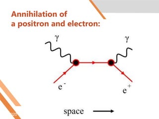
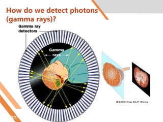
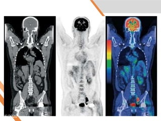
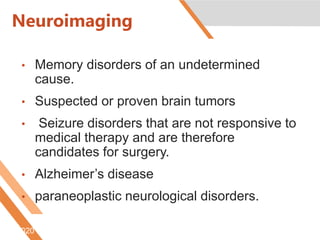
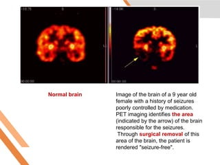
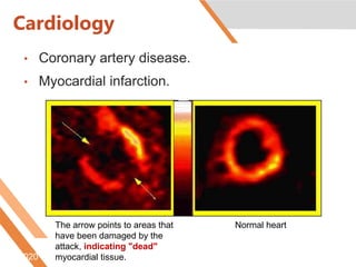
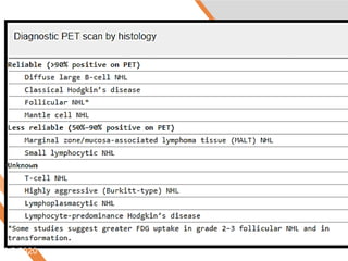
PET/CT scan in
Management of
Hematological
Disorders
6/5/2020
By
Eman Aboelenein
Assistant lecturer of Heamato-
oncology
Internal Medicine Department
Faculty of Medicine
Tanta University
June 5, 2020
Agenda
 What is P.E.T?
 What is a Positron?
 How does P.E.T works?
 Uses.
 Risks and limitations.
 In Hematological Disorders
 Take Home Message
6/5/2020
What is PET
• PET is a noninvasive, diagnostic imaging
technique for measuring the metabolic
activity of cells in the human body.
• It was developed in the mid 1970s and it
was the first scanning method to give
functional information about the brain.
6/5/2020 Htt://www.nucmed.buffalo.edu/petdef.htm
Whole-body PET scan using
18F-FDG:
The normal brain and
kidneys are labeled, and
radioactive urine from
breakdown of the FDG is
seen in the bladder.
In addition, a large
metastatic tumor mass
from colon cancer is seen
in the liver.
6/5/2020
What is a Positron?
• A Positron is an anti-matter electron, it
is identical in mass but has an apposite
charge of +1.
• Positron can come from different
number of sources, but for PET they are
produced by nuclear decay.
6/5/2020 .
Annihilation of
a positron and electron:
6/5/2020 .
How do we detect photons
(gamma rays)?
6/5/2020
6/5/2020
Uses:
• Neuroimaging
• Cardiology
• Oncology
• Infectious Diseases
• Pharmacokinetics
• Musculo-skeletal imaging
6/5/2020 Htt://www.nucmed.buffalo.edu/petdef.htm
Neuroimaging
• Memory disorders of an undetermined
cause.
• Suspected or proven brain tumors
• Seizure disorders that are not responsive to
medical therapy and are therefore
candidates for surgery.
• Alzheimer’s disease
• paraneoplastic neurological disorders.
6/5/2020 http://www.radiologyinfo.org/content/petomography.htm
Normal brain Image of the brain of a 9 year old
female with a history of seizures
poorly controlled by medication.
PET imaging identifies the area
(indicated by the arrow) of the brain
responsible for the seizures.
Through surgical removal of this
area of the brain, the patient is
rendered "seizure-free".
Htt://www.nucmed.buffalo.edu/petdef.htm
6/5/2020
Cardiology
• Coronary artery disease.
• Myocardial infarction.
6/5/2020 http://www.radiologyinfo.org/content/petomography.htm
The arrow points to areas that
have been damaged by the
attack, indicating "dead"
myocardial tissue.
Normal heart
Oncology
• Diagnosis, staging, and monitoring
treatment of cancers, particularly in
Hodgkin's lymphoma, non-Hodgkin
lymphoma, and lung cancer.
• Also, FDOPA PET/CT (or F-18-DOPA
PET/CT), localizing, pheochromocytoma
than the MIBG scan
6/5/2020 http://www.radiologyinfo.org/content/petomography.htm
Image showing malignant breast
mass that was not revealed by
conventional imaging techniques
such as CT, MRI, and
mammogram.
Image of same patient with enlarged left
axillary lymph nodes (indicated by
arrows), which through biopsy were found
to be metastatic (spread from another
location). The whole body scan reveals a
mass in the left breast (indicated by
arrow), that was malignant and
subsequently removed.
Htt://www.nucmed.buffalo.edu/petdef.htm
6/5/2020
What are the risks?
• Because the radioactivity is very short-lived,
your radiation exposure is low.
• The substance amount is so small that it
does not affect the normal processes of the
body.
• The radioactive substance may expose
radiation to the fetus in patients who are
pregnant or the infants of women who are
breast-feeding.
6/5/2020 http://www.radiologyinfo.org/content/petomography.htm
Limitations
• PET can give false results if a patient's
chemical balances are not normal.
diabetic patients or patients who have
eaten within a few hours prior to the
examination.
6/5/2020 http://www.radiologyinfo.org/content/petomography.htm
In Hematological
Disorders
6/5/2020
6/5/2020
6/5/2020
6/5/2020
6/5/2020
• The Deauville 5-point scoring system
for the FDG avidity of a Hodgkin’s
lymphoma or Non-Hodgkin’s lymphoma
tumor mass as seen on PET scan:
6/5/2020
Score 1: No uptake above the background.
Score 2: Uptake ≤ mediastinum.
Score 3: Uptake > mediastinum but ≤ liver.
Score 4: Uptake moderately increased
compared to the liver at any site.
Score 5: Uptake markedly increased compared
to the liver at any site.
Score X: New areas of uptake unlikely to be
related to lymphoma.
6/5/2020
• Scores of 1 and 2 are considered to be
negative
• 4 and 5 are considered to be positive.
• Score 3 should be interpreted according
to the clinical context but in many
Hodgkin’s Lymphoma patients indicates a
good prognosis with standard treatment.
6/5/2020
6/5/2020
6/5/2020
6/5/2020
6/5/2020
6/5/2020
6/5/2020
6/5/2020
6/5/2020
6/5/2020
6/5/2020
6/5/2020
Summary for PET in
Lymphoma
• In Diagnosis, Staging.
• Directing site of lymph node Biopsy.
• Selecting therapy.
• Early Evaluation of treatment.
• Post chemotherapy evaluation of
treatment.
• Follow up and Detection of Relapse.
6/5/2020
6/5/2020
6/5/2020
6/5/2020
6/5/2020
Summary of PET in
Myeloma
• Extramedullary lesions.
• Solitary plasmacytoma from MM.
• Smouldering MM.
• Follow up of MM and response to
treatment.
6/5/2020
6/5/2020
Take Home
Message
6/5/2020
• PET /CT is an old modality with an
everyday rediscovering benefits .
• It needs highly specialized experts to
correctly interpret and a continuous
research.
6/5/2020
June 5, 2020

PET/CT in hematological diseses

  • 1.
    PET/CT scan in Managementof Hematological Disorders 6/5/2020
  • 2.
    By Eman Aboelenein Assistant lecturerof Heamato- oncology Internal Medicine Department Faculty of Medicine Tanta University June 5, 2020
  • 3.
    Agenda  What isP.E.T?  What is a Positron?  How does P.E.T works?  Uses.  Risks and limitations.  In Hematological Disorders  Take Home Message 6/5/2020
  • 4.
    What is PET •PET is a noninvasive, diagnostic imaging technique for measuring the metabolic activity of cells in the human body. • It was developed in the mid 1970s and it was the first scanning method to give functional information about the brain. 6/5/2020 Htt://www.nucmed.buffalo.edu/petdef.htm
  • 5.
    Whole-body PET scanusing 18F-FDG: The normal brain and kidneys are labeled, and radioactive urine from breakdown of the FDG is seen in the bladder. In addition, a large metastatic tumor mass from colon cancer is seen in the liver. 6/5/2020
  • 6.
    What is aPositron? • A Positron is an anti-matter electron, it is identical in mass but has an apposite charge of +1. • Positron can come from different number of sources, but for PET they are produced by nuclear decay. 6/5/2020 .
  • 7.
    Annihilation of a positronand electron: 6/5/2020 .
  • 9.
    How do wedetect photons (gamma rays)? 6/5/2020
  • 10.
  • 12.
    Uses: • Neuroimaging • Cardiology •Oncology • Infectious Diseases • Pharmacokinetics • Musculo-skeletal imaging 6/5/2020 Htt://www.nucmed.buffalo.edu/petdef.htm
  • 13.
    Neuroimaging • Memory disordersof an undetermined cause. • Suspected or proven brain tumors • Seizure disorders that are not responsive to medical therapy and are therefore candidates for surgery. • Alzheimer’s disease • paraneoplastic neurological disorders. 6/5/2020 http://www.radiologyinfo.org/content/petomography.htm
  • 14.
    Normal brain Imageof the brain of a 9 year old female with a history of seizures poorly controlled by medication. PET imaging identifies the area (indicated by the arrow) of the brain responsible for the seizures. Through surgical removal of this area of the brain, the patient is rendered "seizure-free". Htt://www.nucmed.buffalo.edu/petdef.htm 6/5/2020
  • 15.
    Cardiology • Coronary arterydisease. • Myocardial infarction. 6/5/2020 http://www.radiologyinfo.org/content/petomography.htm The arrow points to areas that have been damaged by the attack, indicating "dead" myocardial tissue. Normal heart
  • 16.
    Oncology • Diagnosis, staging,and monitoring treatment of cancers, particularly in Hodgkin's lymphoma, non-Hodgkin lymphoma, and lung cancer. • Also, FDOPA PET/CT (or F-18-DOPA PET/CT), localizing, pheochromocytoma than the MIBG scan 6/5/2020 http://www.radiologyinfo.org/content/petomography.htm
  • 17.
    Image showing malignantbreast mass that was not revealed by conventional imaging techniques such as CT, MRI, and mammogram. Image of same patient with enlarged left axillary lymph nodes (indicated by arrows), which through biopsy were found to be metastatic (spread from another location). The whole body scan reveals a mass in the left breast (indicated by arrow), that was malignant and subsequently removed. Htt://www.nucmed.buffalo.edu/petdef.htm 6/5/2020
  • 18.
    What are therisks? • Because the radioactivity is very short-lived, your radiation exposure is low. • The substance amount is so small that it does not affect the normal processes of the body. • The radioactive substance may expose radiation to the fetus in patients who are pregnant or the infants of women who are breast-feeding. 6/5/2020 http://www.radiologyinfo.org/content/petomography.htm
  • 19.
    Limitations • PET cangive false results if a patient's chemical balances are not normal. diabetic patients or patients who have eaten within a few hours prior to the examination. 6/5/2020 http://www.radiologyinfo.org/content/petomography.htm
  • 20.
  • 25.
  • 27.
  • 28.
  • 29.
  • 30.
  • 31.
    • The Deauville5-point scoring system for the FDG avidity of a Hodgkin’s lymphoma or Non-Hodgkin’s lymphoma tumor mass as seen on PET scan: 6/5/2020
  • 32.
    Score 1: Nouptake above the background. Score 2: Uptake ≤ mediastinum. Score 3: Uptake > mediastinum but ≤ liver. Score 4: Uptake moderately increased compared to the liver at any site. Score 5: Uptake markedly increased compared to the liver at any site. Score X: New areas of uptake unlikely to be related to lymphoma. 6/5/2020
  • 34.
    • Scores of1 and 2 are considered to be negative • 4 and 5 are considered to be positive. • Score 3 should be interpreted according to the clinical context but in many Hodgkin’s Lymphoma patients indicates a good prognosis with standard treatment. 6/5/2020
  • 35.
  • 36.
  • 37.
  • 38.
  • 39.
  • 40.
  • 41.
  • 44.
  • 45.
  • 46.
  • 47.
  • 48.
    Summary for PETin Lymphoma • In Diagnosis, Staging. • Directing site of lymph node Biopsy. • Selecting therapy. • Early Evaluation of treatment. • Post chemotherapy evaluation of treatment. • Follow up and Detection of Relapse. 6/5/2020
  • 49.
  • 50.
  • 51.
  • 52.
  • 53.
    Summary of PETin Myeloma • Extramedullary lesions. • Solitary plasmacytoma from MM. • Smouldering MM. • Follow up of MM and response to treatment. 6/5/2020
  • 54.
  • 55.
  • 56.
    • PET /CTis an old modality with an everyday rediscovering benefits . • It needs highly specialized experts to correctly interpret and a continuous research. 6/5/2020
  • 57.