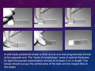
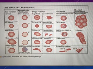
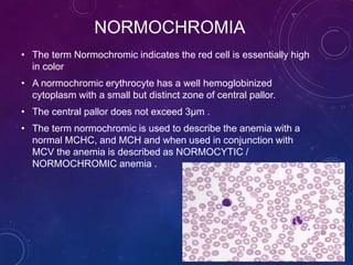
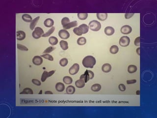
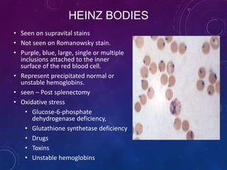
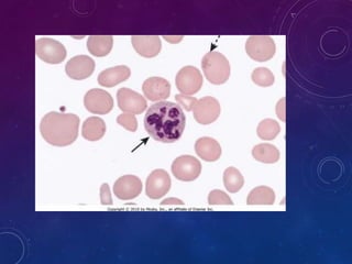
Peripheral blood smear examination
DR. MITHILA
MODERATOR : DR MANJUNATH
INTRODUCTION
• Peripheral blood smear is a very important tool in the
hematology lab
• It provides rapid, reliable access to information about a
variety of hematologic disorders
• Examination of the peripheral blood smear is an
inexpensive but powerful diagnostic tool in both children
and adults
• The smear offers a window into the functional status of
the bone marrow
• Review of the smear is an important adjunct to other
clinical data; in some cases, the peripheral smear alone is
sufficient to establish a diagnosis
INDICATION FOR PERIPHERAL SMEAR
Features suggestive of anemia, unexplained jaundice, or both .
Features suggestive of thrombocytopenia (e.g., petechiae or
abnormal bruising) or neutropenia (e.g., unexpected or severe
infection).
Features suggestive of a lymphoma or leukaemia
lymphadenopathy, splenomegaly, bone pain, and systemic
symptoms such as fever, sweating, and weight loss .
 Features suggestive of a myeloproliferative disease —
splenomegaly, plethora, itching, or weight loss .
General ill health, often with malaise and fever, suggesting
infectious mononucleosis or other viral infection or
inflammatory or malignant disease .
Suspicion of a bacterial or parasitic disease that can be
diagnosed from a blood smear
 Peripheral Smear Preparation
 Staining of Peripheral Blood Smear
 Peripheral Smear Examination
OBJECTIVES
SMEAR PREPARATION 1. Place a drop of blood, about 2-3 mm in
diameter approximately 1 cm from one
end of slide.
2. Place the slide on a flat surface, and hold
the other end between your left thumb
and forefinger.
3. With your right hand, place the smooth
clean edge of a second (spreader) slide
on the specimen slide, just in front of
the blood drop.
4. Hold the spreader slide at a 30°- 45
angle, and draw it back against the drop
of blood
6. Allow the blood to spread almost to the
edges of the slide.
7. Push the spread forward with one light,
smooth moderate speed. A thin film of
blood in the shape of tongue.
8. Label one edge with patient name, lab id
and date.
9. The slides should be rapidly air dried by
waving the slides or using an electrical fan.
A well made peripheral smear is thick at one end and progressively thinner
at the opposite end. The "zone of morphology" (area of optimal thickness
for light microscopic examination) should be at least 2 cm in length. The
smear should occupy the central area of the slide and be margin-free at
the edges
PBS examination requires a systematic approach in
order to gather all possible information.
In addition, all specimens must be evaluated in the
same manner, to assure that consistent information is
obtained.
SLIDE FIXATION AND STAINING
ROMANOWSKY STAINING
IT INCLUDES:
• MAY-GRUNWALD –GEIMSA STAIN,
• JENNER’S STAIN,
• WRIGHT’S STAIN,
• LEISHMAN’S STAIN AND,
• FIELD’S STAIN.
COLOR RESPONSES OF BLOOD CELLS TO
ROMANOWSKY STAINING
• Cellular component Color
• Nuclei Chromatin Purple
• Nucleoli Light blue
• Erythroblast Dark blue
• Erythrocyte Dark pink
• Reticulocyte Grey–blue
CYTOPLASM COLOR
• Lymphocyte Blue
• Metamyelocyte Pink
• Monocyte Grey–blue
• Myelocyte Pink
• Neutrophil Pink/orange
• Promyelocyte Blue
• Basophil Blue
• 1. Macroscopic view : quality of the smear
• 2.The microscopic analysis
• begins on lower power (10x),
• Determine good distribution of the cells
• Scans the edges for abnormal cells
• Find a optimal area in the smear for detailed
examination.
PBS examination - preliminary
Hi-power (40x) :
•To obtain a WBC estimate.
•All of the detailed analysis of the cellular elements using high
power or oil immersion.
• Evaluate the morphology of the WBC and record any
abnormalities such as toxic granulation or Dohle bodies.
• The WBC estimate can be performed using a factor which
is based on the fact that WBC seen in 40x is approx
equivalent to 2000 cells /micro litre.
• For example if the average number of WBC counted per
high power field is 5, the estimate WBC is 5 x 2000 = 10000
OIL IMMERSION
• Perform a 100 WBC differential count , counting is done in
zig zag motion.
• All WBC have to included until a total of 100 have been
counted
• Evaluate RBC for anisocytosis , poikilocytosis ,
hypochromasia , polychromasia, and inclusions.
• Perform platelet estimate and platelet morphology
• Count the number of platelets in 10 OIF.
• Divide by 10
(a) Ten microscopic fields are examined in a vertical direction
from bottom to top or top to bottom
(b) slide is horizontally moved to the next field
(c) Ten microscopic fields are counted vertically.
(d) procedure is repeated until 100 WBCS have been
counted (zig zag motion)
Scanning technique for WBC differential
count and morphologic evaluation
1. RBC
• Size
• Shape
• Color
• Arrangement
• Inclusions
• Abnormal cells
2. WBC
• Total counts
• Differential counts
• Abnormal WBC
3. Platelets
• Counts
• Abnormality
4. Parasites
Evaluation of PBS
RED CELL MORPHOLOGY
Morphology of Normal Red Blood Cells
 Biconcave disc
 Diameter : 7 ~ 8 μm
 Average volume : 90 fl.
 Central pallor occupy 1/3 rd of total size
 Approx same as nucleus of mature lymphocyte
RED CELL ABNORMALITY
• Normal MCV is -80-100 fl
• Microcytes –MCV<80 fl
• Macrocytes – MCV> 100 fl
• Anisocytosis - variation in the size of the
RBC
• Poikilocytosis – Variation in the shape of
RBC
VARIATION IN SIZE
• Anisocytosis- Variation in size of the red blood
cells
• The severity of the variation corresponds to increased
RDW.
• Anisocytosis results from the abnormal cell
development ( deficiency of iron , B12, Folic acid)
• Normal MCV is -80-100 fl
• Microcytes ( MCV <80 fl)
• Macrocytes (MCV >100fl)
MICROCYTES
• A Microcyte is a small cell having
a diameter less <7 & MCV < 80fl.
• Anemia associated with
microcytes is said to microcytic
• Expanded central zone of pallor
anemia
• Decreased or defective globin
synthesis also presents as
Microcytic hypochromic anemia.
MORPHOLOGY - MICROCYTE
IRIRON
DEFICIENCY
THALASSEMIA SIDEROBLASTIC
LEAD
POISONING
CHRONIC
DIESEASE
POPOSSIBLE PATHOLOGY
MACROCYTES
• When MCV of RBC is Increased(>100fl)
• The common cause of macrocytes is due
to the impaired DNA synthesis, RNA
synthesis is unaffected resulting in the
asynchrony between the cytoplasmic
and nuclear maturation .
• Neutrophillic hypersegmentation is
typically seen.
MORPHOLOGY - MACROCYTE
MEGALOBLASTIC
PROCESS
HIGH
RETICULOCYTE
COUNT
LIVER
DIESEASE
HYPOTHYROIDISM
POST
SPLENECTOMY
OVAL
MACROCYRTES
HEMOLYTIC
ANEMIA / ACUTE
BLOOD LOSS
CHRONIC
ALCOHOLISM
FOLATE AND B12
DEFICIENCY
NORMOCYTES
• The average size of the erythrocyte is indicated by
the measurement of the MCV
• A Normal MCV would corresponds to the MCV
reference range ( 80 -100 )fl
• Subsequent review of the peripheral smear reveals
no abnormality in the size variation.
• This scenario is referred to as NORMOCYTIC and red
cells are referred as normocytes.
HEMOGLOBIN CONTENT – COLOR
VARIATION
• NORMOCHROMIA
• HYPOCHROMIA
• HYPERCHROMIA
• POLYCHROMASIA
NORMOCHROMIA
• The term Normochromic indicates the red cell is essentially high
in color
• A normochromic erythrocyte has a well hemoglobinized
cytoplasm with a small but distinct zone of central pallor.
• The central pallor does not exceed 3µm .
• The term normochromic is used to describe the anemia with a
normal MCHC, and MCH and when used in conjunction with
MCV the anemia is described as NORMOCYTIC /
NORMOCHROMIC anemia .
HYPOCHROMIA
• Any RBC having a central area of pallor of greater
than 3µm is said to be hypochromic
• There is a direct relationship between the amount of
hemoglobin deposited in the RBC and the
appearance of red cell when stained.
• The term Hypochromia indicates low color and
indicates that the cells have less hemoglobin.
• MCHC < 32% the anemic process is described as
hypochromic.
HYPOCHROMIA GRADING
1 + AREA OF CENTRAL PALLOR IS ONE HALF OF CELL DIAMETER
2 + AREA OF CENTRAL PALLOR IS TWO THIRDS OF CELL DIAMETER
3 + AREA OF CENTRAL PALLOR IS OF THREE QUARTERS
4 + THIN RIM OF HEMOGLOBIN
POLYCHROMASIA
When RBC are delivered to the peripheral circulation
prematurely appearing diffusely basophilic and are gray
blue in color and usually larger than normal red cell.
The basophilic color is due to the RNA residue involved in
hemoglobin synthesis.
Polychromatic cells are actually reticulocytes.
Any clinical condition in which marrow is stimulated
particularly RBC regeneration will produce a
polychromatic blood picture .
The degree of polychromasia is a excellent indicator of
therapeutic effectiveness when patient is given iron or
vitamin therapy as treatment of anemia
POIKILOCYTOSIS
• Variation In shape is called Poikilocytosis.
• It is of following types-
• Spherocytes
• Elliotocytes
• Target cells
• Schistocytes
• Acanthocytes
• Keratocytes
• Echinocytes
• Bite cells
• Howel jolly bodies
Spherocytosis
• Spherocytes are small dense spheroidal RBC with absence of central pallor .
• Because of their density they are easily seen in the peripheral smear.
• This abnormality is due to the abnormality of the red cell membrane .
• The detailed mechanism for sphering is the congenital condition known as
hereditary spherocytosis.
• This is an inherited , autosomal dominant condition and is due to the deficiency
of the membrane proteins , spectrin and ankyrin .
• Acquired causes of spherocytes are
ABO incompatibility
Autoimmune hemolytic anemia (warm antibody type)
Infections (e.g., EBV, CMV, E. coli, Sepsis/ sepsis)
Severe burns
DIC and HUS
Elliptocytes or ovalocytes
Ovalocytes / elliptocytes are due to the result of
morphological abnormality due to the result of
mechanical weakness or fragility of the
membrane skeleton that may be acquired or
hereditary.
STOMATOCYTES
Red cells with central
biconcave area appears
slit like in dried film.
Wet film it appears as
cup-shaped.
The abnormal morphology
is due to the Membrane
defect.
Seen in
Artifact
Hereditary
stomatocytosis
liver disease,
Alcoholic cirrhosis
Hemolytic anemia
Tear drop cells / dacrocytes
 Tear drop cells appear in
the peripheral circulation
as tear drop or pear
shaped red cells.
 Exact mechanism not
known.
 It is seen in :
 Myelofibrosis
 Bone marrow infiltrated
with hematological or
non-hematological
malignancies
 Iron deficiency anemia
 megaloblastic anemia
TARGET CELLS
Cells in which central
round stained area and
peripheral rim of
cytoplasm.
Seen in Thalassaemia
Chronic liver disease
Hereditary hypo-
betalipoproteinemia
Iron deficiency anemia
Hemoglobinopathies
(Hb C, Hb H, Sickel cell
anemia
Post splenectomy
Acanthocytes or spur cells, are spherical cells with blunt-tipped
or club-shaped spicules of different lengths projecting from their surface at
irregular intervals.
Acanthocytes
 Acanthocytes are seen in
 Hereditary Abetalipoproteinemia
Hereditary acanthocytosis
 End stage liver disease
 Micro angiopathic hemolytic anemia
 Malnutrition
 Post splenectomy
 it is the hallmark in the diagnosis of
the neuro acanthocytosis syndrome
such as
 Chorea-acanthosis and Mcleod
syndrome
SCHISTOCYTES
 These are fragmented erythrocytes.
 Smaller than normal red cells and of varying shape
resulting from some trauma to the cell membrane.
 Triggering events within the circulation leading to
fragmentation of RBC.
 Fluid alteration results in development of fibrin
strands, damaged endothelium.
 The flow of the blood in the circulation sweep the
RBC through the fibrin strands splitting the red cells
 Acquired disorder of RBC formation
 Megaloblastic
 Dyserythropoietic
 Mechanical stress MAHA
 DIC
 Heart valve surgery
 HUS / renal graft rejection
 Direct thermal injury / Severe burns/
SICKLE CELL
• Cells are sickle (boat
shape) or crescent
shape
• Present in film of patient
with homozygosity for
Hb S.
• Usually absent in
neonates and rare in
patients with high Hb F
percentage
RED CELL INCLUSION
• Basophilic stippling (Punctate basophilia)
• Howell – jolly Bodies
• Heinz body
• Cabot Rings
• Protozoan inclusions
• Rouleaux formation
ROULEAUX
• Rouleaux is a condition in which red
cells appear as stacks of coins on the
peripheral smear .
• The stacks of RBC are evenly
distributed through out the smear ,
rouleaux formation is the result of
elevated globulins or fibrinogens in the
plasma where the RBC has been
“bathed “ in the abnormal plasma giving
sticky consistency.
• It is seen in multiple myeloma and
Waldenstroms macroglobulinemia, intra
venous administration of plasma volume
expanders like dextran.
Howell Jolly bodies
 Howell-Jolly bodies are small round
bodies composed of DNA, about 1
µm in diameter, usually single and
in the periphery of a red cell.
 They are readily visible on the
Wright-Giemsa-stained smear.
 The spleen is responsible for the
removal of nuclear material in the
red cells, so in absence of a
functional spleen, nuclear material
is removed ineffectively.
 Howell-Jolly bodies are seen in :
 Post splenectomy
 Functional asplenia
 Anatomical absence of spleen
• Presence of irregular basophilic granules with in Rbc
which are variable in size .
• Stain deep blue with Wright’s stain
• Fine stippling seen with
• Increased polychromatophilia
• Increased production of red cells.
• Coarse stippling
• Lead and heavy metal poisoning
• Disturbed erythropoiesis
• Megaloblastic anemia
• Thalassaemia
• infection
• liver disease
• Unstable Hb
• Pyrimidine-5’-nucleotidase defiency
BASOPHILIC STIPPLING
HEINZ BODIES
• Seen on supravital stains
• Not seen on Romanowsky stain.
• Purple, blue, large, single or multiple
inclusions attached to the inner
surface of the red blood cell.
• Represent precipitated normal or
unstable hemoglobins.
• seen – Post splenectomy
• Oxidative stress
• Glucose-6-phosphate
dehydrogenase deficiency,
• Glutathione synthetase deficiency
• Drugs
• Toxins
• Unstable hemoglobins
CABOT RINGS
• These are Ring shaped
figure of eight or loop
shaped
• Red or Reddish purple
with Wright’s stain and
have no internal structure
• Observed rarely in
• Pernicious anemia,
• Lead poisoning,
WHITE BLOOD CELLS
TYPES
Granulocyte
(polymorphonuclear)
Agranulocyte (mononuclear)
Contain membrane bound granules,
which stains differently with stains
Apparently absent granules, but
contain non specific azurophilic
granules
E.g.
Neutrophils
Basophil
Eosionophil
E.g.
Lymphocyte
Monocyte
Macrophage
POLYMORPHONUCLEAR
NEUTROPHILS
• 40 to 80 percent of total WBC
count(2.0–7.0 ×10⁹/l )
• Diameter - 13 µm
• segmented nucleus and
pink/orange cytoplasm with
fine granulation(0.2-0.3µm)
stain tan to pink with Wright’s
• Lobes -2-5
• Neutrophils usually have
trilobed nucleus.
• small percent has four lobes
and occasionally five lobes.
BAND FORMS
• neutrophils has either a
strand of nuclear material
thicker than a filament
connecting the lobes, or a U-
shaped nucleus of uniform
thickness.
• Up to 8% of circulating
neutrophils are
unsegmented or
partly segmented (‘band’
forms)
• Band cells constitute <5-10% of white blood cells
• An increase in number of band cell and other
immature neutrophils is called a “ shift to left” can be
seen in
• Severe infections, sepsis
• Non infectious inflammatory disease
• Pregnancy
GRANULES
• Toxic granulation-
increase in staining
density and number of
granules
• Seen with Bacterial
infections and other
inflammation
• Administration of G-CSF
• Anaplastic anemia
DOHLE BODIES
• Small, round or oval, pale blue-grey
structure
• Found at periphery of neutrophil.
• Contains Ribosomes and
Endoplasmic reticulum
• Seen in – Bacterial infection
• inflammation
• administration of G-CSF
• during pregnancy
• Pernicious anemia
• Myeloproliferative disorders
• Myelodysplastic disorders
• Cancer chemothrapy
EOSINOPHILS
• Normally 1-6%( 0.02–0.5 ×
109
/l)
• Size- 12–17 µm
• Nucleus- Bilobed
(spectacle shaped)
• Cytoplasm- Pale blue
• Granules - Coarse
spherical gold/orange
Eosinopenia- seen with prolonged steroid
administration.
• Eosinophilia- allergic conditions hay fever, asthma
• severe eosinophilia- parasitic infection
• reactive eosinophilia
• Eosinophilic leukaemia
• Idiopathic hypereosinophilic syndrome
• T-cell lymphoma, B-cell lymphoma
and acute lymphoblastic leukaemia.
BASOPHILS
• Rarest <1%
• Nucleus segments fold up on each other
resulting compact irregular dense
nucleus(closed lotus flower like)
• Granules-large, variable size dark blue
or purple often obscure the nucleus
• Granules are rich in histamine,
serotonin and heparin
• Increase in myeloproliferative disorder-
CML
MONOCYTES
• 2-10% of total wbc count
• Size- largest circulating leucocyte, 15–
18µm in diameter
• Cytoplasm- grey blue
• Nucleus- large , curved , horse shoe
shape
• No segmentation occur
• Chromatin- fine evenly distributed
• Increase in chronic infections and
inflammatory conditions such as
• Tuberculosis and Crohn’s disease,
• Chronic myeloid leukaemias
• Acute leukaemias with a monocytic
component
• Infectious mononucleosis
LYMPHOCYTES
• 20-40% of total WBC count
• It is of two types
1. Small lymphocyte(6-10µm)
2. Large lymphocyte(12-15µm)
• Nucleus-single, sharply
defined, stain dark blue on Wright’s
stain
• Cytoplasm- Pale blue
• Large lymphocytes less densely stain
nuclei & abundant cytoplasm
• Few round purple(azure) granules are
present
• Lymphocytes predominate in the blood
films of infants and young children.
REACTIVE LYMPHOCYTES
• Have slightly larger nuclei
with more open chromatin
• Abundant cytoplasm that
may be irregular.
• Seen in infectious
mononucleosis
• viral infections
PLATELETS MORPHOLOGY
PLATELETS
• Thrombopoiesis take place in
bone marrow
• 1 megakaryocyte produce 4000
platelets
• Normal platelet are about 1.3
micron, blue grey, contain fine,
purple to pink granules
• Red cell to platelet ratio : 10-40:1
• Life span 9-12 days
• Range : 1.5-4.5 lakhs/microL
Platelets
Neubars chamber : count platelets in 64 small
squares
Counts * 250 = total platelets
Normal counts 4.5 to 5.5 lakh
Common Causes of Thrombocytopenia
•Decreased production
−Aplastic anemia
−Acute leukemia
−Viral infections *Parvovirus *CMV
−Amegakaryocytic thrombocytopenia (AMT)
•Increased destruction
−Immune thrombocytopenia
*Idiopathic thrombocytopenic purpura (ITP)
*Neonatal alloimmune thrombocytopenia
(NAITP)
−Disseminated intravascular coagulation (DIC)
−Hypersplenism
Thrombocytosis
• Reactive thrombocytosis
 Post infection
 Inflammation
 Juvenile rheumatoid arthritis
 Collagen vasvular disease
• Essential thrombocythemia
EXAMINATION OF BLOOD
FILMS FOR PARASITES
MALARIA
• Giemsa stain are used, identifies
species and life cycle stages
• Parasitemia is quantifiable
• Threshold of detection thin film: 100
parasites/ L, thick film: 2-20
parasite/L
Thick film Thin film
• Lysed RBCs
• Larger volume
• 0.25microliter / 100 fields blood
element more concentrated
• Good screening for positive or
negative parasitemia and parasite
density difficult to diagnose species
• Fixed RBCs
• Single layer
• Smaller volume
• 0.005 microliter blood required
• Good species differentiation
• Requires more time to ready
A. Peripheral smear
APPEARANCE OF P FALCIPARUM IN THE BLOOD FILMS
Ring or trophozoite
• Many cells infected –
same with more than
one parasite
• Red cell size
unaltered
• Parasite is often
attatch to the margin
of the host cell:
called as accole form
(arrow)
Schizont
 Very rarely seem
except in cerebral
malaria
 A single brown
pigment dot along
with 18-32
merozoites
Gamatocyte
 Sickle shape “cresent”
 Matuer gametocyte is
about 1.5 times larger
than RBC harbouring it
 Microgamatocyte:
Broader, shorter, blunt
ends. Cytoplasm light
blue
 Macrogamatocytes:
Longer, narrower,
pointed ends.
Cytoplasm deep blue
APPEARANCE OF P VIVEX IN FILM
Ring or trophozoite
• Many cells infected –
same with more than
one parasite
• Unoccupied portion by
parasite shows a dotted
or stripped appearance
“Schuffner’s dot”
Schizont
 Represent the full grown
trophozoite
 Contain 12-24 merozoits
 Arranged in the form of
rosette with yellow brown
pigment at the center
Gamatocyte
 Certain schizont get
modified and result in sexual
forms. Merozoite arising
from single schizont are
either all males or females
 Microgamatocyte: Spherical.
Cytoplasm light blue
 Macrogamatocytes:
spherical. Cytoplasm deep
blue
Disadvantages of the Peripheral Blood Smear
Provides information that cannot be obtained from automated
cell counting. However, some limitations are:
• Experience is required to make technically adequate smears.
• There is a non-uniform distribution of white blood cells over
the smear, with larger leukocytes concentrated near the edges
and lymphocytes scattered throughout.
• There is a non-uniform distribution of RBCs over the smear,
with small crowded red blood cells at the thick edge and large
flat red blood cells without central pallor at the feathered edge
THANK YOU

Peripheral smear

  • 1.
    Peripheral blood smearexamination DR. MITHILA MODERATOR : DR MANJUNATH
  • 2.
    INTRODUCTION • Peripheral bloodsmear is a very important tool in the hematology lab • It provides rapid, reliable access to information about a variety of hematologic disorders • Examination of the peripheral blood smear is an inexpensive but powerful diagnostic tool in both children and adults • The smear offers a window into the functional status of the bone marrow • Review of the smear is an important adjunct to other clinical data; in some cases, the peripheral smear alone is sufficient to establish a diagnosis
  • 3.
    INDICATION FOR PERIPHERALSMEAR Features suggestive of anemia, unexplained jaundice, or both . Features suggestive of thrombocytopenia (e.g., petechiae or abnormal bruising) or neutropenia (e.g., unexpected or severe infection). Features suggestive of a lymphoma or leukaemia lymphadenopathy, splenomegaly, bone pain, and systemic symptoms such as fever, sweating, and weight loss .  Features suggestive of a myeloproliferative disease — splenomegaly, plethora, itching, or weight loss .
  • 4.
    General ill health,often with malaise and fever, suggesting infectious mononucleosis or other viral infection or inflammatory or malignant disease . Suspicion of a bacterial or parasitic disease that can be diagnosed from a blood smear
  • 5.
     Peripheral SmearPreparation  Staining of Peripheral Blood Smear  Peripheral Smear Examination OBJECTIVES
  • 6.
    SMEAR PREPARATION 1.Place a drop of blood, about 2-3 mm in diameter approximately 1 cm from one end of slide. 2. Place the slide on a flat surface, and hold the other end between your left thumb and forefinger. 3. With your right hand, place the smooth clean edge of a second (spreader) slide on the specimen slide, just in front of the blood drop. 4. Hold the spreader slide at a 30°- 45 angle, and draw it back against the drop of blood 6. Allow the blood to spread almost to the edges of the slide. 7. Push the spread forward with one light, smooth moderate speed. A thin film of blood in the shape of tongue. 8. Label one edge with patient name, lab id and date. 9. The slides should be rapidly air dried by waving the slides or using an electrical fan.
  • 7.
    A well madeperipheral smear is thick at one end and progressively thinner at the opposite end. The "zone of morphology" (area of optimal thickness for light microscopic examination) should be at least 2 cm in length. The smear should occupy the central area of the slide and be margin-free at the edges
  • 8.
    PBS examination requiresa systematic approach in order to gather all possible information. In addition, all specimens must be evaluated in the same manner, to assure that consistent information is obtained.
  • 9.
  • 10.
    ROMANOWSKY STAINING IT INCLUDES: •MAY-GRUNWALD –GEIMSA STAIN, • JENNER’S STAIN, • WRIGHT’S STAIN, • LEISHMAN’S STAIN AND, • FIELD’S STAIN.
  • 11.
    COLOR RESPONSES OFBLOOD CELLS TO ROMANOWSKY STAINING • Cellular component Color • Nuclei Chromatin Purple • Nucleoli Light blue • Erythroblast Dark blue • Erythrocyte Dark pink • Reticulocyte Grey–blue
  • 12.
    CYTOPLASM COLOR • LymphocyteBlue • Metamyelocyte Pink • Monocyte Grey–blue • Myelocyte Pink • Neutrophil Pink/orange • Promyelocyte Blue • Basophil Blue
  • 13.
    • 1. Macroscopicview : quality of the smear • 2.The microscopic analysis • begins on lower power (10x), • Determine good distribution of the cells • Scans the edges for abnormal cells • Find a optimal area in the smear for detailed examination. PBS examination - preliminary
  • 14.
    Hi-power (40x) : •Toobtain a WBC estimate. •All of the detailed analysis of the cellular elements using high power or oil immersion. • Evaluate the morphology of the WBC and record any abnormalities such as toxic granulation or Dohle bodies. • The WBC estimate can be performed using a factor which is based on the fact that WBC seen in 40x is approx equivalent to 2000 cells /micro litre. • For example if the average number of WBC counted per high power field is 5, the estimate WBC is 5 x 2000 = 10000
  • 15.
    OIL IMMERSION • Performa 100 WBC differential count , counting is done in zig zag motion. • All WBC have to included until a total of 100 have been counted • Evaluate RBC for anisocytosis , poikilocytosis , hypochromasia , polychromasia, and inclusions. • Perform platelet estimate and platelet morphology • Count the number of platelets in 10 OIF. • Divide by 10
  • 16.
    (a) Ten microscopicfields are examined in a vertical direction from bottom to top or top to bottom (b) slide is horizontally moved to the next field (c) Ten microscopic fields are counted vertically. (d) procedure is repeated until 100 WBCS have been counted (zig zag motion) Scanning technique for WBC differential count and morphologic evaluation
  • 17.
    1. RBC • Size •Shape • Color • Arrangement • Inclusions • Abnormal cells 2. WBC • Total counts • Differential counts • Abnormal WBC 3. Platelets • Counts • Abnormality 4. Parasites Evaluation of PBS
  • 18.
  • 19.
    Morphology of NormalRed Blood Cells  Biconcave disc  Diameter : 7 ~ 8 μm  Average volume : 90 fl.  Central pallor occupy 1/3 rd of total size  Approx same as nucleus of mature lymphocyte
  • 20.
    RED CELL ABNORMALITY •Normal MCV is -80-100 fl • Microcytes –MCV<80 fl • Macrocytes – MCV> 100 fl • Anisocytosis - variation in the size of the RBC • Poikilocytosis – Variation in the shape of RBC
  • 22.
    VARIATION IN SIZE •Anisocytosis- Variation in size of the red blood cells • The severity of the variation corresponds to increased RDW. • Anisocytosis results from the abnormal cell development ( deficiency of iron , B12, Folic acid) • Normal MCV is -80-100 fl • Microcytes ( MCV <80 fl) • Macrocytes (MCV >100fl)
  • 23.
    MICROCYTES • A Microcyteis a small cell having a diameter less <7 & MCV < 80fl. • Anemia associated with microcytes is said to microcytic • Expanded central zone of pallor anemia • Decreased or defective globin synthesis also presents as Microcytic hypochromic anemia.
  • 24.
    MORPHOLOGY - MICROCYTE IRIRON DEFICIENCY THALASSEMIASIDEROBLASTIC LEAD POISONING CHRONIC DIESEASE POPOSSIBLE PATHOLOGY
  • 25.
    MACROCYTES • When MCVof RBC is Increased(>100fl) • The common cause of macrocytes is due to the impaired DNA synthesis, RNA synthesis is unaffected resulting in the asynchrony between the cytoplasmic and nuclear maturation . • Neutrophillic hypersegmentation is typically seen.
  • 26.
  • 27.
    NORMOCYTES • The averagesize of the erythrocyte is indicated by the measurement of the MCV • A Normal MCV would corresponds to the MCV reference range ( 80 -100 )fl • Subsequent review of the peripheral smear reveals no abnormality in the size variation. • This scenario is referred to as NORMOCYTIC and red cells are referred as normocytes.
  • 28.
    HEMOGLOBIN CONTENT –COLOR VARIATION • NORMOCHROMIA • HYPOCHROMIA • HYPERCHROMIA • POLYCHROMASIA
  • 29.
    NORMOCHROMIA • The termNormochromic indicates the red cell is essentially high in color • A normochromic erythrocyte has a well hemoglobinized cytoplasm with a small but distinct zone of central pallor. • The central pallor does not exceed 3µm . • The term normochromic is used to describe the anemia with a normal MCHC, and MCH and when used in conjunction with MCV the anemia is described as NORMOCYTIC / NORMOCHROMIC anemia .
  • 30.
    HYPOCHROMIA • Any RBChaving a central area of pallor of greater than 3µm is said to be hypochromic • There is a direct relationship between the amount of hemoglobin deposited in the RBC and the appearance of red cell when stained. • The term Hypochromia indicates low color and indicates that the cells have less hemoglobin. • MCHC < 32% the anemic process is described as hypochromic.
  • 31.
    HYPOCHROMIA GRADING 1 +AREA OF CENTRAL PALLOR IS ONE HALF OF CELL DIAMETER 2 + AREA OF CENTRAL PALLOR IS TWO THIRDS OF CELL DIAMETER 3 + AREA OF CENTRAL PALLOR IS OF THREE QUARTERS 4 + THIN RIM OF HEMOGLOBIN
  • 32.
    POLYCHROMASIA When RBC aredelivered to the peripheral circulation prematurely appearing diffusely basophilic and are gray blue in color and usually larger than normal red cell. The basophilic color is due to the RNA residue involved in hemoglobin synthesis. Polychromatic cells are actually reticulocytes. Any clinical condition in which marrow is stimulated particularly RBC regeneration will produce a polychromatic blood picture . The degree of polychromasia is a excellent indicator of therapeutic effectiveness when patient is given iron or vitamin therapy as treatment of anemia
  • 34.
    POIKILOCYTOSIS • Variation Inshape is called Poikilocytosis. • It is of following types- • Spherocytes • Elliotocytes • Target cells • Schistocytes • Acanthocytes • Keratocytes • Echinocytes • Bite cells • Howel jolly bodies
  • 35.
    Spherocytosis • Spherocytes aresmall dense spheroidal RBC with absence of central pallor . • Because of their density they are easily seen in the peripheral smear. • This abnormality is due to the abnormality of the red cell membrane . • The detailed mechanism for sphering is the congenital condition known as hereditary spherocytosis. • This is an inherited , autosomal dominant condition and is due to the deficiency of the membrane proteins , spectrin and ankyrin . • Acquired causes of spherocytes are ABO incompatibility Autoimmune hemolytic anemia (warm antibody type) Infections (e.g., EBV, CMV, E. coli, Sepsis/ sepsis) Severe burns DIC and HUS
  • 36.
    Elliptocytes or ovalocytes Ovalocytes/ elliptocytes are due to the result of morphological abnormality due to the result of mechanical weakness or fragility of the membrane skeleton that may be acquired or hereditary.
  • 37.
    STOMATOCYTES Red cells withcentral biconcave area appears slit like in dried film. Wet film it appears as cup-shaped. The abnormal morphology is due to the Membrane defect. Seen in Artifact Hereditary stomatocytosis liver disease, Alcoholic cirrhosis Hemolytic anemia
  • 38.
    Tear drop cells/ dacrocytes  Tear drop cells appear in the peripheral circulation as tear drop or pear shaped red cells.  Exact mechanism not known.  It is seen in :  Myelofibrosis  Bone marrow infiltrated with hematological or non-hematological malignancies  Iron deficiency anemia  megaloblastic anemia
  • 39.
    TARGET CELLS Cells inwhich central round stained area and peripheral rim of cytoplasm. Seen in Thalassaemia Chronic liver disease Hereditary hypo- betalipoproteinemia Iron deficiency anemia Hemoglobinopathies (Hb C, Hb H, Sickel cell anemia Post splenectomy
  • 40.
    Acanthocytes or spurcells, are spherical cells with blunt-tipped or club-shaped spicules of different lengths projecting from their surface at irregular intervals. Acanthocytes  Acanthocytes are seen in  Hereditary Abetalipoproteinemia Hereditary acanthocytosis  End stage liver disease  Micro angiopathic hemolytic anemia  Malnutrition  Post splenectomy  it is the hallmark in the diagnosis of the neuro acanthocytosis syndrome such as  Chorea-acanthosis and Mcleod syndrome
  • 41.
    SCHISTOCYTES  These arefragmented erythrocytes.  Smaller than normal red cells and of varying shape resulting from some trauma to the cell membrane.  Triggering events within the circulation leading to fragmentation of RBC.  Fluid alteration results in development of fibrin strands, damaged endothelium.  The flow of the blood in the circulation sweep the RBC through the fibrin strands splitting the red cells  Acquired disorder of RBC formation  Megaloblastic  Dyserythropoietic  Mechanical stress MAHA  DIC  Heart valve surgery  HUS / renal graft rejection  Direct thermal injury / Severe burns/
  • 42.
    SICKLE CELL • Cellsare sickle (boat shape) or crescent shape • Present in film of patient with homozygosity for Hb S. • Usually absent in neonates and rare in patients with high Hb F percentage
  • 43.
    RED CELL INCLUSION •Basophilic stippling (Punctate basophilia) • Howell – jolly Bodies • Heinz body • Cabot Rings • Protozoan inclusions • Rouleaux formation
  • 44.
    ROULEAUX • Rouleaux isa condition in which red cells appear as stacks of coins on the peripheral smear . • The stacks of RBC are evenly distributed through out the smear , rouleaux formation is the result of elevated globulins or fibrinogens in the plasma where the RBC has been “bathed “ in the abnormal plasma giving sticky consistency. • It is seen in multiple myeloma and Waldenstroms macroglobulinemia, intra venous administration of plasma volume expanders like dextran.
  • 45.
    Howell Jolly bodies Howell-Jolly bodies are small round bodies composed of DNA, about 1 µm in diameter, usually single and in the periphery of a red cell.  They are readily visible on the Wright-Giemsa-stained smear.  The spleen is responsible for the removal of nuclear material in the red cells, so in absence of a functional spleen, nuclear material is removed ineffectively.  Howell-Jolly bodies are seen in :  Post splenectomy  Functional asplenia  Anatomical absence of spleen
  • 46.
    • Presence ofirregular basophilic granules with in Rbc which are variable in size . • Stain deep blue with Wright’s stain • Fine stippling seen with • Increased polychromatophilia • Increased production of red cells. • Coarse stippling • Lead and heavy metal poisoning • Disturbed erythropoiesis • Megaloblastic anemia • Thalassaemia • infection • liver disease • Unstable Hb • Pyrimidine-5’-nucleotidase defiency BASOPHILIC STIPPLING
  • 47.
    HEINZ BODIES • Seenon supravital stains • Not seen on Romanowsky stain. • Purple, blue, large, single or multiple inclusions attached to the inner surface of the red blood cell. • Represent precipitated normal or unstable hemoglobins. • seen – Post splenectomy • Oxidative stress • Glucose-6-phosphate dehydrogenase deficiency, • Glutathione synthetase deficiency • Drugs • Toxins • Unstable hemoglobins
  • 48.
    CABOT RINGS • Theseare Ring shaped figure of eight or loop shaped • Red or Reddish purple with Wright’s stain and have no internal structure • Observed rarely in • Pernicious anemia, • Lead poisoning,
  • 49.
  • 50.
    TYPES Granulocyte (polymorphonuclear) Agranulocyte (mononuclear) Contain membranebound granules, which stains differently with stains Apparently absent granules, but contain non specific azurophilic granules E.g. Neutrophils Basophil Eosionophil E.g. Lymphocyte Monocyte Macrophage
  • 52.
    POLYMORPHONUCLEAR NEUTROPHILS • 40 to80 percent of total WBC count(2.0–7.0 ×10⁹/l ) • Diameter - 13 µm • segmented nucleus and pink/orange cytoplasm with fine granulation(0.2-0.3µm) stain tan to pink with Wright’s • Lobes -2-5 • Neutrophils usually have trilobed nucleus. • small percent has four lobes and occasionally five lobes.
  • 53.
    BAND FORMS • neutrophilshas either a strand of nuclear material thicker than a filament connecting the lobes, or a U- shaped nucleus of uniform thickness. • Up to 8% of circulating neutrophils are unsegmented or partly segmented (‘band’ forms)
  • 54.
    • Band cellsconstitute <5-10% of white blood cells • An increase in number of band cell and other immature neutrophils is called a “ shift to left” can be seen in • Severe infections, sepsis • Non infectious inflammatory disease • Pregnancy
  • 55.
    GRANULES • Toxic granulation- increasein staining density and number of granules • Seen with Bacterial infections and other inflammation • Administration of G-CSF • Anaplastic anemia
  • 56.
    DOHLE BODIES • Small,round or oval, pale blue-grey structure • Found at periphery of neutrophil. • Contains Ribosomes and Endoplasmic reticulum • Seen in – Bacterial infection • inflammation • administration of G-CSF • during pregnancy • Pernicious anemia • Myeloproliferative disorders • Myelodysplastic disorders • Cancer chemothrapy
  • 57.
    EOSINOPHILS • Normally 1-6%(0.02–0.5 × 10⁹ /l) • Size- 12–17 µm • Nucleus- Bilobed (spectacle shaped) • Cytoplasm- Pale blue • Granules - Coarse spherical gold/orange
  • 58.
    Eosinopenia- seen withprolonged steroid administration. • Eosinophilia- allergic conditions hay fever, asthma • severe eosinophilia- parasitic infection • reactive eosinophilia • Eosinophilic leukaemia • Idiopathic hypereosinophilic syndrome • T-cell lymphoma, B-cell lymphoma and acute lymphoblastic leukaemia.
  • 59.
    BASOPHILS • Rarest <1% •Nucleus segments fold up on each other resulting compact irregular dense nucleus(closed lotus flower like) • Granules-large, variable size dark blue or purple often obscure the nucleus • Granules are rich in histamine, serotonin and heparin • Increase in myeloproliferative disorder- CML
  • 60.
    MONOCYTES • 2-10% oftotal wbc count • Size- largest circulating leucocyte, 15– 18µm in diameter • Cytoplasm- grey blue • Nucleus- large , curved , horse shoe shape • No segmentation occur • Chromatin- fine evenly distributed • Increase in chronic infections and inflammatory conditions such as • Tuberculosis and Crohn’s disease, • Chronic myeloid leukaemias • Acute leukaemias with a monocytic component • Infectious mononucleosis
  • 61.
    LYMPHOCYTES • 20-40% oftotal WBC count • It is of two types 1. Small lymphocyte(6-10µm) 2. Large lymphocyte(12-15µm) • Nucleus-single, sharply defined, stain dark blue on Wright’s stain • Cytoplasm- Pale blue • Large lymphocytes less densely stain nuclei & abundant cytoplasm • Few round purple(azure) granules are present • Lymphocytes predominate in the blood films of infants and young children.
  • 62.
    REACTIVE LYMPHOCYTES • Haveslightly larger nuclei with more open chromatin • Abundant cytoplasm that may be irregular. • Seen in infectious mononucleosis • viral infections
  • 63.
  • 64.
    PLATELETS • Thrombopoiesis takeplace in bone marrow • 1 megakaryocyte produce 4000 platelets • Normal platelet are about 1.3 micron, blue grey, contain fine, purple to pink granules • Red cell to platelet ratio : 10-40:1 • Life span 9-12 days • Range : 1.5-4.5 lakhs/microL
  • 65.
    Platelets Neubars chamber :count platelets in 64 small squares Counts * 250 = total platelets Normal counts 4.5 to 5.5 lakh Common Causes of Thrombocytopenia •Decreased production −Aplastic anemia −Acute leukemia −Viral infections *Parvovirus *CMV −Amegakaryocytic thrombocytopenia (AMT) •Increased destruction −Immune thrombocytopenia *Idiopathic thrombocytopenic purpura (ITP) *Neonatal alloimmune thrombocytopenia (NAITP) −Disseminated intravascular coagulation (DIC) −Hypersplenism Thrombocytosis • Reactive thrombocytosis  Post infection  Inflammation  Juvenile rheumatoid arthritis  Collagen vasvular disease • Essential thrombocythemia
  • 66.
  • 67.
    MALARIA • Giemsa stainare used, identifies species and life cycle stages • Parasitemia is quantifiable • Threshold of detection thin film: 100 parasites/ L, thick film: 2-20 parasite/L Thick film Thin film • Lysed RBCs • Larger volume • 0.25microliter / 100 fields blood element more concentrated • Good screening for positive or negative parasitemia and parasite density difficult to diagnose species • Fixed RBCs • Single layer • Smaller volume • 0.005 microliter blood required • Good species differentiation • Requires more time to ready A. Peripheral smear
  • 68.
    APPEARANCE OF PFALCIPARUM IN THE BLOOD FILMS Ring or trophozoite • Many cells infected – same with more than one parasite • Red cell size unaltered • Parasite is often attatch to the margin of the host cell: called as accole form (arrow) Schizont  Very rarely seem except in cerebral malaria  A single brown pigment dot along with 18-32 merozoites Gamatocyte  Sickle shape “cresent”  Matuer gametocyte is about 1.5 times larger than RBC harbouring it  Microgamatocyte: Broader, shorter, blunt ends. Cytoplasm light blue  Macrogamatocytes: Longer, narrower, pointed ends. Cytoplasm deep blue
  • 69.
    APPEARANCE OF PVIVEX IN FILM Ring or trophozoite • Many cells infected – same with more than one parasite • Unoccupied portion by parasite shows a dotted or stripped appearance “Schuffner’s dot” Schizont  Represent the full grown trophozoite  Contain 12-24 merozoits  Arranged in the form of rosette with yellow brown pigment at the center Gamatocyte  Certain schizont get modified and result in sexual forms. Merozoite arising from single schizont are either all males or females  Microgamatocyte: Spherical. Cytoplasm light blue  Macrogamatocytes: spherical. Cytoplasm deep blue
  • 70.
    Disadvantages of thePeripheral Blood Smear Provides information that cannot be obtained from automated cell counting. However, some limitations are: • Experience is required to make technically adequate smears. • There is a non-uniform distribution of white blood cells over the smear, with larger leukocytes concentrated near the edges and lymphocytes scattered throughout. • There is a non-uniform distribution of RBCs over the smear, with small crowded red blood cells at the thick edge and large flat red blood cells without central pallor at the feathered edge
  • 76.