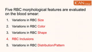
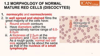
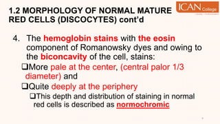
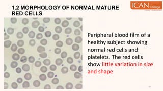
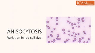
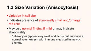
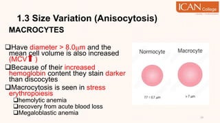
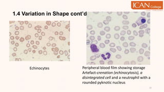
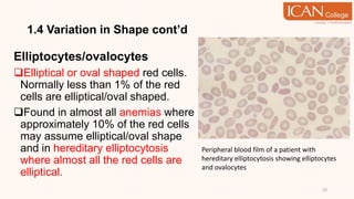
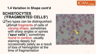
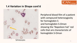
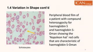
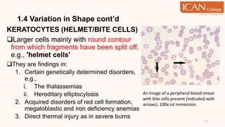
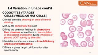
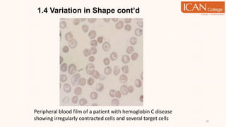
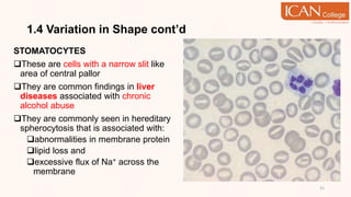
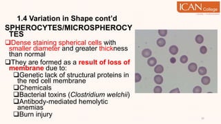
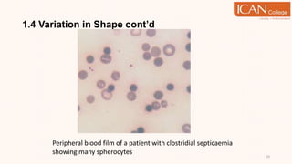
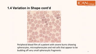
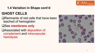
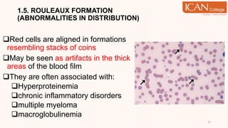
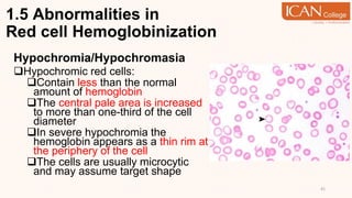
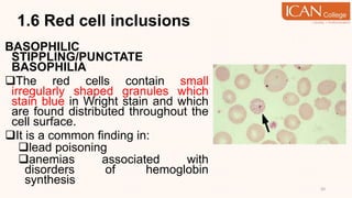
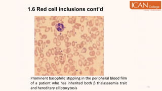
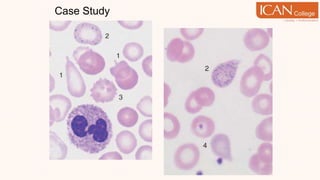
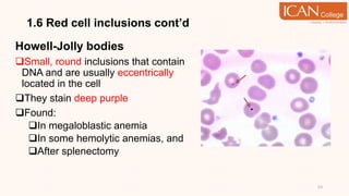
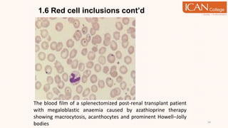
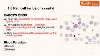
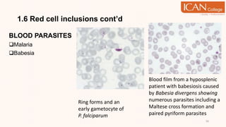
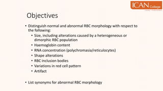
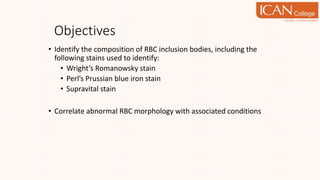
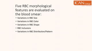
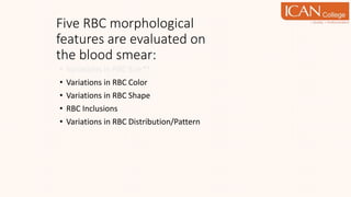
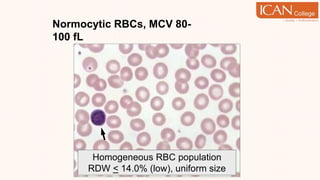
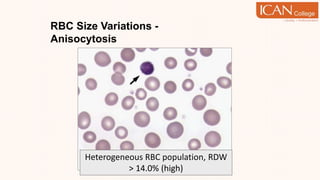
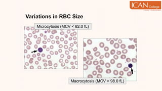
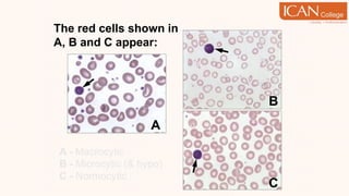
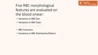
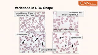
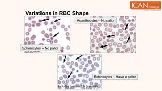
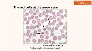
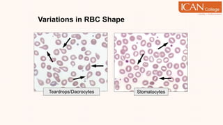
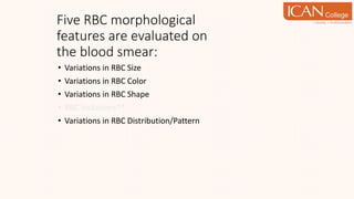
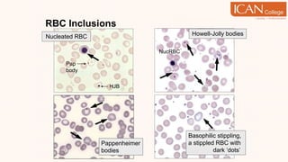
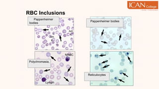
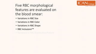
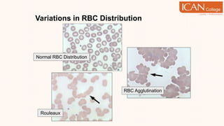
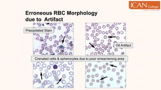
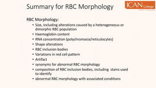
TOPIC 1
RED CELL MORPHOLOGY STUDY
BT2053 HEMATOLOGY II
DIPLOMA IN MEDICAL LABORATORY TECHNOLOGY
SITI BAINUN BINTI MOHD DALI
LEARNING OUTCOME
1. Describe the morphology of normal red cells
2. List and describe the different abnormal erythrocyte forms
3. Assess the morphology of red cells on a stained blood film
4. Describe the grading system for assessing changes in
erythrocyte morphology
1.1 INTRODUCTION
 Evaluation of RBCs morphology can help in
diagnosis of hematologic disorders including:
i. Anemias
ii. Leukemias
iii. Infections
 Need to consider other RBCs parameters such as
HCT, red cell size (MCV) and others at same time.
Blood Film Examination
1. The film should be covered with a cover glass using a
neutral mounting medium
This step is not, however, mandatory
2. The blood film should be inspected under low power
magnification (10X) in order to:
get an idea of the quality of the preparation, i.e.,
whether red cell agglutination or excessive rouleaux is
present
get an idea of the number, distribution and staining of
the leucocytes
find an area where the red cells are evenly distributed
and are not distorted 4
Blood Film Examination
3. Having selected a suitable area, the 40x dry or 100x oil
immersion objective is used to appreciate:
variation in red cell size, shape and staining, and
fine details such as cytoplasmic granules and other red
cell inclusions
5
Five RBC morphological features are evaluated
on the blood smear:
1. Variations in RBC Size
2. Variations in RBC Color
3. Variations in RBC Shape
4. RBC Inclusions
5. Variations in RBC Distribution/Pattern
NORMAL RBCs MORPHOLOGY
1.2 MORPHOLOGY OF NORMAL
MATURE RED CELLS (DISCOCYTES)
1. normocytic and normochromic
2. In well spread and stained films the
great majority of the cells have:
i. Round smooth contours
ii. Have diameters within the
comparatively narrow range of 6.0-
8.0m
iii. A thickness of 2.5m at the
periphery and 1.0m in the center
As a rough guide, normal red cell
size appears to be about the same
as that of the nucleus of a small
lymphocyte
8
1.2 MORPHOLOGY OF NORMAL MATURE
RED CELLS (DISCOCYTES) cont’d
4. The hemoglobin stains with the eosin
component of Romanowsky dyes and owing to
the biconcavity of the cell, stains:
More pale at the center, (central palor 1/3
diameter) and
Quite deeply at the periphery
This depth and distribution of staining in normal
red cells is described as normochromic
9
1.2 MORPHOLOGY OF NORMAL MATURE
RED CELLS
10
Peripheral blood film of a
healthy subject showing
normal red cells and
platelets. The red cells
show little variation in size
and shape
ANISOCYTOSIS
Variation in red cell size
1.3 Size Variation (Anisocytosis)
• Variation in cell size
• Indicates presence of abnormally small and/or large
red cells
• May be a normal finding if mild or may indicate
abnormality
• Spherocytes (appear very small and dense but may have a
normal volume) seen with immune mediated hemolytic
anemia.
1.3 Size Variation (Anisocytosis)
MACROCYTES
Have diameter > 8.0m and the
mean cell volume is also increased
(MCV )
Because of their increased
hemoglobin content they stain darker
than discocytes
Macrocytosis is seen in stress
erythropoiesis
hemolytic anemia
recovery from acute blood loss
Megaloblastic anemia
13
1.3 Size Variation cont’d
14
The film also shows
anisocytosis with
both microcytes and
macrocytes
1.3 Size Variation cont’d
MEGALOCYTES
Large (diameter > 12m), often
oval shaped cells with increased
hemoglobin content
True megalocytes are identified
only if megaloblasts have been
identified in bone marrow
aspirates
seen in vitamin B12 and/or folic
acid deficiency, in association with
some leukemias and in refractory
anemias
15
1.3 Size Variation cont’d
Microcytes
diameter < 6.0m but may appear to
have normal size caused by flattening
of the cells during smear preparation
The MCV less than 80.0fl
Area of central pallor usually
increases because of the coexistent
hypochromia
Are seen in iron deficiency anemia
and a slight degree of microcytosis is
seen in inflammation 16
Microcytosis in a patient
with β thalassaemia
trait
POIKILOCYTOSIS
Variation in red cell shape
1.4 Variation in Shape
(Poikilocytosis)
ACANTHOCYTES ('SPINY
CELLS')
Spheroidal cells with 3-12 spicules
of uneven length irregularly
distributed over the cell surface.
Seen in:
disorders of lipid metabolism
alcoholic liver cirrhosis and
Hepatitis
18
1.4 Variation in Shape cont’d
DACROCYTES ('TEAR DROP
CELLS')
These are tear drop or pear shaped red cells
Could be considered to be discocytes with a
single drawn out spicule.
It is thought that stretching of the cell membrane
beyond a certain limit results in loss of
deformability and ability to revert to normal
discoid shape.
Seen in:
Myelofibrosis,
Myeloid metaplasia
 Tumour metastases to the bone marrow
 Tuberculosis
Drug-induced Heinz body formation
19
Tear drop cells
1.4 Variation in Shape cont’d
DREPANOCYTES ('SICKLE CELLS')
These are crescent shaped red cells
because of the formation of rod-like
polymers of Hb S or some other rare
hemoglobins
Have an increased surface area and
increased mechanical fragility which leads
to hemolysis and hence severe anemia
They are primarily seen in sickle cell
anemia where there is substitution of
valine for glutamic acid at position 6 of the
beta chain in the hemoglobin molecule 20
Sickle cells in a patient with sickle
cell anemia
1.4 Variation in Shape cont’d
ECHINOCYTES (‘BURR CELLS')
Red cells showing numerous, short, evenly
distributed spicules of equal length
These are probably the most common
artifacts in a blood film:
Consistently found in blood samples
that have been stored for some time at
room temperature and
 Because of diffusion of alkaline
substances from the slide into the cells
resulting in an increase in pH and thus
crenation of the cells
In vivo they are seen in uremia, pyruvate
kinase deficiency and neonatal liver
diseases
21
1.4 Variation in Shape cont’d
22
Echinocytes Peripheral blood film showing storage
Artefact-crenation (echinocytosis), a
disintegrated cell and a neutrophil with a
rounded pyknotic nucleus
1.4 Variation in Shape cont’d
Elliptocytes/ovalocytes
Elliptical or oval shaped red cells.
Normally less than 1% of the red
cells are elliptical/oval shaped.
Found in almost all anemias where
approximately 10% of the red cells
may assume elliptical/oval shape
and in hereditary elliptocytosis
where almost all the red cells are
elliptical.
23
Peripheral blood film of a patient with
hereditary elliptocytosis showing elliptocytes
and ovalocytes
1.4 Variation in Shape cont’d
SCHISTOCYTES
('FRAGMENTED CELLS')
Two types can be distinguished:
Small fragments of cells of
varying shape, sometimes
with sharp angles or spines
('spur cells'), sometimes
round in contour, usually
staining deeply but
occasionally palely as a result
of loss of hemoglobin at the
time of fragmentation 24
1.4 Variation in Shape cont’d
25
Schistocytes
Peripheral blood film of a patient
with compound heterozygosity
for hemoglobin S
and hemoglobin S-Oman
showing the ‘Napoleon hat’ red
cells that are characteristic of
hemoglobin S-Oman
1.4 Variation in Shape cont’d
26
Schistocytes
Peripheral blood film of
a patient with compound
heterozygosity for
haemoglobin S
and haemoglobin S-
Oman showing the
‘Napoleon hat’ red cells
that are characteristic of
haemoglobin S-Oman
1.4 Variation in Shape cont’d
KERATOCYTES (HELMET/BITE CELLS)
Larger cells mainly with round contour
from which fragments have been split off,
e.g., 'helmet cells'
They are findings in:
1. Certain genetically determined disorders,
e.g.,
i. The thalassemias
ii. Hereditary elliptocytosis
2. Acquired disorders of red cell formation,
megaloblastic and iron deficiency anemias
3. Direct thermal injury as in severe burns
27
An image of a peripheral blood smear
with bite cells present (indicated with
arrows). 100x oil immersion.
An image of a peripheral blood
smear with bite cells present
(indicated with arrows). 100x oil
immersion.
A peripheral blood smear with a
blister cell present (shown with an
arrow).
1.4 Variation in Shape cont’d
CODOCYTES ('TARGET
CELLS'/'MEXICAN HAT CELLS')
These are cells showing an area of central
staining
They are abnormally thin cells
They are common findings in obstructive
liver diseases where there is accumulation
of cholesterol and lecithin due to inhibition of
plasma LCAT activity by bile salts
Variable numbers are seen in iron deficiency
anemia and thalassemia
There is gross target cell formation after
splenectomy
29
1.4 Variation in Shape cont’d
30
Peripheral blood film of a patient with hemoglobin C disease
showing irregularly contracted cells and several target cells
1.4 Variation in Shape cont’d
STOMATOCYTES
These are cells with a narrow slit like
area of central pallor
They are common findings in liver
diseases associated with chronic
alcohol abuse
They are commonly seen in hereditary
spherocytosis that is associated with:
abnormalities in membrane protein
lipid loss and
excessive flux of Na+ across the
membrane
31
1.4 Variation in Shape cont’d
SPHEROCYTES/MICROSPHEROCY
TES
Dense staining spherical cells with
smaller diameter and greater thickness
than normal
They are formed as a result of loss of
membrane due to:
Genetic lack of structural proteins in
the red cell membrane
Chemicals
Bacterial toxins (Clostridium welchii)
Antibody-mediated hemolytic
anemias
Burn injury 32
1.4 Variation in Shape cont’d
33
Peripheral blood film of a patient with clostridial septicaemia
showing many spherocytes
1.4 Variation in Shape cont’d
34
Peripheral blood film of a patient with severe burns showing
spherocytes, microspherocytes and red cells that appear to be
budding off very small spherocytic fragments
1.4 Variation in Shape cont’d
GHOST CELLS
Remnants of red cells that have been
leached of hemoglobin
See membrane only
Associated with deposition of
complement and intravascular
hemolysis
35
ROULEAUX FORMATION
1.5. ROULEAUX FORMATION
(ABNORMALITIES IN DISTRIBUTION)
Red cells are aligned in formations
resembling stacks of coins
May be seen as artifacts in the thick
areas of the blood film
They are often associated with:
Hyperproteinemia
chronic inflammatory disorders
multiple myeloma
macroglobulinemia
37
Rouleaux formation
38
Peripheral blood film in Multiple Myeloma
Agglutination vs Rouleaux
• Size and shape of clumps of red cells
• Only mature red blood cells form
rouleaux – reticulocytes/
polychomatophils do not
• All stages or erythrocytes may
agglutinate
• Saline test:
1. 1 drop of blood and 2-3 drops of saline
on slide
2. Examine with microscope (40X
objective)
Rouleaux should disperse
Agglutination will not
agglutination
rouleaux
Abnormalities in
Red cell Hemoglobinization
1.5 Abnormalities in
Red cell Hemoglobinization
Hypochromia/Hypochromasia
Hypochromic red cells:
Contain less than the normal
amount of hemoglobin
The central pale area is increased
to more than one-third of the cell
diameter
In severe hypochromia the
hemoglobin appears as a thin rim at
the periphery of the cell
The cells are usually microcytic
and may assume target shape
41
1.5 Abnormalities in Red cell
Hemoglobinization cont’d
42
Hypochromic red cells in a patient with iron-deficiency anemia
1.5 Abnormalities in Red cell
Hemoglobinization cont’d
It is a consistent finding in iron deficiency anemia,
thalassemia and sideroblastic anemia.
In doubtful cases it is wise to compare the staining
of the suspect film with that of a normal film stained at
the same time
Poor drying of the film may cause a 'false
hypochromia‘
This can be distinguished from a true one in that
the change in the central pale area is sudden while
in true hypochromia it is gradual
43
Artefactual changes
produced by 5% water in the
methanol used for fixation.
1.5 Abnormalities in Red cell
Hemoglobinization cont’d
Hyperchromia/Hyperchromasia
Because over-saturation of a red cell can
not take place, true hyperchromia does
not exist
Usually, deep staining of red cells is seen
in:
Macrocytosis when the red cell
thickness is increased and the mean
cell volume also increased
Spherocytes in which the red cell
thickness is greater than normal and
the mean cell hemoglobin concentration
is slightly increased
45
1.5 Abnormalities in Red cell
Hemoglobinization cont’d
Polychromasia/Polychromatophilia
As reticulocytes contain residual
RNA:
They will have the affinity for the
basic component of the
Romanowsky stain, and
Assume a degree of blue staining
proportional to the amount of RNA
An increase in reticulocytes in the
peripheral blood will be seen as a
polychromatic red cell population
which is also macrocytic
46
1.5 Abnormalities in Red cell
Hemoglobinization cont’d
47
Fragments including microspherocytes
in the peripheral blood film of a patient
with the hemolytic uraemic syndrome.
The film also shows polychromasia and
a nucleated red blood cell (NRBC)
A polychromatic cell which is
also larger then a normal cell;
it may be designated a
polychromatic macrocyte
1.5 Abnormalities in Red cell
Hemoglobinization cont’d
Dimorphism/ Anisochromasia
 This is the presence of two populations
of red cells, namely hypochromic and
normochromic, in the same film in
approximately equal proportions
It is a finding in:
Treated iron deficiency anemia
where there is the new
normochromic red cell population and
the original hypochromic population,
and
Patients with hypochromic anemia
who have been transfused
48
A dimorphic peripheral blood film from a
patient with sideroblastic anaemia as a
consequence of a myelodysplastic
syndrome. One population of cells is
normocytic and normochromic while the
other is microcytic and hypochromic
RED CELL INCLUSION
1.6 Red cell inclusions
BASOPHILIC
STIPPLING/PUNCTATE
BASOPHILIA
The red cells contain small
irregularly shaped granules which
stain blue in Wright stain and which
are found distributed throughout the
cell surface.
It is a common finding in:
lead poisoning
anemias associated with
disorders of hemoglobin
synthesis
50
1.6 Red cell inclusions cont’d
51
Prominent basophilic stippling in the peripheral blood film
of a patient who has inherited both β thalassaemia trait
and hereditary elliptocytosis
Case Study
1.6 Red cell inclusions cont’d
Howell-Jolly bodies
Small, round inclusions that contain
DNA and are usually eccentrically
located in the cell
They stain deep purple
Found:
In megaloblastic anemia
In some hemolytic anemias, and
After splenectomy
53
1.6 Red cell inclusions cont’d
54
The blood film of a splenectomized post-renal transplant patient
with megaloblastic anaemia caused by azathioprine therapy
showing macrocytosis, acanthocytes and prominent Howell–Jolly
bodies
1.6 Red cell inclusions cont’d
CABOT'S RINGS
These are incomplete or complete rings, even
figures of '8’
They appear as reddish - violet fine
filamentous configuration in Wright- stained
films
 They are remnants of the microtubules of the
mitotic spindle
Blood Parasites
Malaria
Babesia
55
1.6 Red cell inclusions cont’d
BLOOD PARASITES
Malaria
Babesia
56
Ring forms and an
early gametocyte of
P. falciparum
Blood film from a hyposplenic
patient with babesiosis caused
by Babesia divergens showing
numerous parasites including a
Maltese cross formation and
paired pyriform parasites
REVIEW OF ABNORMAL RBC
MORPHOLOGY
Objectives
• Distinguish normal and abnormal RBC morphology with respect to
the following:
• Size, including alterations caused by a heterogeneous or
dimorphic RBC population
• Haemoglobin content
• RNA concentration (polychromasia/reticulocytes)
• Shape alterations
• RBC inclusion bodies
• Variations in red cell pattern
• Artifact
• List synonyms for abnormal RBC morphology
Objectives
• Identify the composition of RBC inclusion bodies, including the
following stains used to identify:
• Wright’s Romanowsky stain
• Perl’s Prussian blue iron stain
• Supravital stain
• Correlate abnormal RBC morphology with associated conditions
Five RBC morphological
features are evaluated on
the blood smear:
• Variations in RBC Size
• Variations in RBC Color
• Variations in RBC Shape
• RBC Inclusions
• Variations in RBC Distribution/Pattern
Five RBC morphological
features are evaluated on
the blood smear:
• Variations in RBC Size**
• Variations in RBC Color
• Variations in RBC Shape
• RBC Inclusions
• Variations in RBC Distribution/Pattern
Normocytic RBCs, MCV 80-
100 fL
Homogeneous RBC population
RDW < 14.0% (low), uniform size
RBC Size Variations -
Anisocytosis
Heterogeneous RBC population, RDW
> 14.0% (high)
Variations in RBC Size
Microcytosis (MCV < 82.0 fL)
Macrocytosis (MCV > 98.0 fL)
The red cells shown in
A, B and C appear:
A - Macrocytic
B - Microcytic (& hypo)
C - Normocytic
A
B
C
The red cell picture shown
demonstrates:
Anisocytosis with high RDW – RBC population is
heterogeneous with microcytic and macrocytic red cells,
aka dimorphic with two RBC populations
Five RBC morphological
features are evaluated on
the blood smear:
• Variations in RBC Size
• Variations in RBC Color**
• Variations in RBC Shape
• RBC Inclusions
• Variations in RBC Distribution/Pattern
Variations in RBC Size and/or
Hgb Content (Color)
The red cells shown in A and B
exhibit:
A – Hypochromia, B - Polychromasia
B
A
• Variations in RBC Size
• Variations in RBC Color
• Variations in RBC Shape**
• RBC Inclusions
• Variations in RBC Distribution/Pattern
Five RBC morphological
features are evaluated on
the blood smear:
End-stage Liver disease
Spherocytes
Schistocytes
Poikilocytosis - Abnormal RBC
Shape, Rigid RBCs
Normal Discoid Shape –
Deformable Red Cells
Damaged red cells
Variations in RBC Shape
Spherocytes – No pallor
Acanthocytes – No pallor
Variations in RBC Shape
Echinocytes – Have a pallor
Includes crenated & burr cells
The red cells at the arrows are:
Acanthocytes – no pallor area (a
‘spherocyte with projections’)
Variations in RBC Shape
Target Cells/Codocytes
C Crystals
Sickle Cells/Drepanocytes
Variations in RBC Shape
C crystal Hgb C
SC crystals 
Hgb S & Hgb C
Sickle Cells  Hgb S
Note the target cells
The red cells at the arrows are:
a.Sickle cells that contain Hgb S
b.C crystals that contain Hgb C
c. SC crystals that contain Hgb S AND Hgb C
c. SC crystals (bizarre shape)
Variations in RBC Shape
Pencil forms – microcytic & hypochromic
Ovalocytes/Elliptocytes
Normocytic Ovalocytes
Macrocytic Ovalocytes
Variations in RBC Shape
Stomatocytes
Teardrops/Dacrocytes
• Variations in RBC Size
• Variations in RBC Color
• Variations in RBC Shape
• RBC Inclusions**
• Variations in RBC Distribution/Pattern
Five RBC morphological
features are evaluated on
the blood smear:
RBC Inclusions
Basophilic stippling,
a stippled RBC with
punctate dark ‘dots’
Howell-Jolly bodies
Pappenheimer
bodies
NucRBC
Nucleated RBC
HJB
Pap
body
RBC Inclusions
Reticulocytes
Supravital stain
Polychromasia,
Wright’s stain
NRBC
Lymph
Pappenheimer bodies
Prussian blue iron stain
Pappenheimer
bodies Wright’s stain
Jun
k
Jun
k
RBC Inclusions
B
A
A - Diffuse Polychromasia, faint ‘dots’ B -
Stippled RBCs, punctate ‘dots’
Cabot Ring (figure 8)
Supravital stain
Heinz Bodies,
Wright’s stain
(not visible)
Heinz Bodies
Supravital stain
RBC Inclusions
Malarial gamete
form, P. vivax
Malarial gamete form,
P. falciparum
Malarial ring forms, P. falciparum
Platelet on RBC
Cabot rings
(right arrow)
Identify the RBC inclusions at
A, B and C
A – Basophilic stippling
B – Howell-Jolly body
C – Pappenheimer bodies
B
C
A
• Variations in RBC Size
• Variations in RBC Color
• Variations in RBC Shape
• RBC Inclusions**
• Variations in RBC Distribution/Pattern
Five RBC morphological
features are evaluated on
the blood smear:
Variations in RBC Distribution
Rouleaux
RBC Agglutination
Normal RBC Distribution
Erroneous RBC Morphology
due to Artifact
Oil Artifact
Precipitated Stain
Crenated cells & spherocytes due to poor smear/wrong area
Summary for RBC Morphology
RBC Morphology:
• Size, including alterations caused by a heterogeneous or
dimorphic RBC population
• Haemoglobin content
• RNA concentration (polychromasia/reticulocytes)
• Shape alterations
• RBC inclusion bodies
• Variations in red cell pattern
• Artifact
• synonyms for abnormal RBC morphology
• composition of RBC inclusion bodies, including stains used
to identify
• abnormal RBC morphology with associated conditions
Review Questions
1. What parameters of the red cell morphology are appraised in red cell
morphology study on a stained blood film? Supplement your answers
with examples.
89
THANK YOU!

TOPIC 1 HEMATOLOGY RED CELL MORPHOLOGY STUDY.pptx

  • 1.
    TOPIC 1 RED CELLMORPHOLOGY STUDY BT2053 HEMATOLOGY II DIPLOMA IN MEDICAL LABORATORY TECHNOLOGY SITI BAINUN BINTI MOHD DALI
  • 2.
    LEARNING OUTCOME 1. Describethe morphology of normal red cells 2. List and describe the different abnormal erythrocyte forms 3. Assess the morphology of red cells on a stained blood film 4. Describe the grading system for assessing changes in erythrocyte morphology
  • 3.
    1.1 INTRODUCTION  Evaluationof RBCs morphology can help in diagnosis of hematologic disorders including: i. Anemias ii. Leukemias iii. Infections  Need to consider other RBCs parameters such as HCT, red cell size (MCV) and others at same time.
  • 4.
    Blood Film Examination 1.The film should be covered with a cover glass using a neutral mounting medium This step is not, however, mandatory 2. The blood film should be inspected under low power magnification (10X) in order to: get an idea of the quality of the preparation, i.e., whether red cell agglutination or excessive rouleaux is present get an idea of the number, distribution and staining of the leucocytes find an area where the red cells are evenly distributed and are not distorted 4
  • 5.
    Blood Film Examination 3.Having selected a suitable area, the 40x dry or 100x oil immersion objective is used to appreciate: variation in red cell size, shape and staining, and fine details such as cytoplasmic granules and other red cell inclusions 5
  • 6.
    Five RBC morphologicalfeatures are evaluated on the blood smear: 1. Variations in RBC Size 2. Variations in RBC Color 3. Variations in RBC Shape 4. RBC Inclusions 5. Variations in RBC Distribution/Pattern
  • 7.
  • 8.
    1.2 MORPHOLOGY OFNORMAL MATURE RED CELLS (DISCOCYTES) 1. normocytic and normochromic 2. In well spread and stained films the great majority of the cells have: i. Round smooth contours ii. Have diameters within the comparatively narrow range of 6.0- 8.0m iii. A thickness of 2.5m at the periphery and 1.0m in the center As a rough guide, normal red cell size appears to be about the same as that of the nucleus of a small lymphocyte 8
  • 9.
    1.2 MORPHOLOGY OFNORMAL MATURE RED CELLS (DISCOCYTES) cont’d 4. The hemoglobin stains with the eosin component of Romanowsky dyes and owing to the biconcavity of the cell, stains: More pale at the center, (central palor 1/3 diameter) and Quite deeply at the periphery This depth and distribution of staining in normal red cells is described as normochromic 9
  • 10.
    1.2 MORPHOLOGY OFNORMAL MATURE RED CELLS 10 Peripheral blood film of a healthy subject showing normal red cells and platelets. The red cells show little variation in size and shape
  • 11.
  • 12.
    1.3 Size Variation(Anisocytosis) • Variation in cell size • Indicates presence of abnormally small and/or large red cells • May be a normal finding if mild or may indicate abnormality • Spherocytes (appear very small and dense but may have a normal volume) seen with immune mediated hemolytic anemia.
  • 13.
    1.3 Size Variation(Anisocytosis) MACROCYTES Have diameter > 8.0m and the mean cell volume is also increased (MCV ) Because of their increased hemoglobin content they stain darker than discocytes Macrocytosis is seen in stress erythropoiesis hemolytic anemia recovery from acute blood loss Megaloblastic anemia 13
  • 14.
    1.3 Size Variationcont’d 14 The film also shows anisocytosis with both microcytes and macrocytes
  • 15.
    1.3 Size Variationcont’d MEGALOCYTES Large (diameter > 12m), often oval shaped cells with increased hemoglobin content True megalocytes are identified only if megaloblasts have been identified in bone marrow aspirates seen in vitamin B12 and/or folic acid deficiency, in association with some leukemias and in refractory anemias 15
  • 16.
    1.3 Size Variationcont’d Microcytes diameter < 6.0m but may appear to have normal size caused by flattening of the cells during smear preparation The MCV less than 80.0fl Area of central pallor usually increases because of the coexistent hypochromia Are seen in iron deficiency anemia and a slight degree of microcytosis is seen in inflammation 16 Microcytosis in a patient with β thalassaemia trait
  • 17.
  • 18.
    1.4 Variation inShape (Poikilocytosis) ACANTHOCYTES ('SPINY CELLS') Spheroidal cells with 3-12 spicules of uneven length irregularly distributed over the cell surface. Seen in: disorders of lipid metabolism alcoholic liver cirrhosis and Hepatitis 18
  • 19.
    1.4 Variation inShape cont’d DACROCYTES ('TEAR DROP CELLS') These are tear drop or pear shaped red cells Could be considered to be discocytes with a single drawn out spicule. It is thought that stretching of the cell membrane beyond a certain limit results in loss of deformability and ability to revert to normal discoid shape. Seen in: Myelofibrosis, Myeloid metaplasia  Tumour metastases to the bone marrow  Tuberculosis Drug-induced Heinz body formation 19 Tear drop cells
  • 20.
    1.4 Variation inShape cont’d DREPANOCYTES ('SICKLE CELLS') These are crescent shaped red cells because of the formation of rod-like polymers of Hb S or some other rare hemoglobins Have an increased surface area and increased mechanical fragility which leads to hemolysis and hence severe anemia They are primarily seen in sickle cell anemia where there is substitution of valine for glutamic acid at position 6 of the beta chain in the hemoglobin molecule 20 Sickle cells in a patient with sickle cell anemia
  • 21.
    1.4 Variation inShape cont’d ECHINOCYTES (‘BURR CELLS') Red cells showing numerous, short, evenly distributed spicules of equal length These are probably the most common artifacts in a blood film: Consistently found in blood samples that have been stored for some time at room temperature and  Because of diffusion of alkaline substances from the slide into the cells resulting in an increase in pH and thus crenation of the cells In vivo they are seen in uremia, pyruvate kinase deficiency and neonatal liver diseases 21
  • 22.
    1.4 Variation inShape cont’d 22 Echinocytes Peripheral blood film showing storage Artefact-crenation (echinocytosis), a disintegrated cell and a neutrophil with a rounded pyknotic nucleus
  • 23.
    1.4 Variation inShape cont’d Elliptocytes/ovalocytes Elliptical or oval shaped red cells. Normally less than 1% of the red cells are elliptical/oval shaped. Found in almost all anemias where approximately 10% of the red cells may assume elliptical/oval shape and in hereditary elliptocytosis where almost all the red cells are elliptical. 23 Peripheral blood film of a patient with hereditary elliptocytosis showing elliptocytes and ovalocytes
  • 24.
    1.4 Variation inShape cont’d SCHISTOCYTES ('FRAGMENTED CELLS') Two types can be distinguished: Small fragments of cells of varying shape, sometimes with sharp angles or spines ('spur cells'), sometimes round in contour, usually staining deeply but occasionally palely as a result of loss of hemoglobin at the time of fragmentation 24
  • 25.
    1.4 Variation inShape cont’d 25 Schistocytes Peripheral blood film of a patient with compound heterozygosity for hemoglobin S and hemoglobin S-Oman showing the ‘Napoleon hat’ red cells that are characteristic of hemoglobin S-Oman
  • 26.
    1.4 Variation inShape cont’d 26 Schistocytes Peripheral blood film of a patient with compound heterozygosity for haemoglobin S and haemoglobin S- Oman showing the ‘Napoleon hat’ red cells that are characteristic of haemoglobin S-Oman
  • 27.
    1.4 Variation inShape cont’d KERATOCYTES (HELMET/BITE CELLS) Larger cells mainly with round contour from which fragments have been split off, e.g., 'helmet cells' They are findings in: 1. Certain genetically determined disorders, e.g., i. The thalassemias ii. Hereditary elliptocytosis 2. Acquired disorders of red cell formation, megaloblastic and iron deficiency anemias 3. Direct thermal injury as in severe burns 27 An image of a peripheral blood smear with bite cells present (indicated with arrows). 100x oil immersion.
  • 28.
    An image ofa peripheral blood smear with bite cells present (indicated with arrows). 100x oil immersion. A peripheral blood smear with a blister cell present (shown with an arrow).
  • 29.
    1.4 Variation inShape cont’d CODOCYTES ('TARGET CELLS'/'MEXICAN HAT CELLS') These are cells showing an area of central staining They are abnormally thin cells They are common findings in obstructive liver diseases where there is accumulation of cholesterol and lecithin due to inhibition of plasma LCAT activity by bile salts Variable numbers are seen in iron deficiency anemia and thalassemia There is gross target cell formation after splenectomy 29
  • 30.
    1.4 Variation inShape cont’d 30 Peripheral blood film of a patient with hemoglobin C disease showing irregularly contracted cells and several target cells
  • 31.
    1.4 Variation inShape cont’d STOMATOCYTES These are cells with a narrow slit like area of central pallor They are common findings in liver diseases associated with chronic alcohol abuse They are commonly seen in hereditary spherocytosis that is associated with: abnormalities in membrane protein lipid loss and excessive flux of Na+ across the membrane 31
  • 32.
    1.4 Variation inShape cont’d SPHEROCYTES/MICROSPHEROCY TES Dense staining spherical cells with smaller diameter and greater thickness than normal They are formed as a result of loss of membrane due to: Genetic lack of structural proteins in the red cell membrane Chemicals Bacterial toxins (Clostridium welchii) Antibody-mediated hemolytic anemias Burn injury 32
  • 33.
    1.4 Variation inShape cont’d 33 Peripheral blood film of a patient with clostridial septicaemia showing many spherocytes
  • 34.
    1.4 Variation inShape cont’d 34 Peripheral blood film of a patient with severe burns showing spherocytes, microspherocytes and red cells that appear to be budding off very small spherocytic fragments
  • 35.
    1.4 Variation inShape cont’d GHOST CELLS Remnants of red cells that have been leached of hemoglobin See membrane only Associated with deposition of complement and intravascular hemolysis 35
  • 36.
  • 37.
    1.5. ROULEAUX FORMATION (ABNORMALITIESIN DISTRIBUTION) Red cells are aligned in formations resembling stacks of coins May be seen as artifacts in the thick areas of the blood film They are often associated with: Hyperproteinemia chronic inflammatory disorders multiple myeloma macroglobulinemia 37
  • 38.
  • 39.
    Agglutination vs Rouleaux •Size and shape of clumps of red cells • Only mature red blood cells form rouleaux – reticulocytes/ polychomatophils do not • All stages or erythrocytes may agglutinate • Saline test: 1. 1 drop of blood and 2-3 drops of saline on slide 2. Examine with microscope (40X objective) Rouleaux should disperse Agglutination will not agglutination rouleaux
  • 40.
    Abnormalities in Red cellHemoglobinization
  • 41.
    1.5 Abnormalities in Redcell Hemoglobinization Hypochromia/Hypochromasia Hypochromic red cells: Contain less than the normal amount of hemoglobin The central pale area is increased to more than one-third of the cell diameter In severe hypochromia the hemoglobin appears as a thin rim at the periphery of the cell The cells are usually microcytic and may assume target shape 41
  • 42.
    1.5 Abnormalities inRed cell Hemoglobinization cont’d 42 Hypochromic red cells in a patient with iron-deficiency anemia
  • 43.
    1.5 Abnormalities inRed cell Hemoglobinization cont’d It is a consistent finding in iron deficiency anemia, thalassemia and sideroblastic anemia. In doubtful cases it is wise to compare the staining of the suspect film with that of a normal film stained at the same time Poor drying of the film may cause a 'false hypochromia‘ This can be distinguished from a true one in that the change in the central pale area is sudden while in true hypochromia it is gradual 43
  • 44.
    Artefactual changes produced by5% water in the methanol used for fixation.
  • 45.
    1.5 Abnormalities inRed cell Hemoglobinization cont’d Hyperchromia/Hyperchromasia Because over-saturation of a red cell can not take place, true hyperchromia does not exist Usually, deep staining of red cells is seen in: Macrocytosis when the red cell thickness is increased and the mean cell volume also increased Spherocytes in which the red cell thickness is greater than normal and the mean cell hemoglobin concentration is slightly increased 45
  • 46.
    1.5 Abnormalities inRed cell Hemoglobinization cont’d Polychromasia/Polychromatophilia As reticulocytes contain residual RNA: They will have the affinity for the basic component of the Romanowsky stain, and Assume a degree of blue staining proportional to the amount of RNA An increase in reticulocytes in the peripheral blood will be seen as a polychromatic red cell population which is also macrocytic 46
  • 47.
    1.5 Abnormalities inRed cell Hemoglobinization cont’d 47 Fragments including microspherocytes in the peripheral blood film of a patient with the hemolytic uraemic syndrome. The film also shows polychromasia and a nucleated red blood cell (NRBC) A polychromatic cell which is also larger then a normal cell; it may be designated a polychromatic macrocyte
  • 48.
    1.5 Abnormalities inRed cell Hemoglobinization cont’d Dimorphism/ Anisochromasia  This is the presence of two populations of red cells, namely hypochromic and normochromic, in the same film in approximately equal proportions It is a finding in: Treated iron deficiency anemia where there is the new normochromic red cell population and the original hypochromic population, and Patients with hypochromic anemia who have been transfused 48 A dimorphic peripheral blood film from a patient with sideroblastic anaemia as a consequence of a myelodysplastic syndrome. One population of cells is normocytic and normochromic while the other is microcytic and hypochromic
  • 49.
  • 50.
    1.6 Red cellinclusions BASOPHILIC STIPPLING/PUNCTATE BASOPHILIA The red cells contain small irregularly shaped granules which stain blue in Wright stain and which are found distributed throughout the cell surface. It is a common finding in: lead poisoning anemias associated with disorders of hemoglobin synthesis 50
  • 51.
    1.6 Red cellinclusions cont’d 51 Prominent basophilic stippling in the peripheral blood film of a patient who has inherited both β thalassaemia trait and hereditary elliptocytosis
  • 52.
  • 53.
    1.6 Red cellinclusions cont’d Howell-Jolly bodies Small, round inclusions that contain DNA and are usually eccentrically located in the cell They stain deep purple Found: In megaloblastic anemia In some hemolytic anemias, and After splenectomy 53
  • 54.
    1.6 Red cellinclusions cont’d 54 The blood film of a splenectomized post-renal transplant patient with megaloblastic anaemia caused by azathioprine therapy showing macrocytosis, acanthocytes and prominent Howell–Jolly bodies
  • 55.
    1.6 Red cellinclusions cont’d CABOT'S RINGS These are incomplete or complete rings, even figures of '8’ They appear as reddish - violet fine filamentous configuration in Wright- stained films  They are remnants of the microtubules of the mitotic spindle Blood Parasites Malaria Babesia 55
  • 56.
    1.6 Red cellinclusions cont’d BLOOD PARASITES Malaria Babesia 56 Ring forms and an early gametocyte of P. falciparum Blood film from a hyposplenic patient with babesiosis caused by Babesia divergens showing numerous parasites including a Maltese cross formation and paired pyriform parasites
  • 57.
    REVIEW OF ABNORMALRBC MORPHOLOGY
  • 58.
    Objectives • Distinguish normaland abnormal RBC morphology with respect to the following: • Size, including alterations caused by a heterogeneous or dimorphic RBC population • Haemoglobin content • RNA concentration (polychromasia/reticulocytes) • Shape alterations • RBC inclusion bodies • Variations in red cell pattern • Artifact • List synonyms for abnormal RBC morphology
  • 59.
    Objectives • Identify thecomposition of RBC inclusion bodies, including the following stains used to identify: • Wright’s Romanowsky stain • Perl’s Prussian blue iron stain • Supravital stain • Correlate abnormal RBC morphology with associated conditions
  • 60.
    Five RBC morphological featuresare evaluated on the blood smear: • Variations in RBC Size • Variations in RBC Color • Variations in RBC Shape • RBC Inclusions • Variations in RBC Distribution/Pattern
  • 61.
    Five RBC morphological featuresare evaluated on the blood smear: • Variations in RBC Size** • Variations in RBC Color • Variations in RBC Shape • RBC Inclusions • Variations in RBC Distribution/Pattern
  • 62.
    Normocytic RBCs, MCV80- 100 fL Homogeneous RBC population RDW < 14.0% (low), uniform size
  • 63.
    RBC Size Variations- Anisocytosis Heterogeneous RBC population, RDW > 14.0% (high)
  • 64.
    Variations in RBCSize Microcytosis (MCV < 82.0 fL) Macrocytosis (MCV > 98.0 fL)
  • 65.
    The red cellsshown in A, B and C appear: A - Macrocytic B - Microcytic (& hypo) C - Normocytic A B C
  • 66.
    The red cellpicture shown demonstrates: Anisocytosis with high RDW – RBC population is heterogeneous with microcytic and macrocytic red cells, aka dimorphic with two RBC populations
  • 67.
    Five RBC morphological featuresare evaluated on the blood smear: • Variations in RBC Size • Variations in RBC Color** • Variations in RBC Shape • RBC Inclusions • Variations in RBC Distribution/Pattern
  • 68.
    Variations in RBCSize and/or Hgb Content (Color)
  • 69.
    The red cellsshown in A and B exhibit: A – Hypochromia, B - Polychromasia B A
  • 70.
    • Variations inRBC Size • Variations in RBC Color • Variations in RBC Shape** • RBC Inclusions • Variations in RBC Distribution/Pattern Five RBC morphological features are evaluated on the blood smear:
  • 71.
    End-stage Liver disease Spherocytes Schistocytes Poikilocytosis- Abnormal RBC Shape, Rigid RBCs Normal Discoid Shape – Deformable Red Cells Damaged red cells Variations in RBC Shape
  • 72.
    Spherocytes – Nopallor Acanthocytes – No pallor Variations in RBC Shape Echinocytes – Have a pallor Includes crenated & burr cells
  • 73.
    The red cellsat the arrows are: Acanthocytes – no pallor area (a ‘spherocyte with projections’)
  • 74.
    Variations in RBCShape Target Cells/Codocytes C Crystals Sickle Cells/Drepanocytes
  • 75.
    Variations in RBCShape C crystal Hgb C SC crystals  Hgb S & Hgb C Sickle Cells  Hgb S Note the target cells
  • 76.
    The red cellsat the arrows are: a.Sickle cells that contain Hgb S b.C crystals that contain Hgb C c. SC crystals that contain Hgb S AND Hgb C c. SC crystals (bizarre shape)
  • 77.
    Variations in RBCShape Pencil forms – microcytic & hypochromic Ovalocytes/Elliptocytes Normocytic Ovalocytes Macrocytic Ovalocytes
  • 78.
    Variations in RBCShape Stomatocytes Teardrops/Dacrocytes
  • 79.
    • Variations inRBC Size • Variations in RBC Color • Variations in RBC Shape • RBC Inclusions** • Variations in RBC Distribution/Pattern Five RBC morphological features are evaluated on the blood smear:
  • 80.
    RBC Inclusions Basophilic stippling, astippled RBC with punctate dark ‘dots’ Howell-Jolly bodies Pappenheimer bodies NucRBC Nucleated RBC HJB Pap body
  • 81.
    RBC Inclusions Reticulocytes Supravital stain Polychromasia, Wright’sstain NRBC Lymph Pappenheimer bodies Prussian blue iron stain Pappenheimer bodies Wright’s stain Jun k Jun k
  • 82.
    RBC Inclusions B A A -Diffuse Polychromasia, faint ‘dots’ B - Stippled RBCs, punctate ‘dots’ Cabot Ring (figure 8) Supravital stain Heinz Bodies, Wright’s stain (not visible) Heinz Bodies Supravital stain
  • 83.
    RBC Inclusions Malarial gamete form,P. vivax Malarial gamete form, P. falciparum Malarial ring forms, P. falciparum Platelet on RBC Cabot rings (right arrow)
  • 84.
    Identify the RBCinclusions at A, B and C A – Basophilic stippling B – Howell-Jolly body C – Pappenheimer bodies B C A
  • 85.
    • Variations inRBC Size • Variations in RBC Color • Variations in RBC Shape • RBC Inclusions** • Variations in RBC Distribution/Pattern Five RBC morphological features are evaluated on the blood smear:
  • 86.
    Variations in RBCDistribution Rouleaux RBC Agglutination Normal RBC Distribution
  • 87.
    Erroneous RBC Morphology dueto Artifact Oil Artifact Precipitated Stain Crenated cells & spherocytes due to poor smear/wrong area
  • 88.
    Summary for RBCMorphology RBC Morphology: • Size, including alterations caused by a heterogeneous or dimorphic RBC population • Haemoglobin content • RNA concentration (polychromasia/reticulocytes) • Shape alterations • RBC inclusion bodies • Variations in red cell pattern • Artifact • synonyms for abnormal RBC morphology • composition of RBC inclusion bodies, including stains used to identify • abnormal RBC morphology with associated conditions
  • 89.
    Review Questions 1. Whatparameters of the red cell morphology are appraised in red cell morphology study on a stained blood film? Supplement your answers with examples. 89
  • 90.