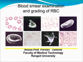
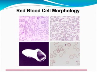
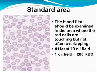
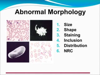
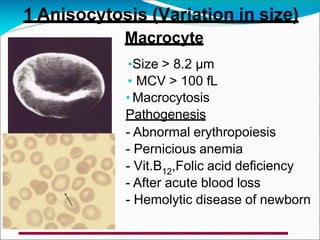
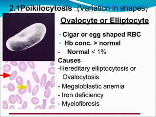
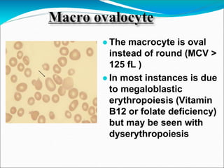
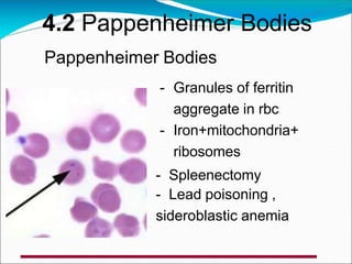
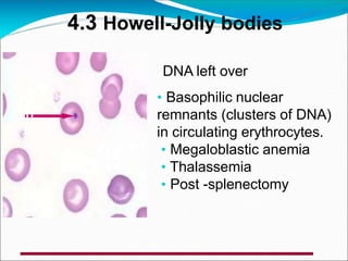
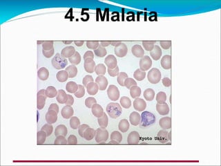
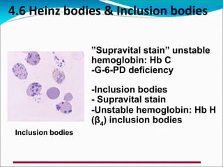
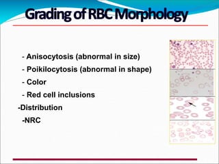
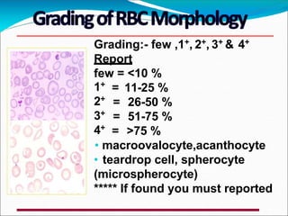
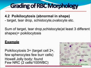
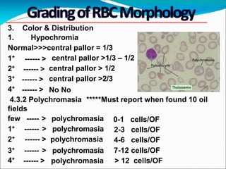
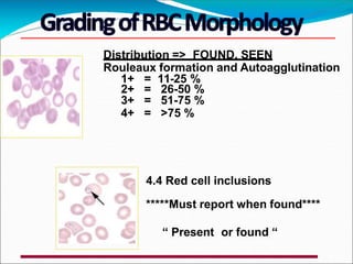
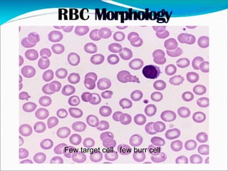
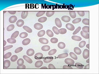
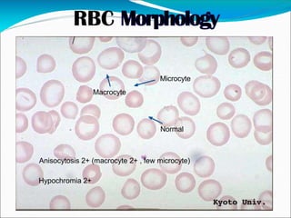
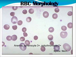
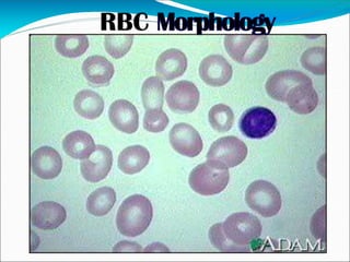
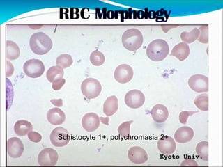
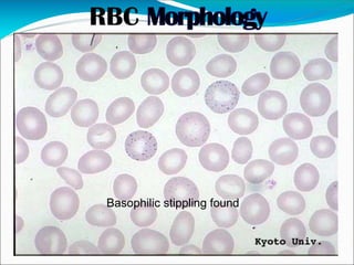
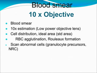
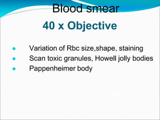
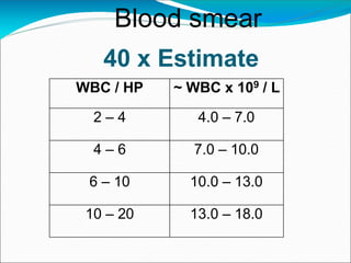
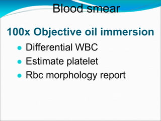
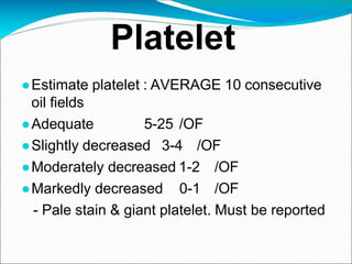
Assoc.Prof. Pornsri Tantiniti
Faculty of Medical Technology
Rangsit University
Blood smear examination
and grading of RBC
●Explain Rbc terminology and pathogenesis
●Detail red blood cells variations in regards to
size, shape, and color etc.
●Grading abnormal red blood cell in patients
●How to examine blood smear
To know about . . . .
Pathogenesis
● Nutritional anemia: Iron deficiency,
Megaloblastic anemia
● Membrane cytoskeletal defect :
Ovalocyte, spherocyte
● Defect metabolite state of rbc :
● E-M pathway (Enz. Deficiency)
● ATP production ( ATP , Na influx),
● Glycolytic Enz.Defect (G6PD Deficiency)
Pathogenesis
● Defect in Hb synthesis & ab.Hb
● Thalassemia
● HbE
● Defect in Lipid bilayer membrane
● Acanthocyte in abeta-liproteinemia
● Anemia chronic disease
● Leukemia
● SLE
Red Blood Cell Morphology
• The blood film
should be examined
in the area where the
red cells are
touching but not
often overlapping.
• At least 10 oil field
• 1 oil field ~ 200 RBC
Standard area
Red cell terminology
• Anisocytosis : variation of red cell sizes
• Poikilocytosis : variation of red cell shapes
• Staining
- Normochromic : red cell with normal color
- Normocytic : red cell with normal size
• Dimorphic red cell : presence of two
different populations of red cell (different in
size , Hb.content , shape ) e.g.
Hypo micro rbc VS. Macrocytic red cell
Red cell terminology
• Inclusion in rbc
• Distribution
• NRC : nucleated red cell
1. Size
2. Shape
3. Staining
4. Inclusion
5. Distribution
6. NRC
Abnormal Morphology
1. Size
Normal RBC (7 µm) >>>
• Variation in size >>>> Anisocytosis
• Macrocyte and Microcyte in blood
smear
Normocyte
• Normal red cells or
erythrocytes show only slight
variation in size and shape.
•Biconcave
•7 µm(6.2-8.2)
•Central pallor 1/3
1. Size
1 Anisocytosis (Variation in size)
Microcyte
•Size <6.2 µm
•MCV < 80 fL
• Microcytosis
Causes
• Thalassemia, sideroblastic
anemia Pyruvate-kinase
deficiency
Macrocyte
•Size > 8.2 µm
• MCV > 100 fL
• Macrocytosis
Pathogenesis
- Abnormal erythropoiesis
- Pernicious anemia
- Vit.B12,Folic acid deficiency
- After acute blood loss
- Hemolytic disease of newborn
1 Anisocytosis (Variation in size)
Normal RBC >>> Biconcave shape
round shape
• Variation in shape >>>> Poikilocytosis
2.1Poikilocytosis (Variation in shapes)
Ovalocyte or Elliptocyte
• Cigar or egg shaped RBC
• Hb conc. > normal
- Normal < 1%
Causes
-Hereditary elliptocytosis or
Ovalocytosis
- Megaloblastic anemia
- Iron deficiency
- Myelofibrosis
●The macrocyte is oval
instead of round (MCV >
125 fL )
●In most instances is due
to megaloblastic
erythropoiesis (Vitamin
B12 or folate deficiency)
but may be seen with
dyserythropoiesis
-Small, spherical cells lacking central pallor
Pathogenesis
-Result from partial removal of RBC
membrane by monocyte-macrophage
system
- Decreased membrane surface
area:cytoplasmic volume ratio
Causes
- Hereditary spherocytosis,
immuno-hemolytic anemia
2. Poikilocytosis or Variation in shape
Spherocyte
2.2 Poikilocytosis
Target cell
-Bull’s eye appearance,Hb
accumulation in center and periphery
with clear interventing
- May be microcyte, nomocyte,
macrocyte.
- Pathogenesis
- Area membrane phospholipids
- Membrane cholesterol
Causes
Liver disorders, iron deficiency,
thalassemia, hemoglobinopathies,
2.3 Poikilocytosis
Teardrop Cell
-Tear drop-shaped cells. Pathogenesis:
pitting function of spleen
Causes
- Thalassemia
- Drug induced “Heinz bodies”
- Extramedullary erythropoiesis
- Myeloid metaplasia
“ Myelofibrosis ”
Stomatocyte
-Cup shape,Uniconcave
Causes
- Cationic drugs
Low “ pH “ response
- Liver disease
- Alcohol intake
- Hereditary stomatocytosis
2.4 Poikilocytosis
2.5 Poikilocytosis
Sickle cell
-Elongated and narrow with one or
both ends curved and pointed,
crescent-shaped.
Causes
- Abnormal Hemoglobin S
2.6 Poikilocytosis
Ecchinocytes (Crenated)
Disc shape and are covered with
10-30 short blunt spicules of
regular form
Causes
Storage artifact (EDTA blood,
delayed in making bl. film)
Acanthocyte (spur cell)
-Spherical shape with 2-20 spicules and
unequal length.
Finger-like projections
Pathogenesis
Expansion of the outer leaflet of lipid
bilayer
Cholesterol /Phospholipid ratio
Causes
- Alcoholic liver cirrhosis, - Hepatitis
- Hereditary abetalipoproteinemia
2.7 Poikilocytosis
- Cell with pairs of spiny 2 or 4-6 regular
projections on surface
- Horned cell
Pathogenesis
Rupture or removal of circular area of
apposed, sealed cellular membrane
projections formed
Causes
- Uremia
- MAHA
- DIC
(Disseminated intravascular coagulation)
Keratocyte (burr cell)
2.8 Poikilocytosis
Schistocyte
Triangular or helmet shaped fragments of RBC
with sharp, pointed projections, and irregular
borders
Pathogenesis
Fragmentation from impact with fibrin strands,
walls of diseased vessels, artificial surfaces,
and turbulent blood flow- Physical/Mechanical
stress
Causes
- Drugs
- Chemical
- Thalassemia
- DIC
(Disseminated intravascular coagulation)
2.9 Poikilocytosis
Normochromia
Hypochromia
3. Staining
Polyochrom
asia
- Normochromia
- Hypochromia
- Polychromasia
Hypochromia
●Central pallor > 1/3,hemoglobin
concentration is decreased
• Hemoglobin concentration decrease
can be caused by decreased amounts
of iron available for hemoglobin
production
• Iron deficiency, thalassemia,
hemoglobinopathies
● Reticulocyte
● Larger than Normal RBC
and purple color
● Normal found 0.2-2%
● Iron deficiency
● Thalassemia
● Hemoglobinopathies
Polyochromasia
4. Inclusion
1. Basophilic Stippling
RNA left over
- Aggregation of rRNA in the
cytoplasm of RBC with Purple-blue
granules
- Lead poisoning
- Pyrimidine-5-nucleotide def.
- Disorders of Hb synthesis
- Hb Constant Spring
4.2 Pappenheimer Bodies
Pappenheimer Bodies
- Granules of ferritin
aggregate in rbc
- Iron+mitochondria+
ribosomes
- Spleenectomy
- Lead poisoning ,
sideroblastic anemia
4.3 Howell-Jolly bodies
DNA left over
• Basophilic nuclear
remnants (clusters of DNA)
in circulating erythrocytes.
• Megaloblastic anemia
• Thalassemia
• Post -splenectomy
“Spindle fibers”
microtubules of
mitotic spindle
- Severe anemia
- Dyserythropoiesis
- Splenectomy
4.4 Carbot’s ring
4.5 Malaria
4.6 Heinz bodies & Inclusion bodies
”Supravital stain” unstable
hemoglobin: Hb C
-G-6-PD deficiency
-Inclusion bodies
- Supravital stain
-Unstable hemoglobin: Hb H
(β4) inclusion bodies
Inclusion bodies
5. Distribution
1. Rouleaux Formation
•Hyperproteinemia
•Macroglobulinemia
- Multiple myeloma
5.2 Agglutination
Agglutination
- Antigen - Antibody
- Cold hemagglutination
(AIHA)
- Paroxysmal cold
hemoglobinuria
Agglutination 3+
6. Nucleated rbc
-Immature rbc mostly are
polychromatic normoblast and
orthochromatic normoblast
- report : no of NRC /100 wbc
ideal area
Screen with objective 10x for good area
- Anisocytosis (abnormal in size)
- Poikilocytosis (abnormal in shape)
- Color
- Red cell inclusions
-Distribution
-NRC
Grading:- few ,1+, 2+, 3+ & 4+
Report
few = <10 %
= 11-25 %
= 26-50 %
= 51-75 %
1+
2+
3+
4+
= >75 %
• macroovalocyte,acanthocyte
• teardrop cell, spherocyte
(microspherocyte)
***** If found you must reported
1. Anisocytosis (Abnormal in size)
- Microcyte
- Macrocyte
Sum of microcyte and macrocyte <,= anisocytosis
Example
Anisocytosis 2+ (microcyte 1+, macrocyte some)
4.2 Poikilocytosis (abnormal in shape)
- target, tear drop, schistocyte,ovalocyte etc.
Sum of target, tear drop,schistocyte(at least 3 different
shapes)= poikilocytosis
Example
Poikilocytosis 3+ (target cell 2+,
few spherocytes few burr cells)
Howell Jolly body: found
Few NRC (2 cells/100WBC)
3. Color & Distribution
1. Hypochromia
Normal>>>central pallor = 1/3
1+
2+
3+
4+
------ >
------ >
------ >
------ >
central pallor >1/3 – 1/2
central pallor > 1/2
central pallor >2/3
No No
4.3.2 Polychromasia *****Must report when found 10 oil
fields
0-1 cells/OF
2-3 cells/OF
4-6 cells/OF
few ----- > polychromasia
1+ ------ > polychromasia
2+ ------ > polychromasia
3+ ------ > polychromasia
4+
------ > polychromasia
7-12 cells/OF
> 12 cells/OF
Distribution => FOUND, SEEN
Rouleaux formation and Autoagglutination
1+ = 11-25 %
2+ = 26-50 %
3+ = 51-75 %
4+ = >75 %
4.4 Red cell inclusions
*****Must report when found****
“ Present or found “
Few target cell , few burr cell
Ovalocytosis 3+
Normal
Microcyte
Macrocyte
Anisocytosis 2+ (macrocyte 2+ , few microcyte)
Hypochromia 2+
Anemia, spherocyte 3+, polychromasia 2+
Schistocyte 1+, polychromasia 2+
Basophilic stippling found
Howell Jolly bodies found
Rouleaux formation 1+
Malaria P.vivax , schiszont found
10 x Objective
● Blood smear
● 10x estimation (Low power objective lens)
● Cell distribution, ideal area (std area)
● RBC agglutination, Rouleaux formation
● Scan abnormal cells (granulocyte precursors,
NRC)
Blood smear
40 x Objective
● Variation of Rbc size,shape, staining
● Scan toxic granules, Howell jolly bodies
● Pappenheimer body
Blood smear
40 x Estimate
WBC / HP ~ WBC x 109 / L
2 – 4 4.0 – 7.0
4 – 6 7.0 – 10.0
6 – 10 10.0 – 13.0
10 – 20 13.0 – 18.0
Blood smear
100x Objective oil immersion
● Differential WBC
● Estimate platelet
● Rbc morphology report
Blood smear
Platelet
●Estimate platelet : AVERAGE 10 consecutive
oil fields
●Adequate 5-25 /OF
●Slightly decreased 3-4 /OF
●Moderately decreased 1-2 /OF
●Markedly decreased 0-1 /OF
- Pale stain & giant platelet. Must be reported
1. BAIN J B, BLOOD CELLS A PRACTICAL GUIDE.
BLACKWELL SCIENCE, 2002
2. นันทรัตน โฆมานะสิน, นพมาศ เข็มทองหลาง, มณเฑียร
พันธุ เมธากุล. การทดสอบพื้นฐานทางห องปฏิบัติการโลหิต
วิทยา ภาค วิชาจุลทรรศน คลินิก คณะเทคนิคการแพทย
มหาวิทยาลัย ขอนแก น. พิมพคร ััังที่ 2. หจก. โรงพิมพคล
ัังนานาวิทยา, 2544.
3. สุภินันท สเป ค-สายเชื้อ. ภาพสีประกอบ โลหิตวิทยา.พิมพ
ครั้งที่ 4.
กรุงเทพ: เอช ที พี เพรส, 2544.

Blood smear examination 15.ppt.pptx

  • 1.
    Assoc.Prof. Pornsri Tantiniti Facultyof Medical Technology Rangsit University Blood smear examination and grading of RBC
  • 2.
    ●Explain Rbc terminologyand pathogenesis ●Detail red blood cells variations in regards to size, shape, and color etc. ●Grading abnormal red blood cell in patients ●How to examine blood smear To know about . . . .
  • 3.
    Pathogenesis ● Nutritional anemia:Iron deficiency, Megaloblastic anemia ● Membrane cytoskeletal defect : Ovalocyte, spherocyte ● Defect metabolite state of rbc : ● E-M pathway (Enz. Deficiency) ● ATP production ( ATP , Na influx), ● Glycolytic Enz.Defect (G6PD Deficiency)
  • 4.
    Pathogenesis ● Defect inHb synthesis & ab.Hb ● Thalassemia ● HbE ● Defect in Lipid bilayer membrane ● Acanthocyte in abeta-liproteinemia ● Anemia chronic disease ● Leukemia ● SLE
  • 5.
    Red Blood CellMorphology
  • 6.
    • The bloodfilm should be examined in the area where the red cells are touching but not often overlapping. • At least 10 oil field • 1 oil field ~ 200 RBC Standard area
  • 7.
    Red cell terminology •Anisocytosis : variation of red cell sizes • Poikilocytosis : variation of red cell shapes • Staining - Normochromic : red cell with normal color - Normocytic : red cell with normal size • Dimorphic red cell : presence of two different populations of red cell (different in size , Hb.content , shape ) e.g. Hypo micro rbc VS. Macrocytic red cell
  • 8.
    Red cell terminology •Inclusion in rbc • Distribution • NRC : nucleated red cell
  • 9.
    1. Size 2. Shape 3.Staining 4. Inclusion 5. Distribution 6. NRC Abnormal Morphology
  • 10.
    1. Size Normal RBC(7 µm) >>> • Variation in size >>>> Anisocytosis • Macrocyte and Microcyte in blood smear
  • 11.
    Normocyte • Normal redcells or erythrocytes show only slight variation in size and shape. •Biconcave •7 µm(6.2-8.2) •Central pallor 1/3 1. Size
  • 12.
    1 Anisocytosis (Variationin size) Microcyte •Size <6.2 µm •MCV < 80 fL • Microcytosis Causes • Thalassemia, sideroblastic anemia Pyruvate-kinase deficiency
  • 13.
    Macrocyte •Size > 8.2µm • MCV > 100 fL • Macrocytosis Pathogenesis - Abnormal erythropoiesis - Pernicious anemia - Vit.B12,Folic acid deficiency - After acute blood loss - Hemolytic disease of newborn 1 Anisocytosis (Variation in size)
  • 14.
    Normal RBC >>>Biconcave shape round shape • Variation in shape >>>> Poikilocytosis
  • 15.
    2.1Poikilocytosis (Variation inshapes) Ovalocyte or Elliptocyte • Cigar or egg shaped RBC • Hb conc. > normal - Normal < 1% Causes -Hereditary elliptocytosis or Ovalocytosis - Megaloblastic anemia - Iron deficiency - Myelofibrosis
  • 16.
    ●The macrocyte isoval instead of round (MCV > 125 fL ) ●In most instances is due to megaloblastic erythropoiesis (Vitamin B12 or folate deficiency) but may be seen with dyserythropoiesis
  • 17.
    -Small, spherical cellslacking central pallor Pathogenesis -Result from partial removal of RBC membrane by monocyte-macrophage system - Decreased membrane surface area:cytoplasmic volume ratio Causes - Hereditary spherocytosis, immuno-hemolytic anemia 2. Poikilocytosis or Variation in shape Spherocyte
  • 18.
    2.2 Poikilocytosis Target cell -Bull’seye appearance,Hb accumulation in center and periphery with clear interventing - May be microcyte, nomocyte, macrocyte. - Pathogenesis - Area membrane phospholipids - Membrane cholesterol Causes Liver disorders, iron deficiency, thalassemia, hemoglobinopathies,
  • 19.
    2.3 Poikilocytosis Teardrop Cell -Teardrop-shaped cells. Pathogenesis: pitting function of spleen Causes - Thalassemia - Drug induced “Heinz bodies” - Extramedullary erythropoiesis - Myeloid metaplasia “ Myelofibrosis ”
  • 20.
    Stomatocyte -Cup shape,Uniconcave Causes - Cationicdrugs Low “ pH “ response - Liver disease - Alcohol intake - Hereditary stomatocytosis 2.4 Poikilocytosis
  • 21.
    2.5 Poikilocytosis Sickle cell -Elongatedand narrow with one or both ends curved and pointed, crescent-shaped. Causes - Abnormal Hemoglobin S
  • 22.
    2.6 Poikilocytosis Ecchinocytes (Crenated) Discshape and are covered with 10-30 short blunt spicules of regular form Causes Storage artifact (EDTA blood, delayed in making bl. film)
  • 23.
    Acanthocyte (spur cell) -Sphericalshape with 2-20 spicules and unequal length. Finger-like projections Pathogenesis Expansion of the outer leaflet of lipid bilayer Cholesterol /Phospholipid ratio Causes - Alcoholic liver cirrhosis, - Hepatitis - Hereditary abetalipoproteinemia 2.7 Poikilocytosis
  • 24.
    - Cell withpairs of spiny 2 or 4-6 regular projections on surface - Horned cell Pathogenesis Rupture or removal of circular area of apposed, sealed cellular membrane projections formed Causes - Uremia - MAHA - DIC (Disseminated intravascular coagulation) Keratocyte (burr cell) 2.8 Poikilocytosis
  • 25.
    Schistocyte Triangular or helmetshaped fragments of RBC with sharp, pointed projections, and irregular borders Pathogenesis Fragmentation from impact with fibrin strands, walls of diseased vessels, artificial surfaces, and turbulent blood flow- Physical/Mechanical stress Causes - Drugs - Chemical - Thalassemia - DIC (Disseminated intravascular coagulation) 2.9 Poikilocytosis
  • 26.
  • 27.
    Hypochromia ●Central pallor >1/3,hemoglobin concentration is decreased • Hemoglobin concentration decrease can be caused by decreased amounts of iron available for hemoglobin production • Iron deficiency, thalassemia, hemoglobinopathies
  • 28.
    ● Reticulocyte ● Largerthan Normal RBC and purple color ● Normal found 0.2-2% ● Iron deficiency ● Thalassemia ● Hemoglobinopathies Polyochromasia
  • 29.
    4. Inclusion 1. BasophilicStippling RNA left over - Aggregation of rRNA in the cytoplasm of RBC with Purple-blue granules - Lead poisoning - Pyrimidine-5-nucleotide def. - Disorders of Hb synthesis - Hb Constant Spring
  • 30.
    4.2 Pappenheimer Bodies PappenheimerBodies - Granules of ferritin aggregate in rbc - Iron+mitochondria+ ribosomes - Spleenectomy - Lead poisoning , sideroblastic anemia
  • 31.
    4.3 Howell-Jolly bodies DNAleft over • Basophilic nuclear remnants (clusters of DNA) in circulating erythrocytes. • Megaloblastic anemia • Thalassemia • Post -splenectomy
  • 32.
    “Spindle fibers” microtubules of mitoticspindle - Severe anemia - Dyserythropoiesis - Splenectomy 4.4 Carbot’s ring
  • 33.
  • 34.
    4.6 Heinz bodies& Inclusion bodies ”Supravital stain” unstable hemoglobin: Hb C -G-6-PD deficiency -Inclusion bodies - Supravital stain -Unstable hemoglobin: Hb H (β4) inclusion bodies Inclusion bodies
  • 35.
    5. Distribution 1. RouleauxFormation •Hyperproteinemia •Macroglobulinemia - Multiple myeloma
  • 36.
    5.2 Agglutination Agglutination - Antigen- Antibody - Cold hemagglutination (AIHA) - Paroxysmal cold hemoglobinuria Agglutination 3+
  • 37.
    6. Nucleated rbc -Immaturerbc mostly are polychromatic normoblast and orthochromatic normoblast - report : no of NRC /100 wbc
  • 38.
    ideal area Screen withobjective 10x for good area
  • 39.
    - Anisocytosis (abnormalin size) - Poikilocytosis (abnormal in shape) - Color - Red cell inclusions -Distribution -NRC
  • 40.
    Grading:- few ,1+,2+, 3+ & 4+ Report few = <10 % = 11-25 % = 26-50 % = 51-75 % 1+ 2+ 3+ 4+ = >75 % • macroovalocyte,acanthocyte • teardrop cell, spherocyte (microspherocyte) ***** If found you must reported
  • 41.
    1. Anisocytosis (Abnormalin size) - Microcyte - Macrocyte Sum of microcyte and macrocyte <,= anisocytosis Example Anisocytosis 2+ (microcyte 1+, macrocyte some)
  • 42.
    4.2 Poikilocytosis (abnormalin shape) - target, tear drop, schistocyte,ovalocyte etc. Sum of target, tear drop,schistocyte(at least 3 different shapes)= poikilocytosis Example Poikilocytosis 3+ (target cell 2+, few spherocytes few burr cells) Howell Jolly body: found Few NRC (2 cells/100WBC)
  • 43.
    3. Color &Distribution 1. Hypochromia Normal>>>central pallor = 1/3 1+ 2+ 3+ 4+ ------ > ------ > ------ > ------ > central pallor >1/3 – 1/2 central pallor > 1/2 central pallor >2/3 No No 4.3.2 Polychromasia *****Must report when found 10 oil fields 0-1 cells/OF 2-3 cells/OF 4-6 cells/OF few ----- > polychromasia 1+ ------ > polychromasia 2+ ------ > polychromasia 3+ ------ > polychromasia 4+ ------ > polychromasia 7-12 cells/OF > 12 cells/OF
  • 44.
    Distribution => FOUND,SEEN Rouleaux formation and Autoagglutination 1+ = 11-25 % 2+ = 26-50 % 3+ = 51-75 % 4+ = >75 % 4.4 Red cell inclusions *****Must report when found**** “ Present or found “
  • 45.
    Few target cell, few burr cell
  • 46.
  • 47.
  • 48.
    Anemia, spherocyte 3+,polychromasia 2+
  • 50.
  • 51.
  • 52.
  • 53.
  • 54.
    Malaria P.vivax ,schiszont found
  • 55.
    10 x Objective ●Blood smear ● 10x estimation (Low power objective lens) ● Cell distribution, ideal area (std area) ● RBC agglutination, Rouleaux formation ● Scan abnormal cells (granulocyte precursors, NRC) Blood smear
  • 56.
    40 x Objective ●Variation of Rbc size,shape, staining ● Scan toxic granules, Howell jolly bodies ● Pappenheimer body Blood smear
  • 57.
    40 x Estimate WBC/ HP ~ WBC x 109 / L 2 – 4 4.0 – 7.0 4 – 6 7.0 – 10.0 6 – 10 10.0 – 13.0 10 – 20 13.0 – 18.0 Blood smear
  • 58.
    100x Objective oilimmersion ● Differential WBC ● Estimate platelet ● Rbc morphology report Blood smear
  • 59.
    Platelet ●Estimate platelet :AVERAGE 10 consecutive oil fields ●Adequate 5-25 /OF ●Slightly decreased 3-4 /OF ●Moderately decreased 1-2 /OF ●Markedly decreased 0-1 /OF - Pale stain & giant platelet. Must be reported
  • 60.
    1. BAIN JB, BLOOD CELLS A PRACTICAL GUIDE. BLACKWELL SCIENCE, 2002 2. นันทรัตน โฆมานะสิน, นพมาศ เข็มทองหลาง, มณเฑียร พันธุ เมธากุล. การทดสอบพื้นฐานทางห องปฏิบัติการโลหิต วิทยา ภาค วิชาจุลทรรศน คลินิก คณะเทคนิคการแพทย มหาวิทยาลัย ขอนแก น. พิมพคร ััังที่ 2. หจก. โรงพิมพคล ัังนานาวิทยา, 2544. 3. สุภินันท สเป ค-สายเชื้อ. ภาพสีประกอบ โลหิตวิทยา.พิมพ ครั้งที่ 4. กรุงเทพ: เอช ที พี เพรส, 2544.