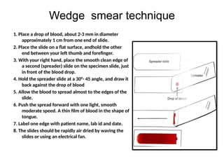
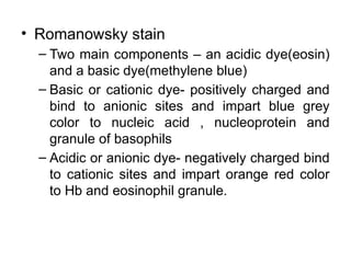
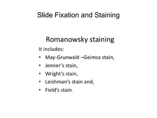
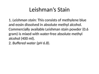
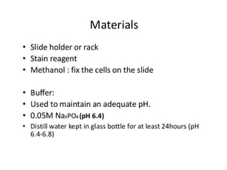
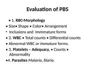
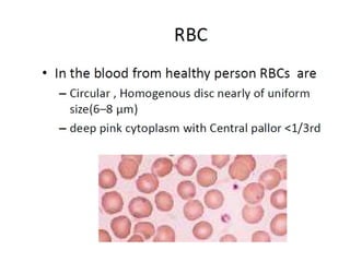
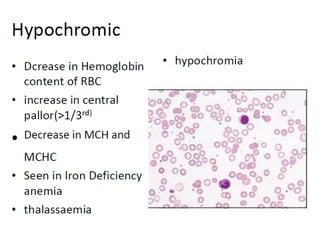
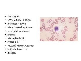
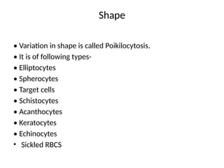
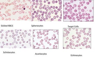
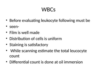
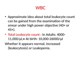
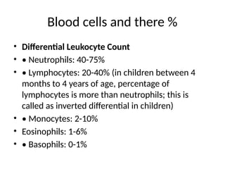
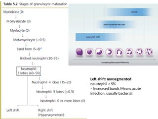
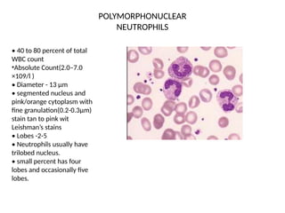
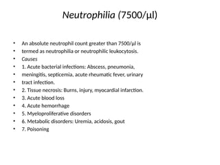
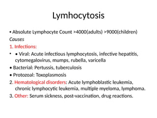
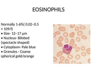
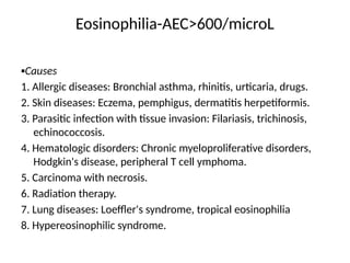
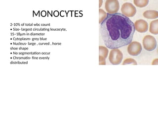
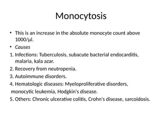
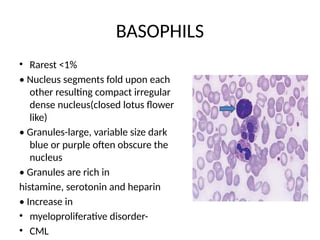
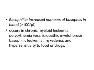
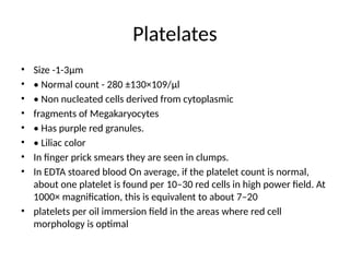
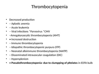
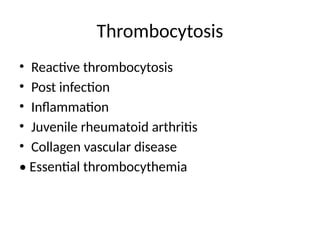
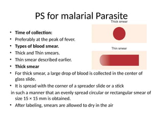
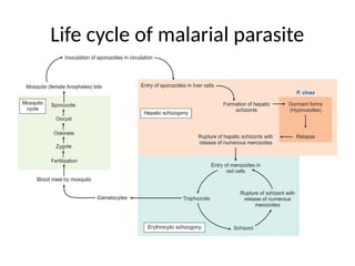
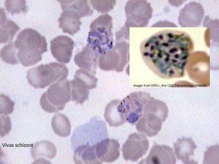
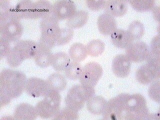
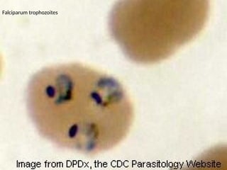
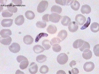
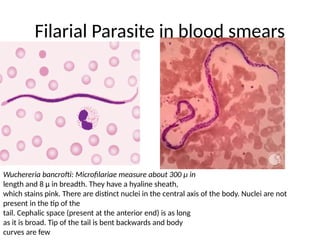
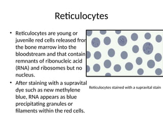
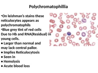
BASIC HAEMATOLOGICAL
TECHNIQUES (PERIPHERAL
BLOOD SMEAR)
Dr. Prajkta Pawar (Gaddewar)
Assistant Professor
Topics to cover
• Practical combined with self directed learning 8:
• Peripheral blood smear examination and staining
• Interpretation and reporting
• Importance of reticulocyte count
• Blood smears of neutrophillic leukococytosis,
eosinophillia, lymphocytosis, leukemoid reaction,
differentiation of leukemoid reaction CML,
• Smears of Malaria and Filaria
Peripheral smear
• Peripheral Smear (PS) : Specimen for
microscopic examination prepared by
spreading a drop of blood across the slide and
staining with romanowsky stain.
INDICATIONS FOR A PERIPHERAL BLOOD FILM
 unexplained cytopenia: anaemia, leucopenia or thrombocytopenia
 unexplained leukocytosis, lymphocytosis or monocytosis
 unexplained jaundice or haemolysis; features of congenital haemolytic
anaemias such as splenomegaly, jaundice or bone pains
 suspected chronic or acute myeloproliferative disease e.g. chronic
myeloid leukaemia, Acute myeloid leukemias
 suspected organ failure such as renal disease, liver failure
 features of hyperviscosity syndrome as in paraproteinaemias,
leukaemic hyperleucocytosis, polycythaemia
 severe bacterial sepsis and parasitic infections
 malignancies with possible bone marrow involvement
 suspected cases of nutritional anaemia
 suspected chronic lymphoproliferative diseases
Peripheral Blood Smear Techniques
• Objective(Technical)
1. Specimen Collection
2. Peripheral Smear Preparation
3. Staining of Peripheral Blood Smear
Specimen Collection
• Venipuncture
– most commonly used method
• Capillary blood (skin puncture) best
– commonly used in infants, children and in
some point of care screening test
• Specimen container
– Glass/ plastic container with added anticoagulant
or additive
• Anticoagulant: Mostly EDTA (Ethylene Diamine
Tetra-acetic Acid (EDTA)
• Plebotomy Procedure
– Verify patient identity
– Check plebotomy tray
– Vein of forearm preferably used
– Follow asepsis, use rubber tourniquet,
– withdraw blood slowly in syringe
– Apply adhesive dressing at puncture site
• Capillary blood (skin puncture)
– Site distal third / forth finger palm, heel pad, great toe
– Lancet is pierced 2-3 mm of depth
– Free spontaneous flow of blood drop is recommended
– Difference -PCV, RBC, Hb concentration
high;conversely platelet count found low in skin prick
blood
• Capillary blood collection
• In adults - ring or middle finger or ear lobe.
• In infants - heel or great toe.
PREPARATION OF A PERIPHERAL BLOOD FILM SLIDE
• Wedge smear technique
• The spun smear
• Cover slip smear technique
And sometimes
• Buffy coat smear for WBCs<1.0 x 10⁹
/ L
• Thick blood smear for blood parasites
Wedge smear technique
1. Place a drop of blood, about 2-3 mm in diameter
approximately 1 cm from one end of slide.
2. Place the slide on a flat surface, andhold the other
end between your left thumb and forefinger.
3. With your right hand, place the smooth clean edge of
a second (spreader) slide on the specimen slide, just
in front of the blood drop.
4. Hold the spreader slide at a 30°- 45 angle, and draw it
back against the drop of blood
5. Allow the blood to spread almost to the edges of the
slide.
6. Push the spread forward with one light, smooth
moderate speed. A thin film of blood in the shape of
tongue.
7. Label one edge with patient name, lab id and date.
8. The slides should be rapidly air dried by waving the
slides or using an electrical fan.
Peripheral Smear Making
• Smear is made by smooth, forward movement
of the 'spreader' along the slide.
• The whole drop should be used up 1 cm before
the end of the slide.
• The length of the smear should be about 3 cm .
• The 'spreader' should not be raised above the
slide surface till whole drop of blood is spread
out.
• Control thickness of the smear by changing the
angle of spreader slide
• Allow the blood film to air-dry completely before
staining.
• Smear is rapidly dried by waving it in the air or keeping
it under an electric fan.
• Slow drying causes shrinkage artifact of red cells.
• Do not blow to dry.
• The moisture from your breath will cause RBC
artifact
Precaution:
Too large drop = too thick smear
Too small drop = too thin smear
Angle correction:
1. In case of Polycythemia:
– high Hematocrite
– angle should be lowered
- ensure that the smear made is not too
thick
2. Too low Hematocrite
– Angle should be raised
A well-spread blood smear
• tongue-shaped with Head,
body and a smooth tail,
• does not cover the entire
area of the slide,
• has both thick and thin
areas with gradual transition
• does not contain any lines or
holes.
Figure 4.1 Blood films made on slides. (A) A well-made film. (B) An irregular patchy
film on a dusty slide. (C) A film that is too thick. (D) A film that has been spread with
inconsistent pressure and using an irregularly edged spreader, resulting in long tails.
(E)A film made on a very greasy slide.
• Romanowsky stain
– Two main components – an acidic dye(eosin)
and a basic dye(methylene blue)
– Basic or cationic dye- positively charged and
bind to anionic sites and impart blue grey
color to nucleic acid , nucleoprotein and
granule of basophils
– Acidic or anionic dye- negatively charged bind
to cationic sites and impart orange red color
to Hb and eosinophil granule.
Slide Fixation and Staining
Leishman’s Stain
1. Leishman stain: This consists of methylene blue
and eosin dissolved in absolute methyl alcohol.
Commercially available Leishman stain powder (0.6
gram) is mixed with water-free absolute methyl
alcohol (400 ml).
2. Buffered water (pH 6.8).
• Method
– Slide fix in methanol for 2-3 minute
– Smear cover with leishman stain for 2 minute
– Add twice the volume of buffered water and
leave for 5 – 7 minute. (A metallic colour scum
apperars over the surface.)
– Wash stain away in a steam of buffer water /
tap water
– Wipe back of slide clean and set it up right to
dry
Leishman stain
• Red cells: pink-red or deep pink
• Polychromatic cells (Reticulocytes):
Gray-blue
• Neutrophils: Pale pink cytoplasm;
mauve-purple granules
• Eosinophils: Pale-pink cytoplasm;
orange-red granules
• Basophils: Blue cytoplasm; dark
blue-violet granules
• Monocytes: Gray-blue cytoplasm;
fine reddish (azurophil) granules
• Small lymphocytes: Dark blue
cytoplasm
• Platelets: Purple
• Nuclei of all cells: Purple-violet
Factors influence smear staining
method
• Blood smear may be under or over stained based
on the following
• Concentration of the stain used
– Low concentration: pale coloured cells (under
staining)
– High concentration: dark stained smear (over stained)
• Time of exposure the stain and the buffer
– Too long: overstaining,
– Too short: understaining
Interpretation of blood smears.
• A peripheral blood smear has three parts:
Head, body, and tail .
• A blood smear should be examined in an
orderly manner.
• Initially observe under low power objective
(10×) to assess
• whether the film is properly spread and
stained,
• Cell distribution, and to select an area for
examination of blood cells.
• Best morphologic details are seen in the area
where red cells are just touching one another.
• Low power view useful to see
• Rouleaux formation,
• autoagglutination of red cells,
• microfilaria.
• High power objective (45×)
is suitable for
• examination of red cell
morphology
• for differential leukocyte
count.
• A rough estimate of total
leukocyte count can be
obtained can crosscheck
the total leukocyte count
done by manual counting
or automated method.
• Oil-immersion objective
(100×) used for more
detailed examination of any
abnormal cells.
Too thick for
examination
Too thin for examination
RBCs are just overlapping adequete for examination
Evaluation of PBS
• 1. RBC-Morphology
• Size• Shape • Color• Arrangement
• Inclusions and Immmature forms
• 2. WBC • Total counts • Differential counts
• Abnormal WBC or immature forms.
• 3. Platelets – Adequacy, • Counts •
Abnormality
•4. Parasites Malaria, filaria.
Variation In Size
• Anisocytosis- Variation in size of the red blood
cells
• Normal MCV is -80-100 fl
• Microcytes ( MCV <80 fl)
• Macrocytes (MCV >100fl)
• Anisocytosis is a feature of most anemias.
Microcytic hypochromic anemia
• Macrocytes
• • When MCV of RBC is
• Increased(>100fl)
• • Macro- ovalocytes are
• seen in Megaloblastic
• anemia
• • Myledysplastic
• syndrome.
• • Round Macrocytes seen
• in Alcoholism, Liver
• disease.
Shape
• Variation in shape is called Poikilocytosis.
• It is of following types-
• Elliptocytes
• Spherocytes
• Target cells
• Schistocytes
• Acanthocytes
• Keratocytes
• Echinocytes
• Sickled RBCS
Sickled RBCS Spherocytes Target Cells
Schistocytes
Acantocytes
Echinocytes
WBCs
• Before evaluating leukocyte following must be
• seen-
• Film is well made
• Distribution of cells is uniform
• Staining is satisfactory
• While scanning estimate the total leucocyte
count
• Differential count is done at oil immersion
Scanning technique for WBC differential count and
morphologic evaluation
• Ten microscopic fields are examined in a vertical
direction from bottom to top or top to bottom
• Slide is horizontally moved to the next field
• Ten microscopic fields are counted vertically.
• Procedure is repeated until 100 WBCS have been
counted (zig zag motion)
Differential Leukocyte Count
start counting the types
of WBCs and go on
entering
Polymorphs (P) ,
Lymphocytes(L),
Monocytes(M)
Eosinophils(E)
Basophils( B )in a box
having 100 cubes
After counting there
number is written as
percentage
Uses of DLC
• 1. To support the diagnosis of infectious,
inflammatory,
• or allergic disorders.
• 2. Diagnosis of malignant blood disorders.
The peripheral smear report is given in this
format
• RBC: Normocytic Normochromic
or there is anisocytosis (variation in size),
anisopoikilocytosis (variation in shape),
anisochromasia (hypochromic, or
hyperchromic should be mentioned). Red cell
inclusions if presents should be mentioned (as
howell jolly bodies)
WBC
• Approximate idea about total leukocyte count
can be gained from the examination of the
smear under high power objective (40× or
45×).
• Total Leukocyte count : In Adults: 4000-
11,000/μl,• At birth: 10,000-26000/μl
Whether it appears normal, increased
(leukocytosis) or Leukopenia.
DLC
• Differential leukocyte count (DLC): DLC refers
to relative proportion of different leukocytes
expressed as a percentage
• P% L%M%E%B% total 100%
• Abnormal findings as shift to left i.e immature
form of neutrophils or activated lymphocytes,
or blast if present should be mentioned.
• Platelets: adequate or increased or depleted
should be mentioned.
• Parasite: seen or not seen should be
mentioned.
One Normal report
• RBC: Normocytic Normochromic
• TLC: Within normal limits
• DLC: P-60%, L-35%, M-3%, E-2%, B-1%
• Platelets: adequate
• Parasite: Not seen
Blood cells and there %
• Differential Leukocyte Count
• • Neutrophils: 40-75%
• • Lymphocytes: 20-40% (in children between 4
months to 4 years of age, percentage of
lymphocytes is more than neutrophils; this is
called as inverted differential in children)
• • Monocytes: 2-10%
• Eosinophils: 1-6%
• • Basophils: 0-1%
Left-shift: nonsegmented
neutrophil > 5%
– Increased bands Means acute
infection, usually bacterial
POLYMORPHONUCLEAR
NEUTROPHILS
• 40 to 80 percent of total
WBC count
•Absolute Count(2.0–7.0
×109/l )
• Diameter - 13 μm
• segmented nucleus and
pink/orange cytoplasm with
fine granulation(0.2-0.3μm)
stain tan to pink wit
Leishman’s stains
• Lobes -2-5
• Neutrophils usually have
trilobed nucleus.
• small percent has four
lobes and occasionally five
lobes.
Neutrophilia (7500/μl)
• An absolute neutrophil count greater than 7500/μl is
• termed as neutrophilia or neutrophilic leukocytosis.
• Causes
• 1. Acute bacterial infections: Abscess, pneumonia,
• meningitis, septicemia, acute rheumatic fever, urinary
• tract infection.
• 2. Tissue necrosis: Burns, injury, myocardial infarction.
• 3. Acute blood loss
• 4. Acute hemorrhage
• 5. Myeloproliferative disorders
• 6. Metabolic disorders: Uremia, acidosis, gout
• 7. Poisoning
Leukemoid reaction
• This refers to the presence of markedly
increased total leukocyte count
(>50,000/cmm) with immature cells in
peripheral blood resembling leukaemia
• but occurring in non-leukemic
disorders .
• Its causes are:
• • Severe bacterial infections, e.g.
septicemia, pneumonia
• • Severe hemorrhage
• • Severe acute hemolysis
• Poisoning
• • Burns
• • Carcinoma metastatic to bone
marrow
• Leukemoid reaction should be
differentiated from
• chronic myeloid leukemia
Leukemoid reaction and Chronic Myeloid
Leukemia
Presentation Leukemoid reaction Chronic myeloid Leukemia
Leukocyte alkaline
phosphatase score
Normal or increased Decreased
Leukemoid reaction showing mostly
neutrophis with few bands and
metamyelocytes
Chronic myeloid leukemia showing
neutrophils with many myelocytes and
basophils
LYMPHOCYTES
• • 20-40% of total wbc count
• • two types
• 1. Small lymphocyte(6-10μm)
• 2. Large lymphocyte(12-15μm)
• • Nucleus-single, sharply
• defined, stain dark blue on
• Wright’s stain
• • Cytoplasm- Pale blue
• • Large lymphocytes less
• densely stain nuclei &
• abundant cytoplasm
• • Few round purple(azure)
• granules are present
Lymhocytosis
▪ Absolute Lymphocyte Count >4000(adults) >9000(children)
Causes
1. Infections:
• • Viral: Acute infectious lymphocytosis, infective hepatitis,
cytomegalovirus, mumps, rubella, varicella
• Bacterial: Pertussis, tuberculosis
• Protozoal: Toxoplasmosis
2. Hematological disorders: Acute lymphoblastic leukemia,
chronic lymphocytic leukemia, multiple myeloma, lymphoma.
3. Other: Serum sickness, post-vaccination, drug reactions.
EOSINOPHILS
Normally 1-6%( 0.02–0.5
× 109/l)
• Size- 12–17 μm
• Nucleus- Bilobed
(spectacle shaped)
• Cytoplasm- Pale blue
• Granules - Coarse
spherical gold/orange
Eosinophilia-AEC>600/microL
▪Causes
1. Allergic diseases: Bronchial asthma, rhinitis, urticaria, drugs.
2. Skin diseases: Eczema, pemphigus, dermatitis herpetiformis.
3. Parasitic infection with tissue invasion: Filariasis, trichinosis,
echinococcosis.
4. Hematologic disorders: Chronic myeloproliferative disorders,
Hodgkin's disease, peripheral T cell ymphoma.
5. Carcinoma with necrosis.
6. Radiation therapy.
7. Lung diseases: Loeffler's syndrome, tropical eosinophilia
8. Hypereosinophilic syndrome.
MONOCYTES
2-10% of total wbc count
• Size- largest circulating leucocyte,
15–18μm in diameter
• Cytoplasm- grey blue
• Nucleus- large , curved , horse
shoe shape
• No segmentation occur
• Chromatin- fine evenly
distributed
Monocytosis
• This is an increase in the absolute monocyte count above
1000/μl.
• Causes
1. Infections: Tuberculosis, subacute bacterial endocarditis,
malaria, kala azar.
2. Recovery from neutropenia.
3. Autoimmune disorders.
4. Hematologic diseases: Myeloproliferative disorders,
monocytic leukemia, Hodgkin's disease.
5. Others: Chronic ulcerative colitis, Crohn's disease, sarcoidosis.
BASOPHILS
• Rarest <1%
• Nucleus segments fold upon each
other resulting compact irregular
dense nucleus(closed lotus flower
like)
• Granules-large, variable size dark
blue or purple often obscure the
nucleus
• Granules are rich in
histamine, serotonin and heparin
• Increase in
• myeloproliferative disorder-
• CML
• Basophilia: Increased numbers of basophils in
blood (>100/μl)
• occurs in chronic myeloid leukemia,
polycythemia vera, idiopathic myelofibrosis,
basophilic leukemia, myxedema, and
hypersensitivity to food or drugs.
Platelates
• Size -1-3μm
• • Normal count - 280 ±130×109/μl
• • Non nucleated cells derived from cytoplasmic
• fragments of Megakaryocytes
• • Has purple red granules.
• • Liliac color
• In finger prick smears they are seen in clumps.
• In EDTA stoared blood On average, if the platelet count is normal,
about one platelet is found per 10–30 red cells in high power field. At
1000× magnification, this is equivalent to about 7–20
• platelets per oil immersion field in the areas where red cell
morphology is optimal
Thrombocytopenia
• Decreased production
• – Aplastic anemia
• – Acute leukemia
• – Viral infections *Parvovirus *CMV
• −Amegakaryocytic thrombocytopenia (AMT)
• • Increased destruction
• – Immune thrombocytopenia
• – Idiopathic thrombocytopenic purpura (ITP)
• – Neonatal alloimmune thrombocytopenia (NAITP)
• – Disseminated intravascular coagulation (DIC)
• – Hypersplenism
• • Pseudothrombocytopenia- due to clumpping of pltelates in EDTA bulb
• Platelet Satellism Occurs in EDTA stored blood
Thrombocytosis
• Reactive thrombocytosis
• Post infection
• Inflammation
• Juvenile rheumatoid arthritis
• Collagen vascular disease
• Essential thrombocythemia
PS for malarial Parasite
• Time of collection:
• Preferably at the peak of fever.
• Types of blood smear.
• Thick and Thin smears.
• Thin smear described earlier.
• Thick smear
• For thick smear, a large drop of blood is collected in the center of
glass slide.
• It is spread with the corner of a spreader slide or a stick
in such a manner that an evenly spread circular or rectangular smear of
size 15 × 15 mm is obtained.
• After labeling, smears are allowed to dry in the air
Life cycle of malarial parasite
Plasmodium Vivax
Early trophozoit:Blue cytoplasmic ring 1/3rd the
diameter of red cell; one side of ring thicker;
red chromatin at thinner part of ring
Late trophozoit: Irregularly thick the cytoplasmic
ring( ameboid); large red chromatin dot
Schizont:12-24 merozoites arranged rosette-like
granular yellow-brown pigment in center
Gametocyte:Large and spherical
Plasmodium falciparum
Often only ring forms and gametocytes are seen; multiple rings in a
single cell common; red cell size normal; high parasite density.
Early trophozoite: A delicate, small fine cytoplasmic ring with 1-2
small chromatindots; ring may be attached to the red cell margin
(accole form).
Late trophozoite:Compact blue ring with1-2 red chromatin dots
Schizont: Very rarely seen exceptin cerebral malaria; contains 18-32
merozoites which fill2/3rds of a red cell; a single block of dark
brown pigment
Gametocyte: Crescentic or banana shaped; larger than a red cell
Vivax trophozoite
Vivax trophozoite
Vivax schizont
Falciparum trophozoites
Falciparum gametocytes
Filarial Parasite in blood smears
Wuchereria bancrofti: Microfilariae measure about 300 μ in
length and 8 μ in breadth. They have a hyaline sheath,
which stains pink. There are distinct nuclei in the central axis of the body. Nuclei are not
present in the tip of the
tail. Cephalic space (present at the anterior end) is as long
as it is broad. Tip of the tail is bent backwards and body
curves are few
Reticulocytes
• Reticulocytes are young or
juvenile red cells released from
the bone marrow into the
bloodstream and that contain
remnants of ribonucleic acid
(RNA) and ribosomes but no
nucleus.
• After staining with a supravital
dye such as new methylene
blue, RNA appears as blue
precipitating granules or
filaments within the red cells.
Reticulocytes stained with a supravital stain
Polychromatophillia
•On leishman’s stains these
reticulocytes appears as
polychromatophils
•Blue grey tint of red cells
Due to Hb and RNA(Residual) in
young cells.
• Larger than normal and
may lack central pallor.
• Implies Reticulocytosis
• Seen in
• Hemolysis
• Acute blood loss
Reticulocyte Count
• Reticulocyte count is performed to assess erythropoietic activity of the bone
marrow.
• As one of the baseline studies in anemia with no obvious cause.
• To diagnose anemia due to ineffective erythropoiesis.
• In hypoplastic anemia or in ineffective erythropoiesis, reticulocyte count is low as
compared to the degree of anemia. Increased erythropoiesis (e.g. in hemolytic
anemia, blood loss, or specific treatment of nutritional
anemia) is associated with increased reticulocyte count.
• To assess response to specific therapy in iron deficiency and megaloblastic
anemias.
• To assess response to erythropoietin therapy in anemia of chronic renal failure.
• To follow the course of bone marrow transplantation for engraftment
• To assess recovery from myelosuppressive therapy
• To assess anemia in neonate
Questions on this topic
1) Name three romanowsky stain.
2) Three causes of eosiophillia
3) Three causes of reticulocytosis
4) Three causes of leukemoid reactions.
5) Three indications for making peripheral smears.
6) Three causes of neutrophillia.
7) Three causes of lymphocytosis.
8) Two causes of basophilia.
9) Name two blood parasites.
10) Name three causes thrombocytosis.
Practical combined with self directed learning 8
• Instructions
• All students needs to prepare voice over PPT on below mentioned topics
and submit in there respective what’s up group.
• (First four-1st
topic, second four -2 nd topic, Third four- 3rd
topic, Fourth
group of 4 or 5 on fourth topic.)
• 1st
topic- Peripheral smear Indications and making of peripheral smears.
Characteristic of good peripheral smear.
• 2nd
Topic- Types of Romanowskys stain. Leishman’s stain procedure.
Factors affecting staing of peripheral smears.
• 3rd
Topic- 5 each causes of neutrophillia, Eosinophillia, Lymphocytosis,
Monocytosis and basophillia
• 4th
topic- Causes of Leukemoid reaction, differences in leukemoid
reaction and Chronic myeloid leukemia.

2.BASIC HAEMATOLOGICAL TECHNIQUES (PERIPHERAL BLOOD SMEAR) (2).pptx

  • 1.
    BASIC HAEMATOLOGICAL TECHNIQUES (PERIPHERAL BLOODSMEAR) Dr. Prajkta Pawar (Gaddewar) Assistant Professor
  • 2.
    Topics to cover •Practical combined with self directed learning 8: • Peripheral blood smear examination and staining • Interpretation and reporting • Importance of reticulocyte count • Blood smears of neutrophillic leukococytosis, eosinophillia, lymphocytosis, leukemoid reaction, differentiation of leukemoid reaction CML, • Smears of Malaria and Filaria
  • 3.
    Peripheral smear • PeripheralSmear (PS) : Specimen for microscopic examination prepared by spreading a drop of blood across the slide and staining with romanowsky stain.
  • 4.
    INDICATIONS FOR APERIPHERAL BLOOD FILM  unexplained cytopenia: anaemia, leucopenia or thrombocytopenia  unexplained leukocytosis, lymphocytosis or monocytosis  unexplained jaundice or haemolysis; features of congenital haemolytic anaemias such as splenomegaly, jaundice or bone pains  suspected chronic or acute myeloproliferative disease e.g. chronic myeloid leukaemia, Acute myeloid leukemias  suspected organ failure such as renal disease, liver failure  features of hyperviscosity syndrome as in paraproteinaemias, leukaemic hyperleucocytosis, polycythaemia  severe bacterial sepsis and parasitic infections  malignancies with possible bone marrow involvement  suspected cases of nutritional anaemia  suspected chronic lymphoproliferative diseases
  • 5.
    Peripheral Blood SmearTechniques • Objective(Technical) 1. Specimen Collection 2. Peripheral Smear Preparation 3. Staining of Peripheral Blood Smear
  • 6.
    Specimen Collection • Venipuncture –most commonly used method • Capillary blood (skin puncture) best – commonly used in infants, children and in some point of care screening test • Specimen container – Glass/ plastic container with added anticoagulant or additive • Anticoagulant: Mostly EDTA (Ethylene Diamine Tetra-acetic Acid (EDTA)
  • 7.
    • Plebotomy Procedure –Verify patient identity – Check plebotomy tray – Vein of forearm preferably used – Follow asepsis, use rubber tourniquet, – withdraw blood slowly in syringe – Apply adhesive dressing at puncture site • Capillary blood (skin puncture) – Site distal third / forth finger palm, heel pad, great toe – Lancet is pierced 2-3 mm of depth – Free spontaneous flow of blood drop is recommended – Difference -PCV, RBC, Hb concentration high;conversely platelet count found low in skin prick blood
  • 8.
    • Capillary bloodcollection • In adults - ring or middle finger or ear lobe. • In infants - heel or great toe.
  • 9.
    PREPARATION OF APERIPHERAL BLOOD FILM SLIDE • Wedge smear technique • The spun smear • Cover slip smear technique And sometimes • Buffy coat smear for WBCs<1.0 x 10⁹ / L • Thick blood smear for blood parasites
  • 10.
    Wedge smear technique 1.Place a drop of blood, about 2-3 mm in diameter approximately 1 cm from one end of slide. 2. Place the slide on a flat surface, andhold the other end between your left thumb and forefinger. 3. With your right hand, place the smooth clean edge of a second (spreader) slide on the specimen slide, just in front of the blood drop. 4. Hold the spreader slide at a 30°- 45 angle, and draw it back against the drop of blood 5. Allow the blood to spread almost to the edges of the slide. 6. Push the spread forward with one light, smooth moderate speed. A thin film of blood in the shape of tongue. 7. Label one edge with patient name, lab id and date. 8. The slides should be rapidly air dried by waving the slides or using an electrical fan.
  • 11.
    Peripheral Smear Making •Smear is made by smooth, forward movement of the 'spreader' along the slide. • The whole drop should be used up 1 cm before the end of the slide. • The length of the smear should be about 3 cm . • The 'spreader' should not be raised above the slide surface till whole drop of blood is spread out.
  • 12.
    • Control thicknessof the smear by changing the angle of spreader slide • Allow the blood film to air-dry completely before staining. • Smear is rapidly dried by waving it in the air or keeping it under an electric fan. • Slow drying causes shrinkage artifact of red cells. • Do not blow to dry. • The moisture from your breath will cause RBC artifact
  • 13.
    Precaution: Too large drop= too thick smear Too small drop = too thin smear Angle correction: 1. In case of Polycythemia: – high Hematocrite – angle should be lowered - ensure that the smear made is not too thick 2. Too low Hematocrite – Angle should be raised
  • 14.
    A well-spread bloodsmear • tongue-shaped with Head, body and a smooth tail, • does not cover the entire area of the slide, • has both thick and thin areas with gradual transition • does not contain any lines or holes.
  • 15.
    Figure 4.1 Bloodfilms made on slides. (A) A well-made film. (B) An irregular patchy film on a dusty slide. (C) A film that is too thick. (D) A film that has been spread with inconsistent pressure and using an irregularly edged spreader, resulting in long tails. (E)A film made on a very greasy slide.
  • 18.
    • Romanowsky stain –Two main components – an acidic dye(eosin) and a basic dye(methylene blue) – Basic or cationic dye- positively charged and bind to anionic sites and impart blue grey color to nucleic acid , nucleoprotein and granule of basophils – Acidic or anionic dye- negatively charged bind to cationic sites and impart orange red color to Hb and eosinophil granule.
  • 19.
  • 20.
    Leishman’s Stain 1. Leishmanstain: This consists of methylene blue and eosin dissolved in absolute methyl alcohol. Commercially available Leishman stain powder (0.6 gram) is mixed with water-free absolute methyl alcohol (400 ml). 2. Buffered water (pH 6.8).
  • 22.
    • Method – Slidefix in methanol for 2-3 minute – Smear cover with leishman stain for 2 minute – Add twice the volume of buffered water and leave for 5 – 7 minute. (A metallic colour scum apperars over the surface.) – Wash stain away in a steam of buffer water / tap water – Wipe back of slide clean and set it up right to dry
  • 23.
    Leishman stain • Redcells: pink-red or deep pink • Polychromatic cells (Reticulocytes): Gray-blue • Neutrophils: Pale pink cytoplasm; mauve-purple granules • Eosinophils: Pale-pink cytoplasm; orange-red granules • Basophils: Blue cytoplasm; dark blue-violet granules • Monocytes: Gray-blue cytoplasm; fine reddish (azurophil) granules • Small lymphocytes: Dark blue cytoplasm • Platelets: Purple • Nuclei of all cells: Purple-violet
  • 24.
    Factors influence smearstaining method • Blood smear may be under or over stained based on the following • Concentration of the stain used – Low concentration: pale coloured cells (under staining) – High concentration: dark stained smear (over stained) • Time of exposure the stain and the buffer – Too long: overstaining, – Too short: understaining
  • 25.
    Interpretation of bloodsmears. • A peripheral blood smear has three parts: Head, body, and tail . • A blood smear should be examined in an orderly manner. • Initially observe under low power objective (10×) to assess • whether the film is properly spread and stained, • Cell distribution, and to select an area for examination of blood cells. • Best morphologic details are seen in the area where red cells are just touching one another. • Low power view useful to see • Rouleaux formation, • autoagglutination of red cells, • microfilaria.
  • 26.
    • High powerobjective (45×) is suitable for • examination of red cell morphology • for differential leukocyte count. • A rough estimate of total leukocyte count can be obtained can crosscheck the total leukocyte count done by manual counting or automated method. • Oil-immersion objective (100×) used for more detailed examination of any abnormal cells.
  • 27.
    Too thick for examination Toothin for examination RBCs are just overlapping adequete for examination
  • 28.
    Evaluation of PBS •1. RBC-Morphology • Size• Shape • Color• Arrangement • Inclusions and Immmature forms • 2. WBC • Total counts • Differential counts • Abnormal WBC or immature forms. • 3. Platelets – Adequacy, • Counts • Abnormality •4. Parasites Malaria, filaria.
  • 33.
    Variation In Size •Anisocytosis- Variation in size of the red blood cells • Normal MCV is -80-100 fl • Microcytes ( MCV <80 fl) • Macrocytes (MCV >100fl) • Anisocytosis is a feature of most anemias.
  • 34.
  • 35.
    • Macrocytes • •When MCV of RBC is • Increased(>100fl) • • Macro- ovalocytes are • seen in Megaloblastic • anemia • • Myledysplastic • syndrome. • • Round Macrocytes seen • in Alcoholism, Liver • disease.
  • 36.
    Shape • Variation inshape is called Poikilocytosis. • It is of following types- • Elliptocytes • Spherocytes • Target cells • Schistocytes • Acanthocytes • Keratocytes • Echinocytes • Sickled RBCS
  • 37.
    Sickled RBCS SpherocytesTarget Cells Schistocytes Acantocytes Echinocytes
  • 38.
    WBCs • Before evaluatingleukocyte following must be • seen- • Film is well made • Distribution of cells is uniform • Staining is satisfactory • While scanning estimate the total leucocyte count • Differential count is done at oil immersion
  • 39.
    Scanning technique forWBC differential count and morphologic evaluation • Ten microscopic fields are examined in a vertical direction from bottom to top or top to bottom • Slide is horizontally moved to the next field • Ten microscopic fields are counted vertically. • Procedure is repeated until 100 WBCS have been counted (zig zag motion)
  • 40.
    Differential Leukocyte Count startcounting the types of WBCs and go on entering Polymorphs (P) , Lymphocytes(L), Monocytes(M) Eosinophils(E) Basophils( B )in a box having 100 cubes After counting there number is written as percentage
  • 41.
    Uses of DLC •1. To support the diagnosis of infectious, inflammatory, • or allergic disorders. • 2. Diagnosis of malignant blood disorders.
  • 42.
    The peripheral smearreport is given in this format • RBC: Normocytic Normochromic or there is anisocytosis (variation in size), anisopoikilocytosis (variation in shape), anisochromasia (hypochromic, or hyperchromic should be mentioned). Red cell inclusions if presents should be mentioned (as howell jolly bodies)
  • 43.
    WBC • Approximate ideaabout total leukocyte count can be gained from the examination of the smear under high power objective (40× or 45×). • Total Leukocyte count : In Adults: 4000- 11,000/μl,• At birth: 10,000-26000/μl Whether it appears normal, increased (leukocytosis) or Leukopenia.
  • 44.
    DLC • Differential leukocytecount (DLC): DLC refers to relative proportion of different leukocytes expressed as a percentage • P% L%M%E%B% total 100% • Abnormal findings as shift to left i.e immature form of neutrophils or activated lymphocytes, or blast if present should be mentioned.
  • 45.
    • Platelets: adequateor increased or depleted should be mentioned. • Parasite: seen or not seen should be mentioned.
  • 46.
    One Normal report •RBC: Normocytic Normochromic • TLC: Within normal limits • DLC: P-60%, L-35%, M-3%, E-2%, B-1% • Platelets: adequate • Parasite: Not seen
  • 47.
    Blood cells andthere % • Differential Leukocyte Count • • Neutrophils: 40-75% • • Lymphocytes: 20-40% (in children between 4 months to 4 years of age, percentage of lymphocytes is more than neutrophils; this is called as inverted differential in children) • • Monocytes: 2-10% • Eosinophils: 1-6% • • Basophils: 0-1%
  • 48.
    Left-shift: nonsegmented neutrophil >5% – Increased bands Means acute infection, usually bacterial
  • 49.
    POLYMORPHONUCLEAR NEUTROPHILS • 40 to80 percent of total WBC count •Absolute Count(2.0–7.0 ×10⁹/l ) • Diameter - 13 μm • segmented nucleus and pink/orange cytoplasm with fine granulation(0.2-0.3μm) stain tan to pink wit Leishman’s stains • Lobes -2-5 • Neutrophils usually have trilobed nucleus. • small percent has four lobes and occasionally five lobes.
  • 50.
    Neutrophilia (7500/μl) • Anabsolute neutrophil count greater than 7500/μl is • termed as neutrophilia or neutrophilic leukocytosis. • Causes • 1. Acute bacterial infections: Abscess, pneumonia, • meningitis, septicemia, acute rheumatic fever, urinary • tract infection. • 2. Tissue necrosis: Burns, injury, myocardial infarction. • 3. Acute blood loss • 4. Acute hemorrhage • 5. Myeloproliferative disorders • 6. Metabolic disorders: Uremia, acidosis, gout • 7. Poisoning
  • 51.
    Leukemoid reaction • Thisrefers to the presence of markedly increased total leukocyte count (>50,000/cmm) with immature cells in peripheral blood resembling leukaemia • but occurring in non-leukemic disorders . • Its causes are: • • Severe bacterial infections, e.g. septicemia, pneumonia • • Severe hemorrhage • • Severe acute hemolysis • Poisoning • • Burns • • Carcinoma metastatic to bone marrow • Leukemoid reaction should be differentiated from • chronic myeloid leukemia
  • 52.
    Leukemoid reaction andChronic Myeloid Leukemia
  • 53.
    Presentation Leukemoid reactionChronic myeloid Leukemia Leukocyte alkaline phosphatase score Normal or increased Decreased Leukemoid reaction showing mostly neutrophis with few bands and metamyelocytes Chronic myeloid leukemia showing neutrophils with many myelocytes and basophils
  • 54.
    LYMPHOCYTES • • 20-40%of total wbc count • • two types • 1. Small lymphocyte(6-10μm) • 2. Large lymphocyte(12-15μm) • • Nucleus-single, sharply • defined, stain dark blue on • Wright’s stain • • Cytoplasm- Pale blue • • Large lymphocytes less • densely stain nuclei & • abundant cytoplasm • • Few round purple(azure) • granules are present
  • 55.
    Lymhocytosis ▪ Absolute LymphocyteCount >4000(adults) >9000(children) Causes 1. Infections: • • Viral: Acute infectious lymphocytosis, infective hepatitis, cytomegalovirus, mumps, rubella, varicella • Bacterial: Pertussis, tuberculosis • Protozoal: Toxoplasmosis 2. Hematological disorders: Acute lymphoblastic leukemia, chronic lymphocytic leukemia, multiple myeloma, lymphoma. 3. Other: Serum sickness, post-vaccination, drug reactions.
  • 56.
    EOSINOPHILS Normally 1-6%( 0.02–0.5 ×10⁹/l) • Size- 12–17 μm • Nucleus- Bilobed (spectacle shaped) • Cytoplasm- Pale blue • Granules - Coarse spherical gold/orange
  • 57.
    Eosinophilia-AEC>600/microL ▪Causes 1. Allergic diseases:Bronchial asthma, rhinitis, urticaria, drugs. 2. Skin diseases: Eczema, pemphigus, dermatitis herpetiformis. 3. Parasitic infection with tissue invasion: Filariasis, trichinosis, echinococcosis. 4. Hematologic disorders: Chronic myeloproliferative disorders, Hodgkin's disease, peripheral T cell ymphoma. 5. Carcinoma with necrosis. 6. Radiation therapy. 7. Lung diseases: Loeffler's syndrome, tropical eosinophilia 8. Hypereosinophilic syndrome.
  • 58.
    MONOCYTES 2-10% of totalwbc count • Size- largest circulating leucocyte, 15–18μm in diameter • Cytoplasm- grey blue • Nucleus- large , curved , horse shoe shape • No segmentation occur • Chromatin- fine evenly distributed
  • 59.
    Monocytosis • This isan increase in the absolute monocyte count above 1000/μl. • Causes 1. Infections: Tuberculosis, subacute bacterial endocarditis, malaria, kala azar. 2. Recovery from neutropenia. 3. Autoimmune disorders. 4. Hematologic diseases: Myeloproliferative disorders, monocytic leukemia, Hodgkin's disease. 5. Others: Chronic ulcerative colitis, Crohn's disease, sarcoidosis.
  • 60.
    BASOPHILS • Rarest <1% •Nucleus segments fold upon each other resulting compact irregular dense nucleus(closed lotus flower like) • Granules-large, variable size dark blue or purple often obscure the nucleus • Granules are rich in histamine, serotonin and heparin • Increase in • myeloproliferative disorder- • CML
  • 61.
    • Basophilia: Increasednumbers of basophils in blood (>100/μl) • occurs in chronic myeloid leukemia, polycythemia vera, idiopathic myelofibrosis, basophilic leukemia, myxedema, and hypersensitivity to food or drugs.
  • 62.
    Platelates • Size -1-3μm •• Normal count - 280 ±130×109/μl • • Non nucleated cells derived from cytoplasmic • fragments of Megakaryocytes • • Has purple red granules. • • Liliac color • In finger prick smears they are seen in clumps. • In EDTA stoared blood On average, if the platelet count is normal, about one platelet is found per 10–30 red cells in high power field. At 1000× magnification, this is equivalent to about 7–20 • platelets per oil immersion field in the areas where red cell morphology is optimal
  • 63.
    Thrombocytopenia • Decreased production •– Aplastic anemia • – Acute leukemia • – Viral infections *Parvovirus *CMV • −Amegakaryocytic thrombocytopenia (AMT) • • Increased destruction • – Immune thrombocytopenia • – Idiopathic thrombocytopenic purpura (ITP) • – Neonatal alloimmune thrombocytopenia (NAITP) • – Disseminated intravascular coagulation (DIC) • – Hypersplenism • • Pseudothrombocytopenia- due to clumpping of pltelates in EDTA bulb
  • 64.
    • Platelet SatellismOccurs in EDTA stored blood
  • 65.
    Thrombocytosis • Reactive thrombocytosis •Post infection • Inflammation • Juvenile rheumatoid arthritis • Collagen vascular disease • Essential thrombocythemia
  • 66.
    PS for malarialParasite • Time of collection: • Preferably at the peak of fever. • Types of blood smear. • Thick and Thin smears. • Thin smear described earlier. • Thick smear • For thick smear, a large drop of blood is collected in the center of glass slide. • It is spread with the corner of a spreader slide or a stick in such a manner that an evenly spread circular or rectangular smear of size 15 × 15 mm is obtained. • After labeling, smears are allowed to dry in the air
  • 67.
    Life cycle ofmalarial parasite
  • 68.
    Plasmodium Vivax Early trophozoit:Bluecytoplasmic ring 1/3rd the diameter of red cell; one side of ring thicker; red chromatin at thinner part of ring Late trophozoit: Irregularly thick the cytoplasmic ring( ameboid); large red chromatin dot Schizont:12-24 merozoites arranged rosette-like granular yellow-brown pigment in center Gametocyte:Large and spherical
  • 69.
    Plasmodium falciparum Often onlyring forms and gametocytes are seen; multiple rings in a single cell common; red cell size normal; high parasite density. Early trophozoite: A delicate, small fine cytoplasmic ring with 1-2 small chromatindots; ring may be attached to the red cell margin (accole form). Late trophozoite:Compact blue ring with1-2 red chromatin dots Schizont: Very rarely seen exceptin cerebral malaria; contains 18-32 merozoites which fill2/3rds of a red cell; a single block of dark brown pigment Gametocyte: Crescentic or banana shaped; larger than a red cell
  • 70.
  • 71.
  • 72.
  • 73.
  • 74.
  • 75.
  • 76.
    Filarial Parasite inblood smears Wuchereria bancrofti: Microfilariae measure about 300 μ in length and 8 μ in breadth. They have a hyaline sheath, which stains pink. There are distinct nuclei in the central axis of the body. Nuclei are not present in the tip of the tail. Cephalic space (present at the anterior end) is as long as it is broad. Tip of the tail is bent backwards and body curves are few
  • 77.
    Reticulocytes • Reticulocytes areyoung or juvenile red cells released from the bone marrow into the bloodstream and that contain remnants of ribonucleic acid (RNA) and ribosomes but no nucleus. • After staining with a supravital dye such as new methylene blue, RNA appears as blue precipitating granules or filaments within the red cells. Reticulocytes stained with a supravital stain
  • 78.
    Polychromatophillia •On leishman’s stainsthese reticulocytes appears as polychromatophils •Blue grey tint of red cells Due to Hb and RNA(Residual) in young cells. • Larger than normal and may lack central pallor. • Implies Reticulocytosis • Seen in • Hemolysis • Acute blood loss
  • 79.
    Reticulocyte Count • Reticulocytecount is performed to assess erythropoietic activity of the bone marrow. • As one of the baseline studies in anemia with no obvious cause. • To diagnose anemia due to ineffective erythropoiesis. • In hypoplastic anemia or in ineffective erythropoiesis, reticulocyte count is low as compared to the degree of anemia. Increased erythropoiesis (e.g. in hemolytic anemia, blood loss, or specific treatment of nutritional anemia) is associated with increased reticulocyte count. • To assess response to specific therapy in iron deficiency and megaloblastic anemias. • To assess response to erythropoietin therapy in anemia of chronic renal failure. • To follow the course of bone marrow transplantation for engraftment • To assess recovery from myelosuppressive therapy • To assess anemia in neonate
  • 80.
    Questions on thistopic 1) Name three romanowsky stain. 2) Three causes of eosiophillia 3) Three causes of reticulocytosis 4) Three causes of leukemoid reactions. 5) Three indications for making peripheral smears. 6) Three causes of neutrophillia. 7) Three causes of lymphocytosis. 8) Two causes of basophilia. 9) Name two blood parasites. 10) Name three causes thrombocytosis.
  • 81.
    Practical combined withself directed learning 8 • Instructions • All students needs to prepare voice over PPT on below mentioned topics and submit in there respective what’s up group. • (First four-1st topic, second four -2 nd topic, Third four- 3rd topic, Fourth group of 4 or 5 on fourth topic.) • 1st topic- Peripheral smear Indications and making of peripheral smears. Characteristic of good peripheral smear. • 2nd Topic- Types of Romanowskys stain. Leishman’s stain procedure. Factors affecting staing of peripheral smears. • 3rd Topic- 5 each causes of neutrophillia, Eosinophillia, Lymphocytosis, Monocytosis and basophillia • 4th topic- Causes of Leukemoid reaction, differences in leukemoid reaction and Chronic myeloid leukemia.