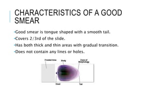
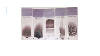
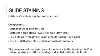
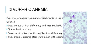
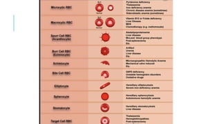
2. ROLE OF PERIPHERAL BLOOD
EXAMINATION
•Evaluation of anemia
•Evaluation of thrombocytopenia/ thrombocytosis
•Identification of abnormal cells
•Inclusions like basophilic stippling, Howell-Jolly
bodies, Cabot ring
•Infections like malaria, microfilaria etc
4. Place a drop of blood, about 2-3 mm in diameter approximately 1 cm
from one end of slide.
Precaution: Too large drop = too thick smear Too small drop = too
thin smear
5. a. Place the slide on a flat surface, and hold the other end
between your left thumb and forefinger.
b. With your right hand, place the smooth clean edge of a
second (spreader) slide on the specimen slide, just in front of
the blood drop.
c. Hold the spreader slide at a 30°- 45° angle, and draw it back
against the drop of blood
d. Allow the blood to spread almost to the edges of the slide
e. Push the spread forward with one light, smooth moderate
speed. Make a thin film of blood in the shape of tongue.
6. Label one edge with lab id
Spread Angle correction:
• High Hct: Angle should be lowered
• Low Hct: Angle should be raised
7. CHARACTERISTICS OF A GOOD
SMEAR
•Good smear is tongue shaped with a smooth tail.
•Covers 2/3rd of the slide.
•Has both thick and thin areas with gradual transition.
•Does not contain any lines or holes.
8.
9. SLIDE STAINING
•Leishman's stain is a polychromatic stain
•Components:
Methanol: fixes cells to slide
Methylene blue stains RNA,DNA: blue-grey color
Eosin stains hemoglobin, eosin granules orange-red color
Eosin + Methylene Blue = thiazine eosinate complex
•The complex will not stain any color unless a buffer is added: 0.05M
sodium phosphate (pH 6.4) and aged distilled water (pH 6.4-6.8)
10. STAINING PROCEDURE
•Thin smear are air dried.
•Flood the smear with stain.
•Stain for 2 minutes.
•Add an equal amount of buffer solution and mix the stain
•Leave the mixture on the slide for 10-15 min.
•Wash off by running water directly to the centre of the slide to
prevent a residue of precipitated stain.
•Stand slide on end, and let dry in air.
13. RBCS ON PERIPHERAL BLOOD FILM
Morphological abnormalities of RBCs can be:
•Anisocytosis: unequal or asymmetrical size
•Poikilocytosis: varied shape
•Hypochromic: decreased hemoglobin content
14. RBC MORPHOLOGY
In the blood from healthy person RBCs are
• Circular, homogenous disc nearly of uniform size (7–8 µm)
• Deep pink cytoplasm with Central pallor <1/3rd
15. MICROCYTIC HYPOCHROMIC RED
CELLS
•RBCs smaller than nucleus of small lymphocyte
•Increase in central pallor (>1/3rd)
•Major causes:
Iron deficiency anemia
Thalassemia minor
Sideroblastic anemia
AOCD
Hemoglobinopathies
16. MACROCYTIC RED CELLS
•RBCs are larger than the nucleus of small lymphocyte.
•Major causes:
Vitamin B12 or folate deficiency
Alcoholism
Liver disease
Hypothyroidism
Drugs impairing DNA synthesis
17. TARGET CELLS
•Characteristic ringed appearance
•Increase in red cell membrane compared to hemoglobin content, so
the excess membrane pools in the middle of the cell.
•Causes of target cells:
Thalassemias
Hemoglobinopathies
Liver disease
Post spelenctomy
Abetalipoproteinemia
18. SCHISTIOCYTES
•Physical assault to erythrocytes within bloodstream can create
fragments called schistiocytes, including helmet cells, triangles,
crescents and microspherocytes.
•Seen in
DIC
MAHA
HUS
TTP
Burns
Prosthetic valves, coarctation of aorta, abnormal cardiac valves
20. ELLIPTOCYTE
•Elliptical and normochromic cell, seen normally in less than 1% of
RBC.
•Causes:
Hereditary elliptocytosis
Iron deficiency anemia (increased with severity)
Megaloblastic anemia
21. BITE CELL OR (DEGMACYTE)
•Appears as a cookie with a bite taken out.
•Oxidative damage of Hb: G6PD enzyme deficiency.
•Bite cells apparently occur when the spleen removes
the Heinz bodies from the RBCs.
22. BURR CELS/ ECHINOCYTES SPUR CELLS/ACANTHOCYTES
10-30 spicules equal in size
and evenly distributed over
RBC surface
Causes:
Renal failure from any cause
Liver diseases, especially when
uremia coexists
Storage artefact
Dehydration
Pyruvate kinase deficiency
Immediately after red cell
transfusion.
5-10 spicules of varying
length. Spicules are irregular in
space and thickness with wide
bases. Appear smaller than
normal cells because they
assume a spheroid shape.
Causes:
Spur cell anemia: alcoholic
cirrhosis,
Postsplenectomy or
hyposplenic state
Hypothyroidism
Abetalipoproteinemia: 50–
100% of cell acanthocytes
23. SICKLE CELLS
•Crescent-shaped sickle cells.
•Causes:
Homozygous for HbS gene
Heterozygous for HbS and either a thalassemia or another abnormal
hemoglobin such as HbC.
•Point mutation: There is substitution of valine for glutamic acid at the
sixth residue of the b chain.
24. POLYCHROMATOPHILIA
•Primitive reticulocytes, are larger than mature erythrocytes.
•Romanowsky preparation: bluish-gray or purple because
of the substantial RNA remnants.
•Polychromatophilia: cells derive its hue from the combination
of blue from the RNA and red from the hemoglobin.
•High level of erythropoietin circulates in a response to anemia.
26. HOWEL-
JOLLY BODIES
• Small, well-defined,round, densely stained inclusions; 1
µm in diameter, eccentric in location that represent DNA
fragment
Post spelenctomy
Megaloblastic anemia
HEINZ BODIES • Denatured hemoglobin caused by oxidation of globin
portion of hemoglobin molecule.
• Removal of Heinz bodies may lead to formation of bite
cells.
SIDEROTIC
GRANULES/
PAPPENHEIME
R BODIES
• Irregular dark blue iron containing granules
demonstrated by Perl’s or Prussian blue stains
• Erythrocytes with Pappenheimer bodies are called
siderocytes.
Lead poisoning
Sideroblastic anemia
27. BASOPHILIC
STIPPLING
• Numerous small, purplish inclusions, which result from RNA
and mitochondrial remnants.
• Seen in
Lead toxicity
Thalassemias
Hemoglobinopathies
Macrocytic anemias
• In their presence iron deficiency can be ruled out.
CABOT
RINGS
• Delicate thread-like inclusions, remnants of the nuclear
membranes in RBC.
• Seen in
Pernicious anemia
Lead poisoning
Alcoholic jaundice
Severe anemia
28. NUCLEATED RBCS
•Dark, dense nucleus in the center of a bluish
(polychromatophilic) or red (orthochromatic) cell periphery.
•They may result in response to marked stimulation of the bone
marrow by erythropoietin in patients with severe anemia.
29. NUCLEATED RBCS
•Causes are:
Newborn (first 3–4 days)
Intense bone marrow stimulation
Acute bleeding severe hemolytic anemia, (e.g. thalassemia, SS
hemoglobinopathy)
Megaloblastic anemia
Postsplenectomy or hyposplenic states: Spleen normally removes
nucleated RBCs
Leukoerythroblastic reaction, seen with extramedullary
hematopoiesis and bone marrow replacement
High WBC count with left shift
30. MACROCYTIC ANEMIA
•Large oval RBCs (macro-ovalocytes) on peripheral smear and
the increased RBC distribution width
•RBCs may show basophilic stippling or howel-jolly bodies
•Neutrophils show nuclear hypersegmentation
•These may present as pancytopenia
31. DIMORPHIC ANEMIA
•Presence of anisocytosis and anisochromia in the same film.
•Seen in
Coexistence of iron deficiency and megaloblastic anaemia
Sideroblastic anemia
Some weeks after iron therapy for iron deficiency anemia
Hypochromic anemia after transfusion with normal cells