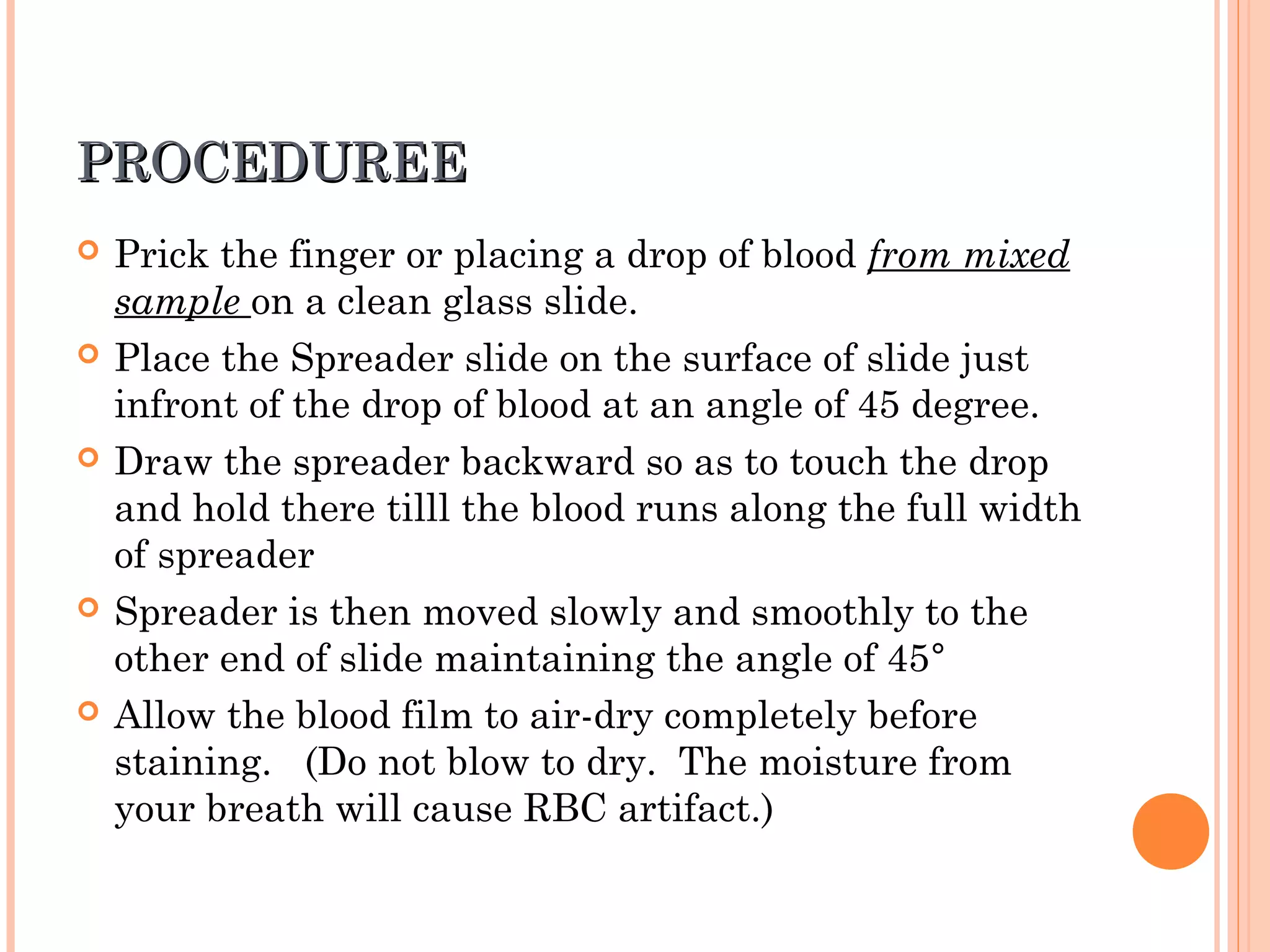
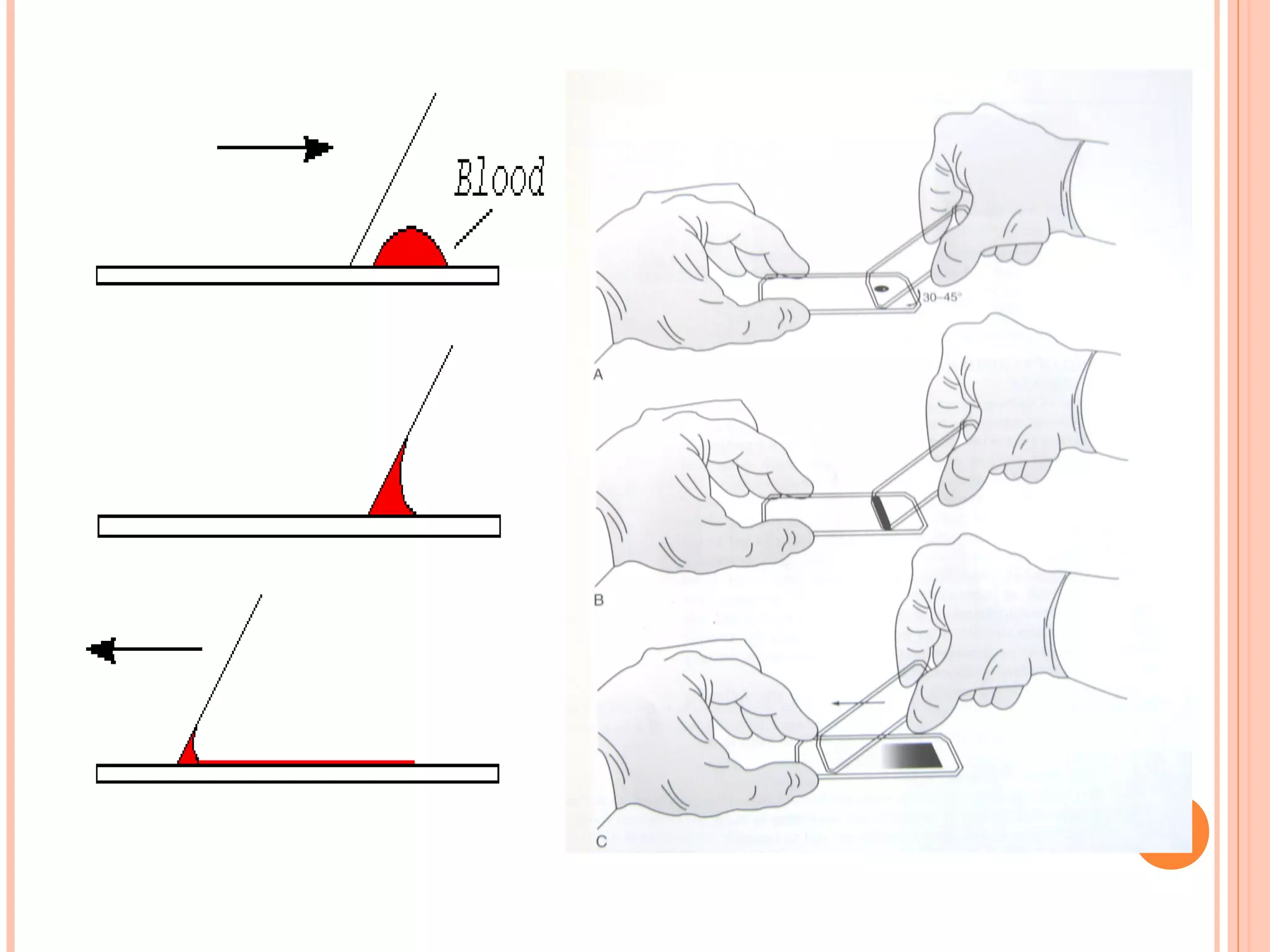
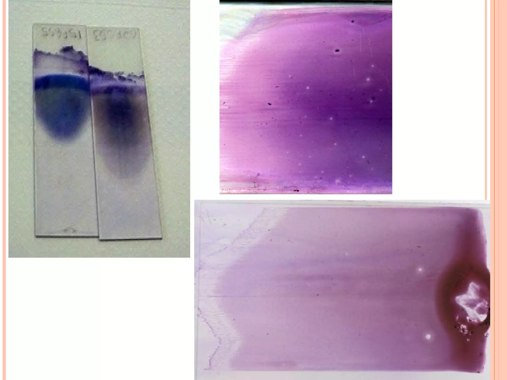
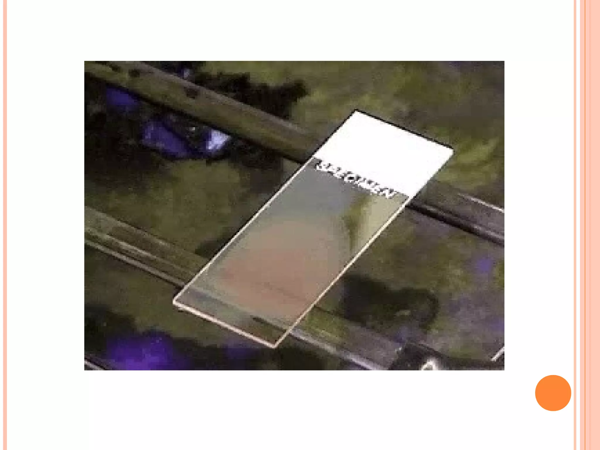
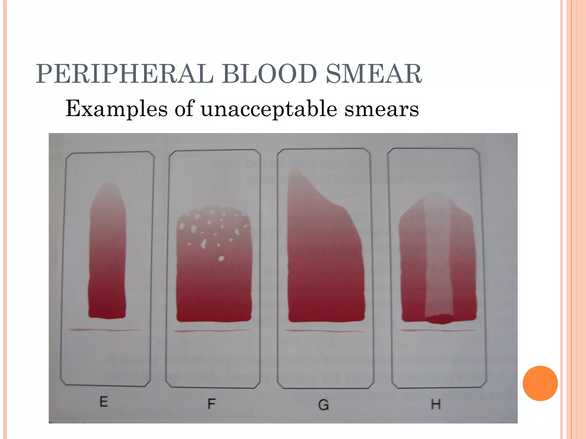
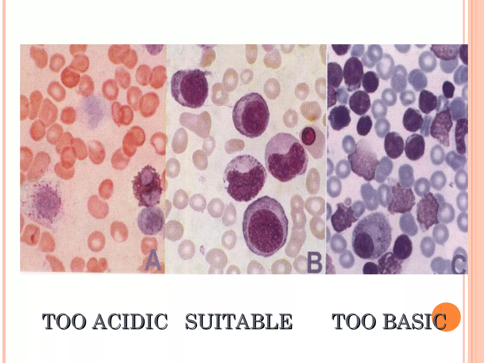
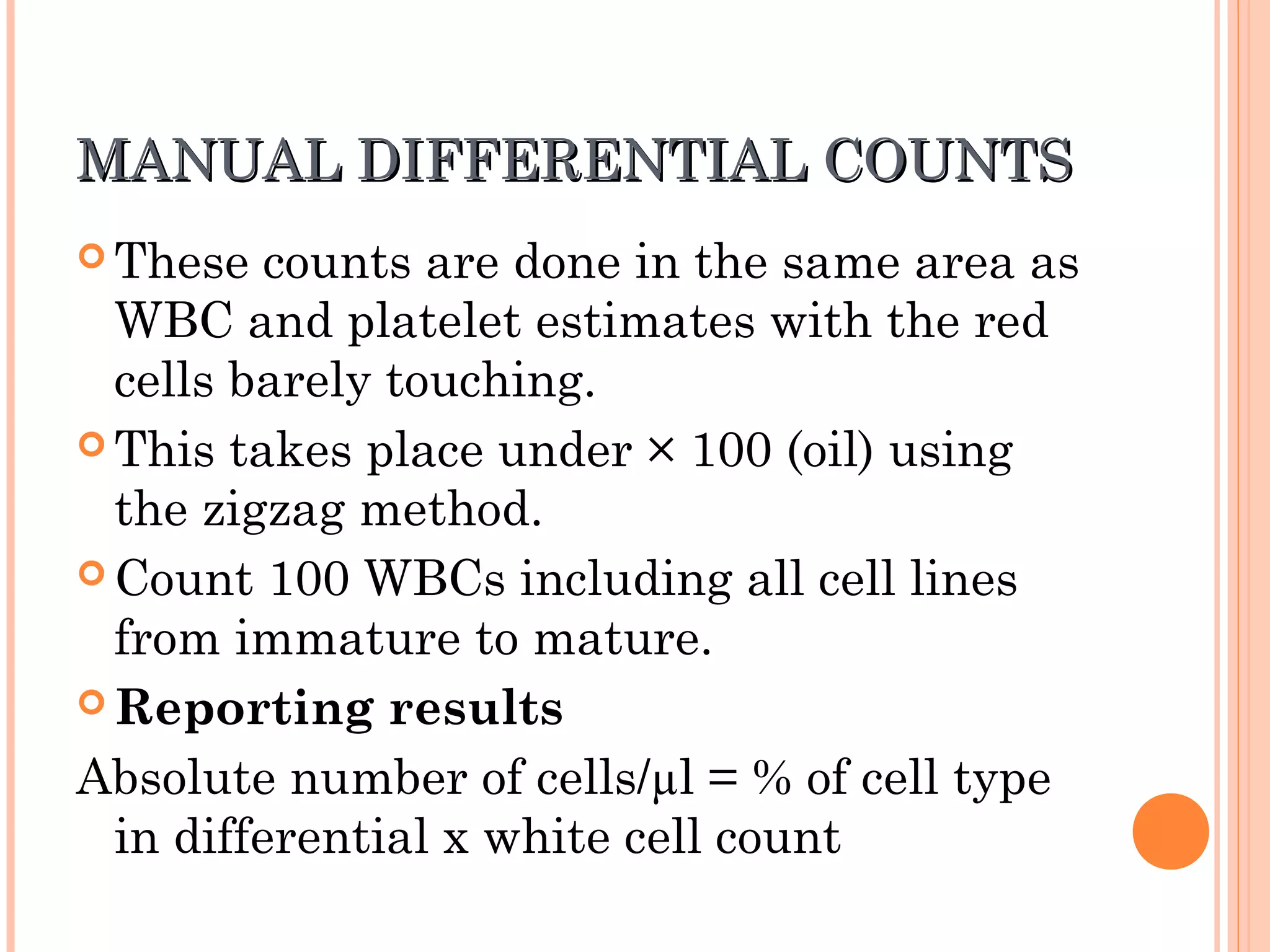
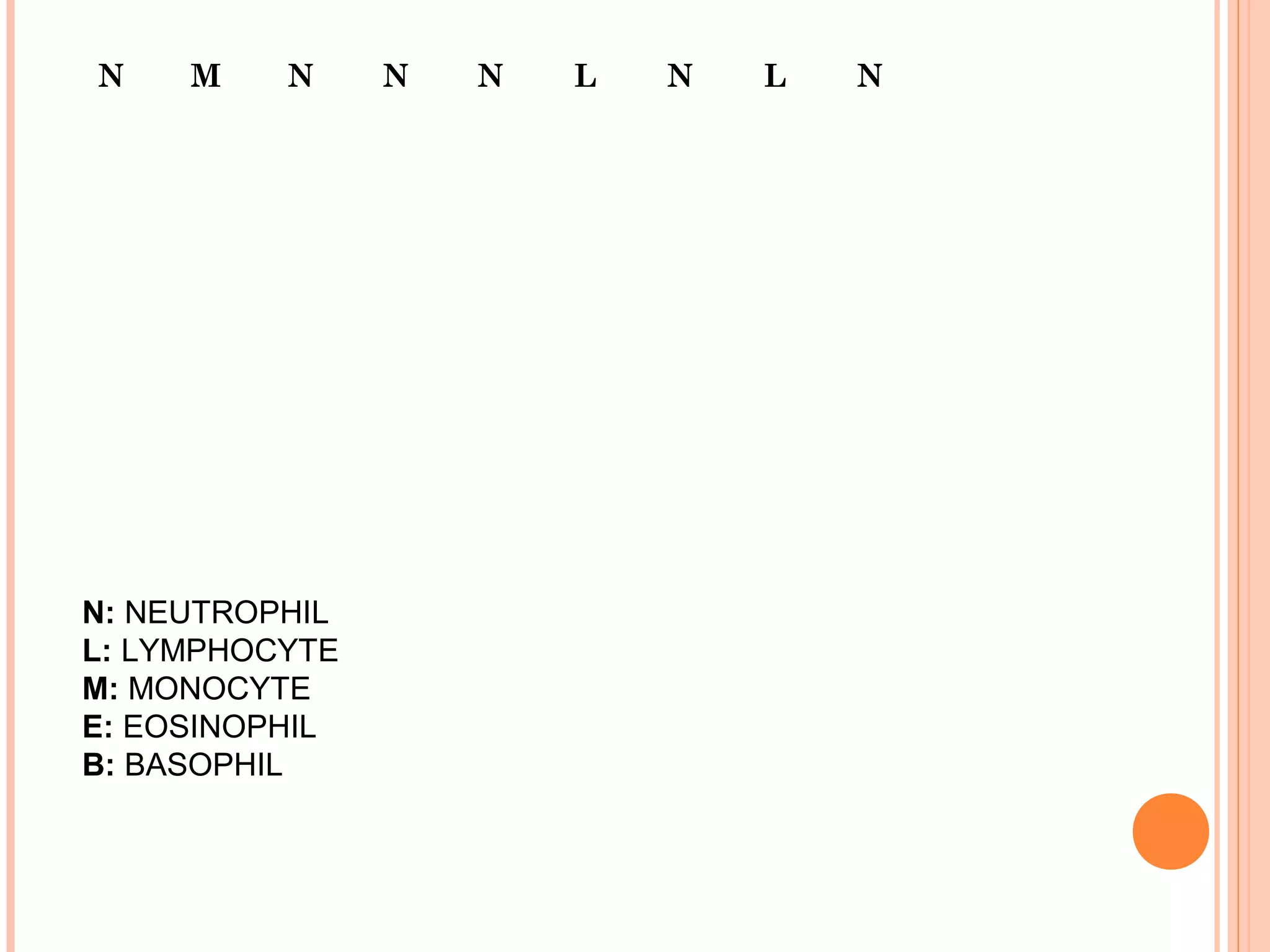
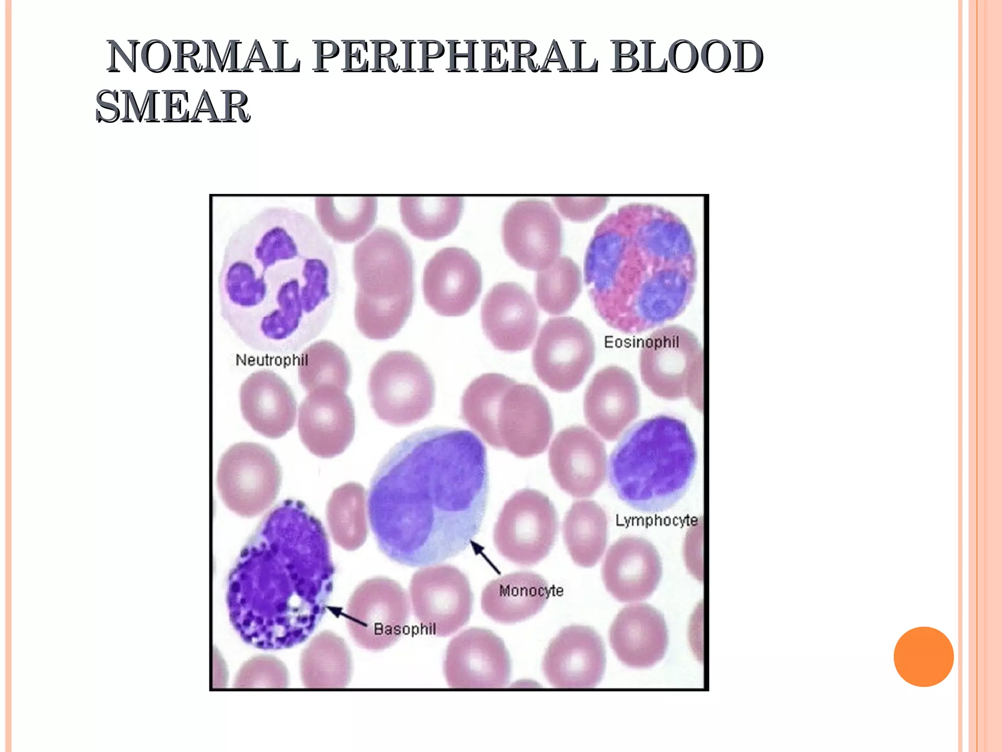
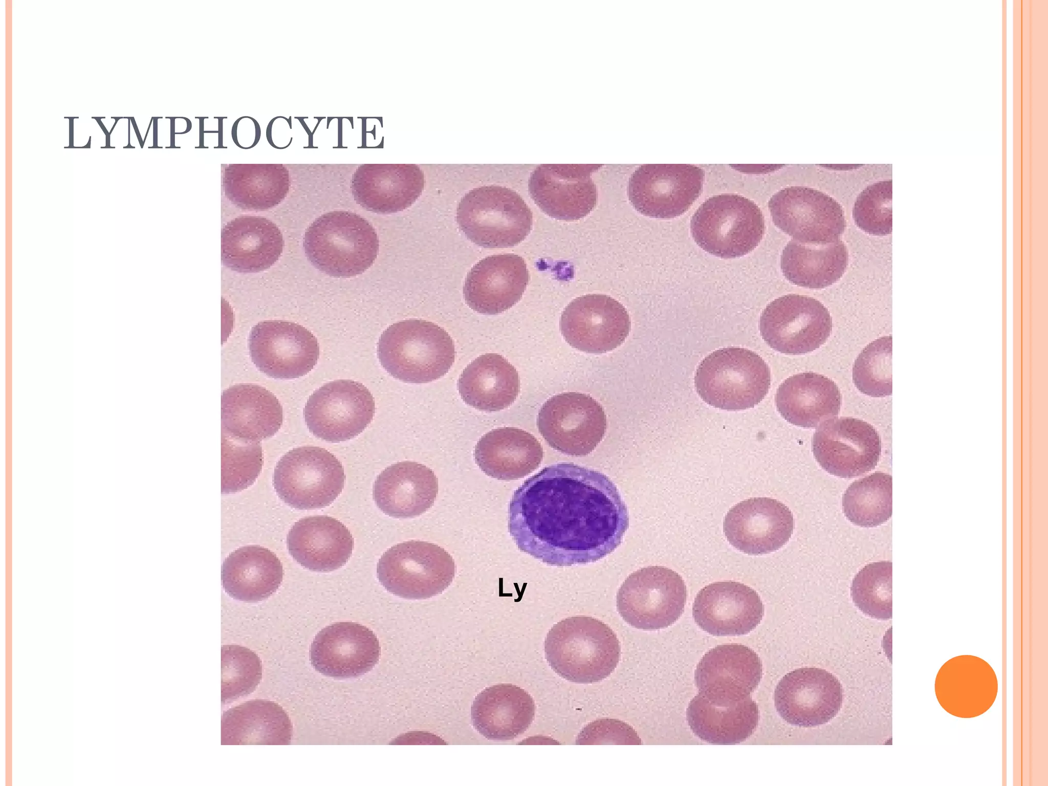
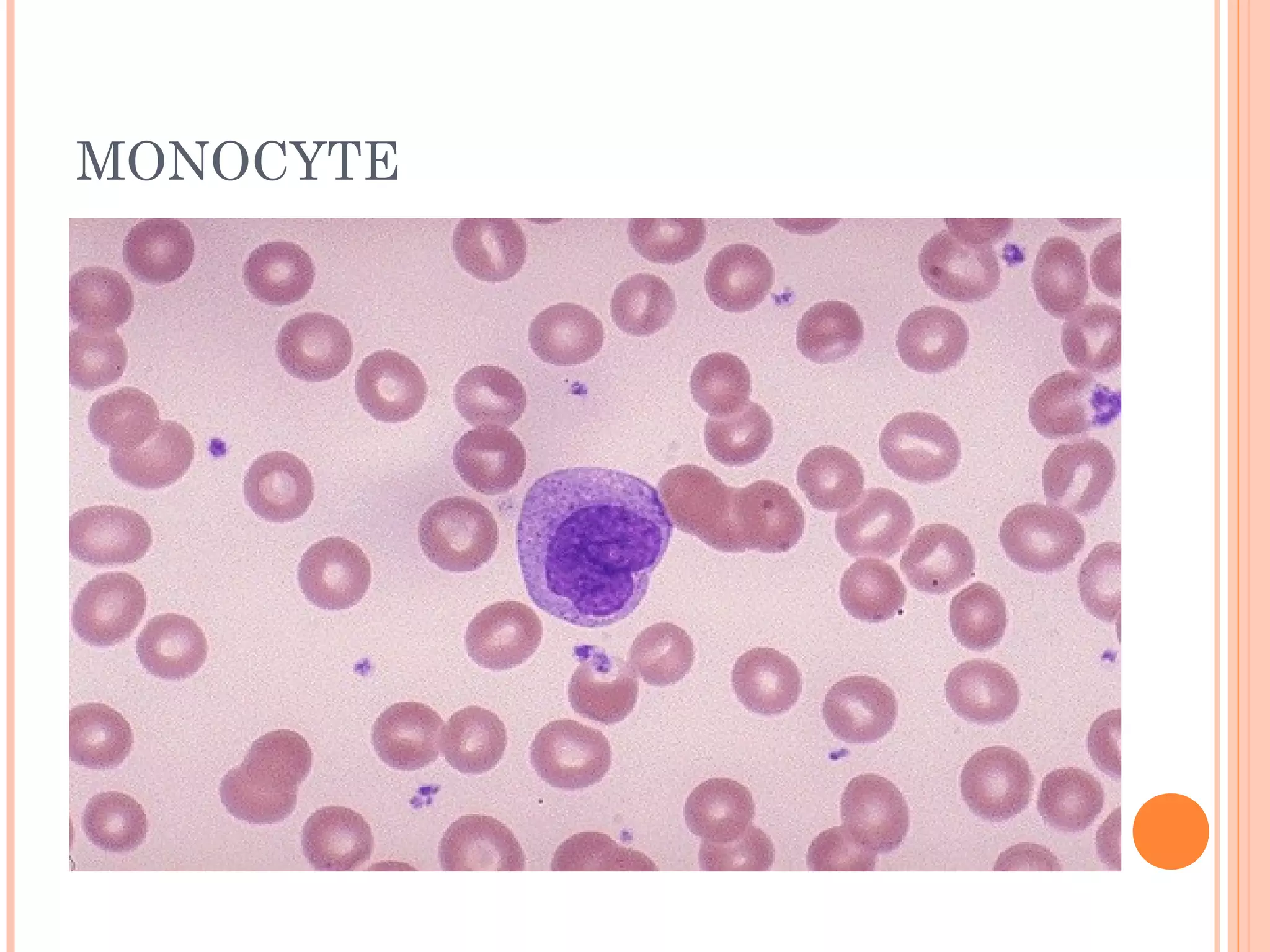
This document provides information about preparing and examining peripheral blood smears. It discusses how to make a good blood smear by ensuring the smear is evenly spread and covers most of the slide. The document also describes staining blood smears using the Leishman's stain method and examining smears under a microscope. Key things to observe during examination include the different types of white blood cells, red blood cell morphology, and any abnormal findings. Performing a manual differential count involves identifying 100 white blood cells and categorizing them by type.


![Peripheral blood smear [autosaved]](https://cdn.slidesharecdn.com/ss_thumbnails/peripheralbloodsmearautosaved-201029200454-thumbnail.jpg?width=640&height=640&fit=bounds)


