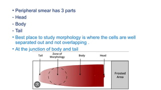
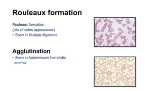
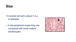
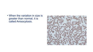
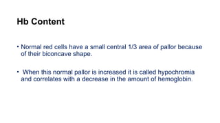
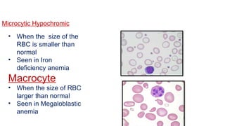
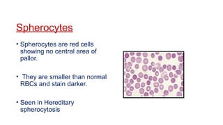
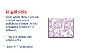
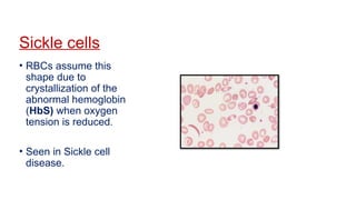
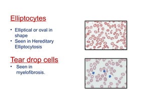
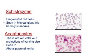
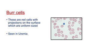
CLIP
DR . NANDITA S
PERIPHERAL SMEAR REPORTING
INDICATIONS OF PERIPHERAL SMEAR
EXAMINATION
To assess the cellularity of the different blood elements
To study the morphology of these blood elements
To look for blood parasites
• To study the response of the body to various disease processes
• Peripheral smear has 3 parts
- Head
- Body
- Tail
• Best place to study morphology is where the cells are well
separated out and not overlapping .
• At the junction of body and tail
LOW POWER
• To get an idea of the quality of the smear
• The number, distribution and staining of the leucocytes
• Red cell agglutination and rouleaux formation
• Extracellular parasites such as microfilaria
• To select a good area where RBCs are just touching each
other
Put a drop of oil and change to oil immersion objective.
Cellular elements are studied in the following order
• RBCs
• WBCs
• Platelets
RBC
• Rouleaux formation/agglutination
• Size
• Hb content
• Shape
• Immature RBC
• Inclusion bodies
• Intracellular parasites
Rouleaux formation
Rouleaux formation
(pile of coins appearance)
• Seen in Multiple Myeloma
Agglutination
• Seen in Autoimmune hemolytic
anemia
Size
• A normal red cell is about 7.2 µ
in diameter
• In the peripheral smear they are
compared with small mature
lymphocytes
• When the variation in size is
greater than normal, it is
called Anisocytosis.
Hb Content
• Normal red cells have a small central 1/3 area of pallor because
of their biconcave shape.
• When this normal pallor is increased it is called hypochromia
and correlates with a decrease in the amount of hemoglobin.
Microcytic Hypochromic
• When the size of the
RBC is smaller than
normal
• Seen in Iron
deficiency anemia
Macrocyte
• When the size of RBC
larger than normal
• Seen in Megaloblastic
anemia
Shape
• Normal red cells are biconcave in shape and appear circular on
a smear as they are flattened out.
• Variation in shape is known as Poikilocytosis.
Spherocytes
• Spherocytes are red cells
showing no central area of
pallor.
• They are smaller than normal
RBCs and stain darker.
• Seen in Hereditary
spherocytosis
Target cells
• Cells which show a central
stained area and a
peripheral stained rim with
unstained cytoplasm in
between.
• They are thinner than
normal cells.
• Seen in Thalassemia
Sickle cells
• RBCs assume this
shape due to
crystallization of the
abnormal hemoglobin
(HbS) when oxygen
tension is reduced.
• Seen in Sickle cell
disease.
Elliptocytes
• Elliptical or oval in
shape
• Seen in Hereditary
Elliptocytosis
Tear drop cells
• Seen in
myelofibrosis.
Schistocytes
• Fragmented red cells
• Seen in Microangiopathic
hemolytic anemia
Acanthocytes
• These are red cells with
projections of varying size
• Seen in
Abetalipoproteinemia
Burr cells
• These are red cells with
projections on the surface
which are uniform sized
• Seen in Uremia.
Immature RBC
Polychromasia
• Term used for red cells which
stain pinkish blue with
Romanowsky stain.
• Larger than normal RBC
• Seen in Hemolysis
Normoblasts
• Normoblasts are seen in
the peripheral circulation
in accelerated
erythropoiesis
• Seen in Hemolytic Anemia
Inclusion bodies
• HowelI Jolly bodies
• Basophilic stippling
• Cabot rings
Howell Jolly bodies
• Howell–Jolly bodies are
nuclear remnants.
• They are small, round
cytoplasmic inclusions that
stain purple on a
Romanowsky stain
• Seen in Megaloblastic anemia
Basophilic stippling
• These are small blue or black
granules in red cells which
are actually aggregates of
ribosomes.
• Seen in Megaloblastic Anemia
Cabot rings
• These are pale staining rings
or figures of eight and are
thought to be remnants of the
nuclear membrane.
• They are seen in
Megaloblastic anemia
Intracellular parasite
Seen in
1. Malaria
2. Bancrofti
WBC
• Count
• Distribution
• Predominant cell
• Immature cells
• Atypical cells
• Normal WBC count is 4000-11,000/cumm
• Leucocytosis-an increase in the total count to values more than
11,000/cumm
• Leucopenia-decrease in the total count to less than 4000
cells/cumm
• The tail end of the smear has to be screened before remarking
that the smear shows leucopenia.
• The differential WBC count should be done under oil immersion
• Differential WBC count is best done in the same area of the
smear where RBC morphology is best
Normal Differential count
Neutrophils 40-70%
Lymphocytes 20-40%
Monocytes 2-10%
Eosinophils 1-6%
Basophils 0-1%
Immature cells
Atypical/activated lymphocytes
• Atypical lymphocytes are seen in
Infectious mononucleosis and
other viral infections
Platelets
• Count
• Distribution: Single / Clumps
• Abnormal forms
Count
• In a normal peripheral smear, every oil immersion field will have
8-25 platelets on an average
• Normal platelet count is 150,000-400,000/µl
• Increase in platelet count more than 4,00000/µL –
Thrombocytosis
• Decrease in platelet count less than 1,50000/ µL-
Thrombocytopenia
Distribution
• Seen in clumps or scattered
Abnormal forms
• Large platelets may be seen
in Immune thrombocytopenic
purpura
THANK YOU

peripheral smear reporting practical-1.pptx

  • 1.
  • 2.
  • 3.
    INDICATIONS OF PERIPHERALSMEAR EXAMINATION To assess the cellularity of the different blood elements To study the morphology of these blood elements To look for blood parasites • To study the response of the body to various disease processes
  • 4.
    • Peripheral smearhas 3 parts - Head - Body - Tail • Best place to study morphology is where the cells are well separated out and not overlapping . • At the junction of body and tail
  • 5.
    LOW POWER • Toget an idea of the quality of the smear • The number, distribution and staining of the leucocytes • Red cell agglutination and rouleaux formation • Extracellular parasites such as microfilaria • To select a good area where RBCs are just touching each other Put a drop of oil and change to oil immersion objective.
  • 6.
    Cellular elements arestudied in the following order • RBCs • WBCs • Platelets
  • 7.
    RBC • Rouleaux formation/agglutination •Size • Hb content • Shape • Immature RBC • Inclusion bodies • Intracellular parasites
  • 8.
    Rouleaux formation Rouleaux formation (pileof coins appearance) • Seen in Multiple Myeloma Agglutination • Seen in Autoimmune hemolytic anemia
  • 9.
    Size • A normalred cell is about 7.2 µ in diameter • In the peripheral smear they are compared with small mature lymphocytes
  • 10.
    • When thevariation in size is greater than normal, it is called Anisocytosis.
  • 11.
    Hb Content • Normalred cells have a small central 1/3 area of pallor because of their biconcave shape. • When this normal pallor is increased it is called hypochromia and correlates with a decrease in the amount of hemoglobin.
  • 12.
    Microcytic Hypochromic • Whenthe size of the RBC is smaller than normal • Seen in Iron deficiency anemia Macrocyte • When the size of RBC larger than normal • Seen in Megaloblastic anemia
  • 13.
    Shape • Normal redcells are biconcave in shape and appear circular on a smear as they are flattened out. • Variation in shape is known as Poikilocytosis.
  • 14.
    Spherocytes • Spherocytes arered cells showing no central area of pallor. • They are smaller than normal RBCs and stain darker. • Seen in Hereditary spherocytosis
  • 15.
    Target cells • Cellswhich show a central stained area and a peripheral stained rim with unstained cytoplasm in between. • They are thinner than normal cells. • Seen in Thalassemia
  • 16.
    Sickle cells • RBCsassume this shape due to crystallization of the abnormal hemoglobin (HbS) when oxygen tension is reduced. • Seen in Sickle cell disease.
  • 17.
    Elliptocytes • Elliptical oroval in shape • Seen in Hereditary Elliptocytosis Tear drop cells • Seen in myelofibrosis.
  • 18.
    Schistocytes • Fragmented redcells • Seen in Microangiopathic hemolytic anemia Acanthocytes • These are red cells with projections of varying size • Seen in Abetalipoproteinemia
  • 19.
    Burr cells • Theseare red cells with projections on the surface which are uniform sized • Seen in Uremia.
  • 20.
    Immature RBC Polychromasia • Termused for red cells which stain pinkish blue with Romanowsky stain. • Larger than normal RBC • Seen in Hemolysis Normoblasts • Normoblasts are seen in the peripheral circulation in accelerated erythropoiesis • Seen in Hemolytic Anemia
  • 21.
    Inclusion bodies • HowelIJolly bodies • Basophilic stippling • Cabot rings
  • 22.
    Howell Jolly bodies •Howell–Jolly bodies are nuclear remnants. • They are small, round cytoplasmic inclusions that stain purple on a Romanowsky stain • Seen in Megaloblastic anemia
  • 23.
    Basophilic stippling • Theseare small blue or black granules in red cells which are actually aggregates of ribosomes. • Seen in Megaloblastic Anemia
  • 24.
    Cabot rings • Theseare pale staining rings or figures of eight and are thought to be remnants of the nuclear membrane. • They are seen in Megaloblastic anemia
  • 25.
  • 26.
    WBC • Count • Distribution •Predominant cell • Immature cells • Atypical cells
  • 27.
    • Normal WBCcount is 4000-11,000/cumm • Leucocytosis-an increase in the total count to values more than 11,000/cumm • Leucopenia-decrease in the total count to less than 4000 cells/cumm • The tail end of the smear has to be screened before remarking that the smear shows leucopenia.
  • 28.
    • The differentialWBC count should be done under oil immersion • Differential WBC count is best done in the same area of the smear where RBC morphology is best
  • 29.
    Normal Differential count Neutrophils40-70% Lymphocytes 20-40% Monocytes 2-10% Eosinophils 1-6% Basophils 0-1%
  • 30.
  • 31.
    Atypical/activated lymphocytes • Atypicallymphocytes are seen in Infectious mononucleosis and other viral infections
  • 32.
    Platelets • Count • Distribution:Single / Clumps • Abnormal forms
  • 33.
    Count • In anormal peripheral smear, every oil immersion field will have 8-25 platelets on an average
  • 34.
    • Normal plateletcount is 150,000-400,000/µl • Increase in platelet count more than 4,00000/µL – Thrombocytosis • Decrease in platelet count less than 1,50000/ µL- Thrombocytopenia
  • 35.
    Distribution • Seen inclumps or scattered Abnormal forms • Large platelets may be seen in Immune thrombocytopenic purpura
  • 36.

Editor's Notes

  • #21 Pappen heimer bodies- small peripherally sited basophilic erythrocyte inclusion.composed of hemosiderin
  • #22 Nuclear . Small round cytoplasmic inclusions remnants- hemolytic anemia,megaloblastic anemia,post splenectomy
  • #23 RNA-lead poisoning,thalassemia . Numerous basophilic granules distributed along cells