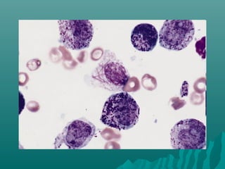
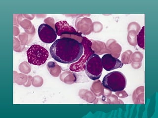
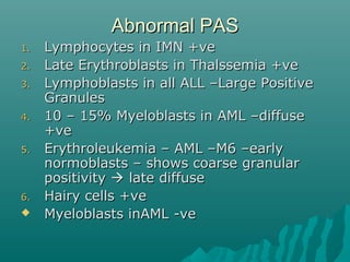
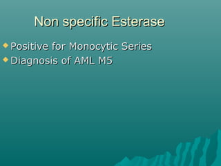
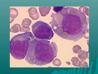
Peripheral smearPeripheral smear
TypesTypes
1.1. WedgeWedge
2.2. Spun – most uniform distribution ofSpun – most uniform distribution of
blood cellsblood cells
3.3. ThickThick
4.4. Buffy layerBuffy layer
5.5. WetWet
Wedge filmWedge film
 Spreading slide 30 – 45 degreeSpreading slide 30 – 45 degree
 Severe anemia - > 60 degreeSevere anemia - > 60 degree
 Polycythemia -< 15 degreePolycythemia -< 15 degree
Thick filmThick film
 ParasitesParasites
 View large volumeView large volume
 4 drops4 drops
 Join them over an area of 1 cm2Join them over an area of 1 cm2
Buffy layer filmBuffy layer film
1.1. Pernicious anemia – demonstratePernicious anemia – demonstrate
megaloblastsmegaloblasts
2.2. AIHA – erythrophagocytosisAIHA – erythrophagocytosis
3.3. Tumor cells in aleukemic leukemiaTumor cells in aleukemic leukemia
4.4. LE cellsLE cells
Wet filmWet film
 Examination of blood cells suspendeExamination of blood cells suspende
in plasma +- supravital stainin plasma +- supravital stain
1.1. Demonstrate parasitesDemonstrate parasites
2.2. Induce red cell sicklingInduce red cell sickling
3.3. Study the red cells by NomarskiStudy the red cells by Nomarski
interference microscopy afterinterference microscopy after
fixation in Gluteraldehydefixation in Gluteraldehyde
Blood smear preparationBlood smear preparation
 EDTAEDTA
 Ideal smearIdeal smear
1.1. Length of the smear shud be 2/3 rd ofLength of the smear shud be 2/3 rd of
the slidethe slide
2.2. Uniformly thickenedUniformly thickened
3.3. Thin – printed matter can be readThin – printed matter can be read
4.4. Margin shud be free and centrally placedMargin shud be free and centrally placed
5.5. Tongue shapedTongue shaped
6.6. Shud have adequate thin area –free fromShud have adequate thin area –free from
vacoule ,serration etcvacoule ,serration etc
Biological causes of poor filmBiological causes of poor film
1.1. Cold agglutininCold agglutinin
2.2. LipemiaLipemia
3.3. RouleauxRouleaux
FixativesFixatives
1.1. Methyl alcohol – Fixative Of ChoiceMethyl alcohol – Fixative Of Choice
2.2. Ethyl alcoholEthyl alcohol
Staining of peripheral smearStaining of peripheral smear
 Romanowsky type dyesRomanowsky type dyes
1.1. LeishmanLeishman
2.2. WrightWright
3.3. May Grunwald GiemsaMay Grunwald Giemsa
4.4. JennerJenner
 Compound dyesCompound dyes
 Neutral dyesNeutral dyes
 Field stain rapid stain for parasitesField stain rapid stain for parasites
EosinEosin
 Cytoplasm , collagen ,muscle fibersCytoplasm , collagen ,muscle fibers
 AcidicAcidic
 H & E staining –H & E staining –
 Cytoplasm pink –orangeCytoplasm pink –orange
 Nuclei – blue / purpleNuclei – blue / purple
 Eosin stains red blood cells intenslyEosin stains red blood cells intensly
redred
 Haemetoxylin – nuclear stainHaemetoxylin – nuclear stain
Methylene blueMethylene blue
 Component of Gram stain .WrightComponent of Gram stain .Wright
stain ,Jenner’s stainstain ,Jenner’s stain
May Grunwald Giemsa stainMay Grunwald Giemsa stain
 Histopathological diagnosis forHistopathological diagnosis for
Malaria and other parasitesMalaria and other parasites
 Fixative – acetone free methylFixative – acetone free methyl
alcoholalcohol
Jenner’s stainJenner’s stain
 Methylene blue eosinateMethylene blue eosinate
Wright stainWright stain
 Eosin + MBEosin + MB
Romanowsky stainRomanowsky stain
 Basic dye forBasic dye for
1.1. Nucleic acidNucleic acid
2.2. NucleoproteinNucleoprotein
3.3. Basophilic granulesBasophilic granules
4.4. Weakly to neutrophil granulesWeakly to neutrophil granules
 Acidic dyes forAcidic dyes for
1.1. HgHg
2.2. Eosinophil granulesEosinophil granules
Leishman’s stainLeishman’s stain
 William Boog Leishman –BritishWilliam Boog Leishman –British
PathologistPathologist
 In India he studies Enteric Fever andIn India he studies Enteric Fever and
Kala AzarKala Azar
 Buffered water PH 6.8Buffered water PH 6.8
 ComponentsComponents
1.1. Methylene Blue – Nuclear stainMethylene Blue – Nuclear stain
2.2. Eosin – cytoplasmic stainEosin – cytoplasmic stain
3.3. Acetone free Methyl Alcohol – solventAcetone free Methyl Alcohol – solvent
and fixativeand fixative
ProcedureProcedure
1.1. Enough stain to cover entire smear-Enough stain to cover entire smear-
keep for 2 min – fixation bykeep for 2 min – fixation by
methanolmethanol
2.2. Add distilled water - double theAdd distilled water - double the
amount—10 min –actual stainingamount—10 min –actual staining
3.3. Keep for 7 – 10 minKeep for 7 – 10 min
4.4. WashWash
5.5. DryDry
Good stainingGood staining
 Pinkish in colourPinkish in colour
 RBC – coppery redRBC – coppery red
 Eosinophil – orange red – withEosinophil – orange red – with
yellowish tinged granulesyellowish tinged granules
 No depositNo deposit
 No cell shrinking or swellingNo cell shrinking or swelling
 If under atained add stain + waterIf under atained add stain + water
for another 5minfor another 5min
 If over stained –add stain onlyIf over stained –add stain only
Special stainingSpecial staining
1.1. Peroxidase stainPeroxidase stain
2.2. Sudan Black B stainSudan Black B stain
3.3. PAS stainPAS stain
4.4. Chloro Acetate esterase stainChloro Acetate esterase stain
5.5. Non Specific EsterasesNon Specific Esterases
6.6. Combined NSE & CAE stainingCombined NSE & CAE staining
7.7. Iron stainIron stain
8.8. Reticulin stainReticulin stain
Peroxidase stainPeroxidase stain
 Enzyme found in Primary andEnzyme found in Primary and
Secondary granules in WBCSecondary granules in WBC
 MPO – localised in the AzurophilicMPO – localised in the Azurophilic
granules of Neutrophils & Monocytesgranules of Neutrophils & Monocytes
 It can be demonstrated inIt can be demonstrated in
Eosinophils and Basophils alsoEosinophils and Basophils also
 MPO occurs onlu in the Primary GranulesMPO occurs onlu in the Primary Granules
of Myeloid cellsof Myeloid cells
 It can be detected from theIt can be detected from the
Promyelocyte upto the granularPromyelocyte upto the granular
stagestage
 Lymphoid series are strongly MPOLymphoid series are strongly MPO
negativenegative
 Reddish brown appearanceReddish brown appearance
 All myelocytic cell line except MyeloblastsAll myelocytic cell line except Myeloblasts
shows Positiveshows Positive
 The more mature the cells become , theThe more mature the cells become , the
weaker the reaction becomesweaker the reaction becomes
 Auer rods are strongly PositiveAuer rods are strongly Positive
Indications for peroxidase stainingIndications for peroxidase staining
 Recognition of Acute Leukemias ofRecognition of Acute Leukemias of
myeloid origin – negative makesmyeloid origin – negative makes
lymphocyte origin – but does notlymphocyte origin – but does not
exclude a myeloid originexclude a myeloid origin
 PositivePositive sub division of Myeloidsub division of Myeloid
Leukemias by FABLeukemias by FAB
ResultResult
 Cells of Myeloid series positive withCells of Myeloid series positive with
increasing intensity and no. of granules asincreasing intensity and no. of granules as
the cells maturethe cells mature
 Early blasts negativeEarly blasts negative
 Lymphoid cells are negativeLymphoid cells are negative
 Erythroid cells and RBC may be Positive –Erythroid cells and RBC may be Positive –
due to Non enzymatic Reaction betweendue to Non enzymatic Reaction between
Hb and stainHb and stain
 Imp to distinguish AML from ALLImp to distinguish AML from ALL
 Neutrophils show decreased staining inNeutrophils show decreased staining in
congenital MPO deficiency andcongenital MPO deficiency and
Myelodysplastic syndromesMyelodysplastic syndromes
Sudan Black B stainSudan Black B stain
 Fat soluble substanceFat soluble substance
 StainsStains Fat cells ,Macrophages andFat cells ,Macrophages and
GranulocytesGranulocytes
 Positive is dark brown to black in thePositive is dark brown to black in the
cytoplasm of the cell containing lipidcytoplasm of the cell containing lipid
 Granulocytes +ve inc with maturityGranulocytes +ve inc with maturity
 Cells of Burkitts’ Lymphoma +veCells of Burkitts’ Lymphoma +ve
 Large vacoulated ALL –L 3 cells +veLarge vacoulated ALL –L 3 cells +ve
 Myeloblasts –veMyeloblasts –ve
 Lumphoblasts and Lymphocytes areLumphoblasts and Lymphocytes are
–ve–ve
 Some cases of AML and CML –ve orSome cases of AML and CML –ve or
reduced sudanophiliareduced sudanophilia
PASPAS
 Positive -Diffuse / granular pinkish colourPositive -Diffuse / granular pinkish colour
in cytoplasmin cytoplasm
 Normal positiveNormal positive
1.1. GranulocytesGranulocytes
2.2. MonocytesMonocytes
3.3. MegakaryocytesMegakaryocytes
4.4. LymphocytesLymphocytes
 Normal negativeNormal negative
1.1. Erythroid seriesErythroid series
Abnormal PASAbnormal PAS
1.1. Lymphocytes in IMN +veLymphocytes in IMN +ve
2.2. Late Erythroblasts in Thalssemia +veLate Erythroblasts in Thalssemia +ve
3.3. Lymphoblasts in all ALL –Large PositiveLymphoblasts in all ALL –Large Positive
GranulesGranules
4.4. 10 – 15% Myeloblasts in AML –diffuse10 – 15% Myeloblasts in AML –diffuse
+ve+ve
5.5. Erythroleukemia – AML –M6 –earlyErythroleukemia – AML –M6 –early
normoblasts – shows coarse granularnormoblasts – shows coarse granular
positivitypositivity  late diffuselate diffuse
6.6. Hairy cells +veHairy cells +ve
 Myeloblasts inAML -veMyeloblasts inAML -ve
Non specific EsteraseNon specific Esterase
 Positive for Monocytic SeriesPositive for Monocytic Series
 Diagnosis of AML M5Diagnosis of AML M5
Iron stainIron stain
 PEARL’S PRUSSIAN BLUE StainPEARL’S PRUSSIAN BLUE Stain
 Absent in Fe def. AnemiaAbsent in Fe def. Anemia
 Increased inIncreased in
1.1. Transfusion HaemosiderosisTransfusion Haemosiderosis
2.2. HaemochromatosisHaemochromatosis
3.3. Refractory Sideroblastic AnemiaRefractory Sideroblastic Anemia
4.4. Aplastic AnemiaAplastic Anemia
5.5. Pernicious anemiaPernicious anemia
 Nucleus and Cytoplasm pinkNucleus and Cytoplasm pink
 Hemosiderin – bright blue – greenHemosiderin – bright blue – green
granulesgranules
PS reportingPS reporting
 Size of the RBC is compared to smallSize of the RBC is compared to small
lymphocyteslymphocytes
 Microcytes –MCV<80 flMicrocytes –MCV<80 fl
 Macrocytes – MCV> 96 flMacrocytes – MCV> 96 fl
 AnisocytosisAnisocytosis
 PoikilocytosisPoikilocytosis
SpherocytesSpherocytes
 Dark stained central pallor region –Dark stained central pallor region –
hyperchromasiahyperchromasia
1.1. Heriditary SpherocytosisHeriditary Spherocytosis
2.2. AIHAAIHA
3.3. Chemical & Bacterial ToxinsChemical & Bacterial Toxins
Peripheral smear
Peripheral smear
Peripheral smear
Peripheral smear
Peripheral smear
Peripheral smear
Peripheral smear
Peripheral smear
Peripheral smear
Peripheral smear
Peripheral smear
Peripheral smear
Peripheral smear
Peripheral smear
Peripheral smear
Peripheral smear
Peripheral smear
Peripheral smear
Peripheral smear
Peripheral smear
Peripheral smear
Peripheral smear
Peripheral smear
Peripheral smear
Peripheral smear
Peripheral smear
Peripheral smear
Peripheral smear
Peripheral smear
Peripheral smear
Peripheral smear
Peripheral smear
Peripheral smear
Peripheral smear
Peripheral smear
Peripheral smear
Peripheral smear
Peripheral smear
Peripheral smear
Peripheral smear
Peripheral smear
Peripheral smear
Peripheral smear
Peripheral smear
Peripheral smear
Peripheral smear
Peripheral smear
Peripheral smear
Peripheral smear
Peripheral smear
Peripheral smear

Peripheral smear

  • 1.
  • 2.
    TypesTypes 1.1. WedgeWedge 2.2. Spun– most uniform distribution ofSpun – most uniform distribution of blood cellsblood cells 3.3. ThickThick 4.4. Buffy layerBuffy layer 5.5. WetWet
  • 3.
    Wedge filmWedge film Spreading slide 30 – 45 degreeSpreading slide 30 – 45 degree  Severe anemia - > 60 degreeSevere anemia - > 60 degree  Polycythemia -< 15 degreePolycythemia -< 15 degree
  • 4.
    Thick filmThick film ParasitesParasites  View large volumeView large volume  4 drops4 drops  Join them over an area of 1 cm2Join them over an area of 1 cm2
  • 5.
    Buffy layer filmBuffylayer film 1.1. Pernicious anemia – demonstratePernicious anemia – demonstrate megaloblastsmegaloblasts 2.2. AIHA – erythrophagocytosisAIHA – erythrophagocytosis 3.3. Tumor cells in aleukemic leukemiaTumor cells in aleukemic leukemia 4.4. LE cellsLE cells
  • 6.
    Wet filmWet film Examination of blood cells suspendeExamination of blood cells suspende in plasma +- supravital stainin plasma +- supravital stain 1.1. Demonstrate parasitesDemonstrate parasites 2.2. Induce red cell sicklingInduce red cell sickling 3.3. Study the red cells by NomarskiStudy the red cells by Nomarski interference microscopy afterinterference microscopy after fixation in Gluteraldehydefixation in Gluteraldehyde
  • 7.
    Blood smear preparationBloodsmear preparation  EDTAEDTA  Ideal smearIdeal smear 1.1. Length of the smear shud be 2/3 rd ofLength of the smear shud be 2/3 rd of the slidethe slide 2.2. Uniformly thickenedUniformly thickened 3.3. Thin – printed matter can be readThin – printed matter can be read 4.4. Margin shud be free and centrally placedMargin shud be free and centrally placed 5.5. Tongue shapedTongue shaped 6.6. Shud have adequate thin area –free fromShud have adequate thin area –free from vacoule ,serration etcvacoule ,serration etc
  • 9.
    Biological causes ofpoor filmBiological causes of poor film 1.1. Cold agglutininCold agglutinin 2.2. LipemiaLipemia 3.3. RouleauxRouleaux
  • 10.
    FixativesFixatives 1.1. Methyl alcohol– Fixative Of ChoiceMethyl alcohol – Fixative Of Choice 2.2. Ethyl alcoholEthyl alcohol
  • 11.
    Staining of peripheralsmearStaining of peripheral smear  Romanowsky type dyesRomanowsky type dyes 1.1. LeishmanLeishman 2.2. WrightWright 3.3. May Grunwald GiemsaMay Grunwald Giemsa 4.4. JennerJenner  Compound dyesCompound dyes  Neutral dyesNeutral dyes  Field stain rapid stain for parasitesField stain rapid stain for parasites
  • 12.
    EosinEosin  Cytoplasm ,collagen ,muscle fibersCytoplasm , collagen ,muscle fibers  AcidicAcidic  H & E staining –H & E staining –  Cytoplasm pink –orangeCytoplasm pink –orange  Nuclei – blue / purpleNuclei – blue / purple  Eosin stains red blood cells intenslyEosin stains red blood cells intensly redred  Haemetoxylin – nuclear stainHaemetoxylin – nuclear stain
  • 13.
    Methylene blueMethylene blue Component of Gram stain .WrightComponent of Gram stain .Wright stain ,Jenner’s stainstain ,Jenner’s stain
  • 14.
    May Grunwald GiemsastainMay Grunwald Giemsa stain  Histopathological diagnosis forHistopathological diagnosis for Malaria and other parasitesMalaria and other parasites  Fixative – acetone free methylFixative – acetone free methyl alcoholalcohol
  • 15.
    Jenner’s stainJenner’s stain Methylene blue eosinateMethylene blue eosinate
  • 16.
    Wright stainWright stain Eosin + MBEosin + MB
  • 17.
    Romanowsky stainRomanowsky stain Basic dye forBasic dye for 1.1. Nucleic acidNucleic acid 2.2. NucleoproteinNucleoprotein 3.3. Basophilic granulesBasophilic granules 4.4. Weakly to neutrophil granulesWeakly to neutrophil granules  Acidic dyes forAcidic dyes for 1.1. HgHg 2.2. Eosinophil granulesEosinophil granules
  • 18.
    Leishman’s stainLeishman’s stain William Boog Leishman –BritishWilliam Boog Leishman –British PathologistPathologist  In India he studies Enteric Fever andIn India he studies Enteric Fever and Kala AzarKala Azar  Buffered water PH 6.8Buffered water PH 6.8  ComponentsComponents 1.1. Methylene Blue – Nuclear stainMethylene Blue – Nuclear stain 2.2. Eosin – cytoplasmic stainEosin – cytoplasmic stain 3.3. Acetone free Methyl Alcohol – solventAcetone free Methyl Alcohol – solvent and fixativeand fixative
  • 19.
    ProcedureProcedure 1.1. Enough stainto cover entire smear-Enough stain to cover entire smear- keep for 2 min – fixation bykeep for 2 min – fixation by methanolmethanol 2.2. Add distilled water - double theAdd distilled water - double the amount—10 min –actual stainingamount—10 min –actual staining 3.3. Keep for 7 – 10 minKeep for 7 – 10 min 4.4. WashWash 5.5. DryDry
  • 20.
    Good stainingGood staining Pinkish in colourPinkish in colour  RBC – coppery redRBC – coppery red  Eosinophil – orange red – withEosinophil – orange red – with yellowish tinged granulesyellowish tinged granules  No depositNo deposit  No cell shrinking or swellingNo cell shrinking or swelling  If under atained add stain + waterIf under atained add stain + water for another 5minfor another 5min  If over stained –add stain onlyIf over stained –add stain only
  • 21.
    Special stainingSpecial staining 1.1.Peroxidase stainPeroxidase stain 2.2. Sudan Black B stainSudan Black B stain 3.3. PAS stainPAS stain 4.4. Chloro Acetate esterase stainChloro Acetate esterase stain 5.5. Non Specific EsterasesNon Specific Esterases 6.6. Combined NSE & CAE stainingCombined NSE & CAE staining 7.7. Iron stainIron stain 8.8. Reticulin stainReticulin stain
  • 22.
    Peroxidase stainPeroxidase stain Enzyme found in Primary andEnzyme found in Primary and Secondary granules in WBCSecondary granules in WBC  MPO – localised in the AzurophilicMPO – localised in the Azurophilic granules of Neutrophils & Monocytesgranules of Neutrophils & Monocytes  It can be demonstrated inIt can be demonstrated in Eosinophils and Basophils alsoEosinophils and Basophils also
  • 23.
     MPO occursonlu in the Primary GranulesMPO occurs onlu in the Primary Granules of Myeloid cellsof Myeloid cells  It can be detected from theIt can be detected from the Promyelocyte upto the granularPromyelocyte upto the granular stagestage  Lymphoid series are strongly MPOLymphoid series are strongly MPO negativenegative  Reddish brown appearanceReddish brown appearance  All myelocytic cell line except MyeloblastsAll myelocytic cell line except Myeloblasts shows Positiveshows Positive  The more mature the cells become , theThe more mature the cells become , the weaker the reaction becomesweaker the reaction becomes  Auer rods are strongly PositiveAuer rods are strongly Positive
  • 24.
    Indications for peroxidasestainingIndications for peroxidase staining  Recognition of Acute Leukemias ofRecognition of Acute Leukemias of myeloid origin – negative makesmyeloid origin – negative makes lymphocyte origin – but does notlymphocyte origin – but does not exclude a myeloid originexclude a myeloid origin  PositivePositive sub division of Myeloidsub division of Myeloid Leukemias by FABLeukemias by FAB
  • 25.
    ResultResult  Cells ofMyeloid series positive withCells of Myeloid series positive with increasing intensity and no. of granules asincreasing intensity and no. of granules as the cells maturethe cells mature  Early blasts negativeEarly blasts negative  Lymphoid cells are negativeLymphoid cells are negative  Erythroid cells and RBC may be Positive –Erythroid cells and RBC may be Positive – due to Non enzymatic Reaction betweendue to Non enzymatic Reaction between Hb and stainHb and stain  Imp to distinguish AML from ALLImp to distinguish AML from ALL  Neutrophils show decreased staining inNeutrophils show decreased staining in congenital MPO deficiency andcongenital MPO deficiency and Myelodysplastic syndromesMyelodysplastic syndromes
  • 29.
    Sudan Black BstainSudan Black B stain  Fat soluble substanceFat soluble substance  StainsStains Fat cells ,Macrophages andFat cells ,Macrophages and GranulocytesGranulocytes  Positive is dark brown to black in thePositive is dark brown to black in the cytoplasm of the cell containing lipidcytoplasm of the cell containing lipid  Granulocytes +ve inc with maturityGranulocytes +ve inc with maturity  Cells of Burkitts’ Lymphoma +veCells of Burkitts’ Lymphoma +ve  Large vacoulated ALL –L 3 cells +veLarge vacoulated ALL –L 3 cells +ve
  • 30.
     Myeloblasts –veMyeloblasts–ve  Lumphoblasts and Lymphocytes areLumphoblasts and Lymphocytes are –ve–ve  Some cases of AML and CML –ve orSome cases of AML and CML –ve or reduced sudanophiliareduced sudanophilia
  • 33.
    PASPAS  Positive -Diffuse/ granular pinkish colourPositive -Diffuse / granular pinkish colour in cytoplasmin cytoplasm  Normal positiveNormal positive 1.1. GranulocytesGranulocytes 2.2. MonocytesMonocytes 3.3. MegakaryocytesMegakaryocytes 4.4. LymphocytesLymphocytes  Normal negativeNormal negative 1.1. Erythroid seriesErythroid series
  • 34.
    Abnormal PASAbnormal PAS 1.1.Lymphocytes in IMN +veLymphocytes in IMN +ve 2.2. Late Erythroblasts in Thalssemia +veLate Erythroblasts in Thalssemia +ve 3.3. Lymphoblasts in all ALL –Large PositiveLymphoblasts in all ALL –Large Positive GranulesGranules 4.4. 10 – 15% Myeloblasts in AML –diffuse10 – 15% Myeloblasts in AML –diffuse +ve+ve 5.5. Erythroleukemia – AML –M6 –earlyErythroleukemia – AML –M6 –early normoblasts – shows coarse granularnormoblasts – shows coarse granular positivitypositivity  late diffuselate diffuse 6.6. Hairy cells +veHairy cells +ve  Myeloblasts inAML -veMyeloblasts inAML -ve
  • 36.
    Non specific EsteraseNonspecific Esterase  Positive for Monocytic SeriesPositive for Monocytic Series  Diagnosis of AML M5Diagnosis of AML M5
  • 39.
    Iron stainIron stain PEARL’S PRUSSIAN BLUE StainPEARL’S PRUSSIAN BLUE Stain  Absent in Fe def. AnemiaAbsent in Fe def. Anemia  Increased inIncreased in 1.1. Transfusion HaemosiderosisTransfusion Haemosiderosis 2.2. HaemochromatosisHaemochromatosis 3.3. Refractory Sideroblastic AnemiaRefractory Sideroblastic Anemia 4.4. Aplastic AnemiaAplastic Anemia 5.5. Pernicious anemiaPernicious anemia
  • 41.
     Nucleus andCytoplasm pinkNucleus and Cytoplasm pink  Hemosiderin – bright blue – greenHemosiderin – bright blue – green granulesgranules
  • 46.
    PS reportingPS reporting Size of the RBC is compared to smallSize of the RBC is compared to small lymphocyteslymphocytes  Microcytes –MCV<80 flMicrocytes –MCV<80 fl  Macrocytes – MCV> 96 flMacrocytes – MCV> 96 fl  AnisocytosisAnisocytosis  PoikilocytosisPoikilocytosis
  • 47.
    SpherocytesSpherocytes  Dark stainedcentral pallor region –Dark stained central pallor region – hyperchromasiahyperchromasia 1.1. Heriditary SpherocytosisHeriditary Spherocytosis 2.2. AIHAAIHA 3.3. Chemical & Bacterial ToxinsChemical & Bacterial Toxins

Editor's Notes

  • #27 AML M0- MPO negative ,,,Neutrophil is Positive
  • #28 AML M 1 – strongly MPO positive
  • #29 AML M2 – MPO positive
  • #33 ALL L 3…………Burkkitt’s Lymphoma
  • #38 AML M 5
  • #39 Erythrophagocytosis
  • #41 Hemosiderin – Bright blue green granules – Hemochromatosis in cardiac muscle
  • #45 Erythroleukemia
  • #51 Sickle cells / drepanocytes
  • #52 Target cells
  • #55 Acanthocytes
  • #56 Burr
  • #57 Schistocytes
  • #58 Pencil / cigar shaped cells
  • #59 Blister cells –single or multiple vacoules at the periphery
  • #60 Keratocytes / Horn cells
  • #61 Reticulocytes / Polychromatic cells
  • #62 Nucleated RBC
  • #63 Schuffner’s dots
  • #64 Maurer’s dot
  • #65 Howell Jolly bodies – Nuclear remennts
  • #66 Cabot ring
  • #67 Basophilic stippling – aggregated ribosomes
  • #68 Heinz bodies – denatured hb bound to cell membrane
  • #69 Pappenheimer bodies – ferric iron complexed with Protein
  • #70 Siderocytes
  • #76 Toxic granules
  • #77 Dohle or Amato bodies – blue staining cytoplasmic inclusions
  • #78 Alder Rielly anomaly – course dark azurophilic granules
  • #79 Chediak Higashi – giant lysosomes
  • #80 May Hegglin –irregular sized inclusions
  • #81 Smudge cells – degenerated lymphocytes
  • #82 Jordan’s anomaly Vacoulation
  • #83 Pelger Huet Anomaly – bilobed nucleated beutrophil
  • #84 Macropolycytes
  • #85 Nuclear appendages
  • #86 Myeloblast
  • #93 Myeloblast with Auer rod
  • #95 Glanzman’s disease