SlideShare a Scribd company logo
Krishnakumar , Tony
2010 MBBS Page 1
X – RAYS
 X ray pelvis showing both hips
o VIEWS: AP, lateral, oblique views
 HIP DISLOCATION
Anterior (rare)
 blow to knee with hip widely abducted
 clinically: limb fixed, externally rotated and abducted
 femoral head tends to migrate superiorly
 attempt closed reduction under GA
 then CT of hip to assess joint congruity
Krishnakumar , Tony
2010 MBBS Page 2
Posterior
 severe forces to knee with hip flexed and adducted (e.g. knee
into dashboard in MVA)
 clinically: limb shortened, internally rotated and adducted
 femoral head tends to migrate inferiorly/medially
 +/– fracture of posterior lip of acetabulum or intra-articular
fracture
 sciatic nerve injury common especially with associated
acetabular fracture
 assess knee, femoral shaft for other injuries/fractures
 +/– fracture of posterior lip of acetabulum or intra-articular
fracture
 attempt closed reduction under GA +/– image intensifier
 then CT to assess congruity and acetabular integrity
 traction x 6 weeks, then ROM
 ORIF if unstable, intra-articular fragments, or posterior wall
fractures
Central
 associated with acetabular fracture
After Total Hip Arthroplasty (THA)
 occurs in 1-4% of primary THA and 16% in revision cases
Krishnakumar , Tony
2010 MBBS Page 3
 about 74% of THA dislocations are posterior, 16% anterior and
8% central
 THA are unstable in the position of flexion and internal rotation
Treatment
 complete muscle relaxation is key – conscious sedation (IV
fentanyl and versed) or spinal or GA
 assistant applies downward pressure to pelvis
 reduction for posterior dislocation – fully flex hip, abduct and
externally rotate hip,
 apply upward traction on femur
 reduction for anterior dislocation – fully flex hip , adduct and
internally rotate hip,
 apply downward pressure on femur

Complications
 post-traumatic arthritis due to cartilage injury or intra-articular
loose body
 femoral head injury including osteonecrosis + fracture; 100% if
nothing12 hours before reduction
 sciatic nerve palsy in 25% (10% permanent)
 fracture of femoral shaft or neck
 heterotopic ossification
 coxa magna (occurs in up to 50% of children after a hip
dislocation)
 sciatic nerve palsy in 25% (10% permanent)
 fracture of femoral shaft or neck
 knee injury (posterior cruciate ligament (PCL) tear with
dashboard injury)
DISLOCATED KNEE (anterior)
Krishnakumar , Tony
2010 MBBS Page 4
 bad high energy injury
 associated injuries
• popliteal artery intimal tear or disruption 35-50%
• capsular, ligamentous and common peroneal nerve injury
Investigations
 angiogram
Treatment
 closed reduction, above knee cylinder cast x 4 weeks
 alternately, external fixation especially if vascular repair
• surgical repair of all ligaments if high demand patient
SUPRACONDYLAR FRACTURE
 usually in children
 fall on outstretched hand
 type of fracture is based on the distal segment .extension type
common
Krishnakumar , Tony
2010 MBBS Page 5
 Treatment
 children
o closed reduction +/– percutaneous pinning in O.R. with
fluoroscopy
o cast in flexion x 3 weeks
 adult
o undisplaced fracture, may be treated in cast
o displaced fracture, ORIF since closed reduction usually
inadequate
 Complications
 stiffnes most common
RADIAL HEAD FRACTURE
 mechanism: fall on outstretched hand (FOOSH)
 clinically: progressive pain due to hemarthrosis with loss of
ROM and pain on lateral side of elbow
 aggravated by forearm pronation or supination
 careful, may not be seen radiographically
 look for “sail sign” of anterior fat pad or the prescence of a
posterior fat pad on x-ray to detect occult
 radial head fractures
Mason Classification
Type 1: undisplaced segmental fracture, usually normal ROM
Type 2: displaced segmental fracture, ROM compromised
Type 3: comminuted fracture
Type 4: Type 3 with posterior dislocation
Treatment
 Type 1: elbow slab, sling 3-5 days, early ROM
 Type 2: ORIF radial head
 Type 3/4: excision of radial head +/– prosthesis
OLECRANON FRACTURE
 fall on point of elbow with avulsion by triceps or fall on
outstretched arm
Krishnakumar , Tony
2010 MBBS Page 6
 active extension absent
 gross displacement can not be reduced closed because of pull of
triceps
Treatment
 undisplaced: above elbow cast 2 weeks, early ROM
 displaced: ORIF, above elbow slab x 1 week, early ROM
ELBOW DISLOCATION
 usually young people in sporting events or high speed MVA
 > 90% are posterior or posterior-lateral
 fall on outstretched hand
 rule out concurrent radial head or coracoid process fractures
Treatment of Posterior Dislocation
 closed reduction: traction then flexion
 ❏ above elbow backslab with elbow 90 degrees and wrist
pronated
 ❏ open reduction if unstable or loose body (unusual)
Complications
 stiffness
 intra-articular loose body
• usually from joint surface cartilage
• not obvious on x-ray
Krishnakumar , Tony
2010 MBBS Page 7
• occasionally medial epicondyle is pulled into joint, especially
in children
 heterotopic ossification (bone formation)
 • prevented by indomethacin immediately following surgery
RADIAL HEAD FRACTURE
 mechanism: fall on outstretched hand (FOOSH)
 clinically: progressive pain due to hemarthrosis with loss of
ROM and pain on lateral side of elbow
 aggravated by forearm pronation or supination
 careful, may not be seen radiographically
 look for “sail sign” of anterior fat pad or the prescence of a
posterior fat pad on x-ray to detect occult
 radial head fractures
 Mason Classification
 Type 1: undisplaced segmental fracture, usually normal ROM
 Type 2: displaced segmental fracture, ROM compromised
 Type 3: comminuted fracture
 Type 4: Type 3 with posterior dislocation
Treatment
 ❏ Type 1: elbow slab, sling 3-5 days, early ROM
 ❏ Type 2: ORIF radial head
 ❏ Type 3/4: excision of radial head +/– prosthesis
Krishnakumar , Tony
2010 MBBS Page 8
GALEAZZI FRACTURE
 fracture of distal radius
 dislocation of distal radio-ulnar joint (DRUJ) at wrist
 treatment: immobilize in supination to reduce DRUJ, ORIF
MONTEGGIA FRACTURE
 fracture of ulna with associated dislocation of radial head
 treatment: ORIF is recommended- open reduction of the ulna is
usually followed by
 indirect reduction of the radius
Krishnakumar , Tony
2010 MBBS Page 9
COLLES' FRACTURE
Etiology
 most common wrist fracture
 fall on outstretched hand (FOOSH)
 most common in osteoporotic bone

Diagnosis
clinical
o swelling, ecchymosis, tenderness
o “dinner fork” deformity (Figure 14)
o assess neurovascular status (carpal tunnel syndrome)
X-ray: distal fragment is
Krishnakumar , Tony
2010 MBBS Page 10
 dorsally displaced with dorsal comminution
 dorsally tilted fragment with apex of fracture volar
 supinated
 radially deviated
 shortened (radial styloid normally 1cm distal to ulna) +/–
fracture of ulnar styloid
Treatment
 nondisplaced
o short arm cast applied to wrist under gentle traction
o neutral wrist position
 displaced
 anesthesia - hematoma block commonly used
 disimpaction - axial traction with increasing force over 2
minutes
 (pull on thumb and ring finger, with countertraction at the
elbow)
 reduce by pulling hand into
o slight flexion
o full pronation
o full ulnar deviation
 maintain reduction with direct pressure to fracture site, apply
well moulded dorsal-radial slab (splint)
 post-reduction x-ray (AP/lateral), goal to correct dorsal
angulation and regain radial length
 check arm after 24 hours for swelling, neurovascular status
 circular cast after 1-2 weeks; check cast at 1, 2, 6 weeks; cast off
after 6 weeks, physiotherapy
 (ROM, grip strength)
 if inadequate reduction at any time
o try closed reduction under GA
o ORIF
Krishnakumar , Tony
2010 MBBS Page 11
ANTERIOR SHOULDER DISLOCATION
 over 90% of all shoulder dislocations, usually traumatic
 may be of two general types:
• involuntary: traumatic, unidirectional, Bankart lesion, responds
to surgery
• voluntary: atraumatic, multidirectional, bilateral, rehab,
surgery is last resort
occurs when abducted arm is externally rotated or
hyperextended
recurrence rate depends on age of first dislocation
Ossification around elbow
Capitulum ………………………… 1 year
Radial head………………………… 3 year
Internal epicondyle……………... 5 year (last to fuse – 16 years )
Trochlea……………………………... 7 year
Olecranon…………………………… 1year
External epicondyle……………… 11 year
Krishnakumar , Tony
2010 MBBS Page 12
• at age 20: 80%; at age 21-40: 60-70%; at age 40-60: 40-60%;
at age > 60: < 10%
associated with Hill-Sachs and Bankart lesion
• indentation of humeral head after impaction on glenoid rim
SHOULDER . . . CONT.
 avulsion of capsule when shoulder dislocates
 associated bony avulsion called "Bony Bankart Lesion"
 occurs in 85% of all anterior dislocations
 axillary nerve and musculocutaneous nerve at risk
 some associated injuries more common in elderly
 vascular injury and fracture of greater tuberosity
Physical Examination
 humeral head can be palpated anteriorly
 arm held in slight abduction and external rotation
 ❏ loss of internal rotation with anterioinferior humeral head
 axillary nerve may be damaged, therefore check sensation and
contraction over lateral deltoid;
 for musculocutaneous nerve check sensation of lateral forearm
and contraction of biceps
 apprehension test: for recurrent shoulder instability
 with patient supine, gently abduct and externally rotate patient’s
arm to a position where it may
 easily dislocate; if shoulder is dislocatable, patient will have a
look of apprehension on face
X-Rays
 humeral head anterior (to Mercedes Benz sign) in trans-
scapular view
 axillary view is diagnostic
 AP view may show Hill-Sachs lesion if recurrent
 rule out associated humeral neck fracture
Treatment
 intravenous sedation and muscle relaxation
 gentle longitudinal traction and countertraction
 +/– alternating internal and external rotation
Krishnakumar , Tony
2010 MBBS Page 13
 Hippocratic Method - foot used in axilla for countertraction
(not recommended - risk of nerve damage)
 Stimsons’s method - patient prone with arm hanging over edge
of table, weight hung on wrist
 (typically 5 lbs for 15-20 mins)
 X-Ray to verify reduction and check neurologic status
❏ sling x 3 weeks with movement of elbow, wrist, fingers
 • rehabilitation aimed at strengthening dynamic stabilizers and
avoiding the unstable position
(i.e. external rotation and abduction)
 recurrent instability and dislocations may require surgery
COLD ORTHOPEDICSBONE
TUMOURS
❏ primary bone tumours are rare after 3rd decade
❏ metastases to bone are relatively common after 3rd decade
BENIGN BONE TUMOURS
1. Osteoid Osteoma
❏ age 10-25 years
❏ small, round radiolucent nidus (< 1 cm) surrounded by dense bone
Krishnakumar , Tony
2010 MBBS Page 14
❏ tibia and femur; diaphyseal
❏ produces severe intermittent pain, mostly at night
❏ characteristically relieved by ASA
2. Osteochondroma
❏ metaphysis of long bone
❏ cartilage-capped bony spur on surface of bone (“mushroom” on x-
ray)
❏ may be multiple (hereditary form) - higher risk of malignant
change
❏ generally not painful unless impinging on neurovascular structure
❏ malignant degeneration occurs in 1-2 %
3. Enchondroma
❏ age 20-40 years
❏ 50% occur in the small tubular bones of the hand and foot; others
in femur, humerus, ribs
❏ benign cartilage growth, develops in medullary cavity
❏ single/multiple enlarged rarefied areas in tubular bones
❏ lytic lesion with specks of calcification on x-ray
4. Cystic Lesions
❏ includes unicameral bone cyst, aneurysmal bone cyst, fibrous
cortical defect
❏ children and young adults
❏ local pain, pathological fracture or accidental detection
Krishnakumar , Tony
2010 MBBS Page 15
❏ translucent area on metaphyseal side of growth plate
❏ cortex thinned/expanded; well defined lesion
❏ treatment of unicameral bone cyst with steroid injections +/– bone
graft
Treatment
❏ in general, curettage +/– bone graft
BENIGN AGGRESSIVE BONE TUMOURS
1. Giant Cell Tumours
❏ 80% occur > 20 years, average 35 years
❏ distal femur, proximal tibia, distal radius
❏ pain and swelling
❏ cortex appears thinned, expanded; well demarcated sclerotic
margin
❏ 1/3 benign, 1/3 invasive, 1/3 metastasize
❏ 30% reccur within 2 years of surgery
Soap bubble appearance
Krishnakumar , Tony
2010 MBBS Page 16
2. Osteoblastoma
❏ aggressive tumour forming osteoid
❏ lesions > 2 cm in size and grow rapidly
❏ painful
❏ most frequent in spine and long bones (humerus, femur, tibia)
Treatment
❏ controversial, should do metastatic work up
❏ wide local excision +/– bone graft
1. Osteosarcoma
❏ bimodal age distribution
• ages 10-20 (60%)
• > 50 with history of Paget's disease
❏ invasive, variable histology; frequent metastases
❏ predilection for distal femur (45%), tibia (20%) and proximal
humerus (15%)
❏ history of trauma common
❏ painful, tender, poorly defined swelling
❏ x-ray shows Codman's Triangle: characteristic periosteal elevation
and spicule formation representing tumour
extension into periosteum with calcification
Krishnakumar , Tony
2010 MBBS Page 17
❏ treatment with complete resection (limb salvage, rarely amputation)
adjuvant chemo, radiotherapy
2. Chondrosarcoma
❏ primary: previous normal bone, patient over 40; expands into
cortex to give pain,
pathological fracture, flecks of calcification
❏ secondary: malignant degeneration of preexisting cartilage tumour
such as enchondroma or osteochondroma
❏ occurs in pelvis, femur, ribs, shoulder
❏ x-ray shows large exostosis with calcification in cap
❏ highly resistant to chemotherapy, treat with aggressive surgical
resection
2. Ewing's Sarcoma
❏ thought to be undifferentiated member of a family of neural
tumours distinct form neuroblastoma
❏ most occur between 5 - 20 years old
❏ florid periosteal reaction in diaphysis of long bone; ages 10-20
❏ present with mild fever, anemia, leukocytosis and elevated ESR
❏ moth-eaten appearance with periosteal "onion-skinning"
❏ metastases frequent
❏ treatment: chemotherapy, resection, radiation

More Related Content

What's hot

Ctev.ppt by krr
Ctev.ppt by krrCtev.ppt by krr
Ctev.ppt by krr
ramachandra reddy
 
Colles fracture
Colles fractureColles fracture
Colles fracture
Dr.Monica Dhanani
 
Intertrochanteric fracture
Intertrochanteric fractureIntertrochanteric fracture
Intertrochanteric fracture
MONTHER ALKHAWLANY
 
Shoulder dislocation: Types and Management Methods of Reduction
Shoulder dislocation: Types and Management Methods of ReductionShoulder dislocation: Types and Management Methods of Reduction
Shoulder dislocation: Types and Management Methods of Reduction
Uzair Siddiqui
 
Femur fracture
Femur fractureFemur fracture
Femur fracture
muhammad bilal
 
Baker's cyst
Baker's cystBaker's cyst
Baker's cyst
Siwaporn Khureerung
 
Clinical Examination of Hip
Clinical Examination of HipClinical Examination of Hip
Clinical Examination of Hip
Vivek Mathew Philip
 
Knee examination
Knee examinationKnee examination
Knee examination
Dhananjaya Sabat
 
Cast & slab by dr. ahmed shedeed
Cast & slab by dr. ahmed shedeedCast & slab by dr. ahmed shedeed
Cast & slab by dr. ahmed shedeed
Ahmed-shedeed
 
Sequestrum and its types
Sequestrum and its typesSequestrum and its types
Sequestrum and its types
Anil Kumar Prakash
 
Psoas abscess
Psoas abscessPsoas abscess
Psoas abscess
Dr Sushil Gyawali
 
Non Union
Non UnionNon Union
Non Union
Abdullah Mamun
 
Hip dislocation
Hip dislocationHip dislocation
Hip dislocation
SCGH ED CME
 
Pelvic fractures
Pelvic fracturesPelvic fractures
Pelvic fractures
Madhukar Reddy
 
Cubitus varus and valgus
Cubitus varus and valgusCubitus varus and valgus
Cubitus varus and valgus
BipulBorthakur
 
Shoulder dislocation
Shoulder dislocationShoulder dislocation
Shoulder dislocation
SCGH ED CME
 
Fractures of distal end radius
Fractures of distal end radiusFractures of distal end radius
Fractures of distal end radius
Mahak Jain
 
Giant cell tumor
Giant cell tumorGiant cell tumor
Giant cell tumor
Sudheer Kumar
 
Perthes ’ disease
Perthes ’ diseasePerthes ’ disease
Perthes ’ disease
Mannan Ahmed
 
InterTrochanteric Fractures
InterTrochanteric FracturesInterTrochanteric Fractures
InterTrochanteric Fractures
Kevin Ambadan
 

What's hot (20)

Ctev.ppt by krr
Ctev.ppt by krrCtev.ppt by krr
Ctev.ppt by krr
 
Colles fracture
Colles fractureColles fracture
Colles fracture
 
Intertrochanteric fracture
Intertrochanteric fractureIntertrochanteric fracture
Intertrochanteric fracture
 
Shoulder dislocation: Types and Management Methods of Reduction
Shoulder dislocation: Types and Management Methods of ReductionShoulder dislocation: Types and Management Methods of Reduction
Shoulder dislocation: Types and Management Methods of Reduction
 
Femur fracture
Femur fractureFemur fracture
Femur fracture
 
Baker's cyst
Baker's cystBaker's cyst
Baker's cyst
 
Clinical Examination of Hip
Clinical Examination of HipClinical Examination of Hip
Clinical Examination of Hip
 
Knee examination
Knee examinationKnee examination
Knee examination
 
Cast & slab by dr. ahmed shedeed
Cast & slab by dr. ahmed shedeedCast & slab by dr. ahmed shedeed
Cast & slab by dr. ahmed shedeed
 
Sequestrum and its types
Sequestrum and its typesSequestrum and its types
Sequestrum and its types
 
Psoas abscess
Psoas abscessPsoas abscess
Psoas abscess
 
Non Union
Non UnionNon Union
Non Union
 
Hip dislocation
Hip dislocationHip dislocation
Hip dislocation
 
Pelvic fractures
Pelvic fracturesPelvic fractures
Pelvic fractures
 
Cubitus varus and valgus
Cubitus varus and valgusCubitus varus and valgus
Cubitus varus and valgus
 
Shoulder dislocation
Shoulder dislocationShoulder dislocation
Shoulder dislocation
 
Fractures of distal end radius
Fractures of distal end radiusFractures of distal end radius
Fractures of distal end radius
 
Giant cell tumor
Giant cell tumorGiant cell tumor
Giant cell tumor
 
Perthes ’ disease
Perthes ’ diseasePerthes ’ disease
Perthes ’ disease
 
InterTrochanteric Fractures
InterTrochanteric FracturesInterTrochanteric Fractures
InterTrochanteric Fractures
 

Viewers also liked

orthopedic xray .ppt
orthopedic  xray .pptorthopedic  xray .ppt
orthopedic xray .ppt
معتز فرعون
 
Introduction to musculoskeletal radiology
Introduction to musculoskeletal radiologyIntroduction to musculoskeletal radiology
Introduction to musculoskeletal radiologySubhanjan Das
 
Fracture ppt
Fracture pptFracture ppt
Fracture ppt
missmarimo
 
Fracture ppt
Fracture pptFracture ppt
Fracture ppt
TCardona1
 
Fractures Presentation
Fractures PresentationFractures Presentation
Fractures Presentation
kingsley ubawike
 
Interpretation of musculoskeletal x rays
Interpretation of musculoskeletal x  raysInterpretation of musculoskeletal x  rays
Interpretation of musculoskeletal x raysjatinder12345
 

Viewers also liked (7)

orthopedic xray .ppt
orthopedic  xray .pptorthopedic  xray .ppt
orthopedic xray .ppt
 
Introduction to musculoskeletal radiology
Introduction to musculoskeletal radiologyIntroduction to musculoskeletal radiology
Introduction to musculoskeletal radiology
 
Fracture ppt
Fracture pptFracture ppt
Fracture ppt
 
Fractures
FracturesFractures
Fractures
 
Fracture ppt
Fracture pptFracture ppt
Fracture ppt
 
Fractures Presentation
Fractures PresentationFractures Presentation
Fractures Presentation
 
Interpretation of musculoskeletal x rays
Interpretation of musculoskeletal x  raysInterpretation of musculoskeletal x  rays
Interpretation of musculoskeletal x rays
 

Similar to Ortho xray for mbbs students

Wrist forearm elbow
Wrist forearm elbowWrist forearm elbow
Wrist forearm elbow
sand whale
 
Instability around elbow -1st part
Instability around elbow -1st partInstability around elbow -1st part
Instability around elbow -1st part
Vishnu Raja
 
Apophyseal injuries of elbow , medial epicondyle avulsion fractures
Apophyseal injuries of elbow , medial epicondyle avulsion fractures Apophyseal injuries of elbow , medial epicondyle avulsion fractures
Apophyseal injuries of elbow , medial epicondyle avulsion fractures
ORTHO RIFLE
 
32,Principles of Dislocation Manangment.pptx
32,Principles of Dislocation Manangment.pptx32,Principles of Dislocation Manangment.pptx
32,Principles of Dislocation Manangment.pptx
Bedrumohammed2
 
Thoracolumbar fracture cme
Thoracolumbar fracture cmeThoracolumbar fracture cme
Thoracolumbar fracture cme
Siti Aishah Mohd Zameri
 
Management of Hip Dislocations
Management of Hip DislocationsManagement of Hip Dislocations
Management of Hip Dislocations
ahmedashourful
 
Shoulder dislocation
Shoulder dislocationShoulder dislocation
Shoulder dislocation
ujjalrajbangshi
 
Commen injuries of lower limbs
Commen injuries of lower limbsCommen injuries of lower limbs
Commen injuries of lower limbs
DrHiba M
 
Elbow dislocations
Elbow dislocationsElbow dislocations
Elbow dislocations
Dr. Anurag Mittal
 
L01_Hip-dislocatinos-femoral-head.ppt
L01_Hip-dislocatinos-femoral-head.pptL01_Hip-dislocatinos-femoral-head.ppt
L01_Hip-dislocatinos-femoral-head.ppt
toto798365
 
L01 hip dislocation, pipkin
L01 hip dislocation, pipkinL01 hip dislocation, pipkin
L01 hip dislocation, pipkin
Claudiu Cucu
 
Upper limb fractures (part2)
Upper limb fractures (part2)Upper limb fractures (part2)
Upper limb fractures (part2)
Apoorv Jain
 
Supra condylar humerus fracture in children
Supra condylar humerus fracture in childrenSupra condylar humerus fracture in children
Supra condylar humerus fracture in children
Subodh Pathak
 
Clavicle fracture
Clavicle fractureClavicle fracture
Clavicle fractureHarshita89
 
Conservative treatment for knee injury
Conservative treatment for knee injuryConservative treatment for knee injury
Conservative treatment for knee injurySitanshu Barik
 
SHOULDER DISLOCATION-1.pptx
SHOULDER DISLOCATION-1.pptxSHOULDER DISLOCATION-1.pptx
SHOULDER DISLOCATION-1.pptx
Lawrenceshamboko
 
Orthopedics 5th year, 2nd lecture (Dr. Ariwan)
Orthopedics 5th year, 2nd lecture (Dr. Ariwan)Orthopedics 5th year, 2nd lecture (Dr. Ariwan)
Orthopedics 5th year, 2nd lecture (Dr. Ariwan)
College of Medicine, Sulaymaniyah
 
Presentation 3
Presentation 3Presentation 3
Presentation 3
Toey Sutisa
 
Hip dislocation
 Hip dislocation Hip dislocation
Hip dislocation
MONTHER ALKHAWLANY
 
Distal radius fracture
Distal radius fractureDistal radius fracture
Distal radius fracture
BipulBorthakur
 

Similar to Ortho xray for mbbs students (20)

Wrist forearm elbow
Wrist forearm elbowWrist forearm elbow
Wrist forearm elbow
 
Instability around elbow -1st part
Instability around elbow -1st partInstability around elbow -1st part
Instability around elbow -1st part
 
Apophyseal injuries of elbow , medial epicondyle avulsion fractures
Apophyseal injuries of elbow , medial epicondyle avulsion fractures Apophyseal injuries of elbow , medial epicondyle avulsion fractures
Apophyseal injuries of elbow , medial epicondyle avulsion fractures
 
32,Principles of Dislocation Manangment.pptx
32,Principles of Dislocation Manangment.pptx32,Principles of Dislocation Manangment.pptx
32,Principles of Dislocation Manangment.pptx
 
Thoracolumbar fracture cme
Thoracolumbar fracture cmeThoracolumbar fracture cme
Thoracolumbar fracture cme
 
Management of Hip Dislocations
Management of Hip DislocationsManagement of Hip Dislocations
Management of Hip Dislocations
 
Shoulder dislocation
Shoulder dislocationShoulder dislocation
Shoulder dislocation
 
Commen injuries of lower limbs
Commen injuries of lower limbsCommen injuries of lower limbs
Commen injuries of lower limbs
 
Elbow dislocations
Elbow dislocationsElbow dislocations
Elbow dislocations
 
L01_Hip-dislocatinos-femoral-head.ppt
L01_Hip-dislocatinos-femoral-head.pptL01_Hip-dislocatinos-femoral-head.ppt
L01_Hip-dislocatinos-femoral-head.ppt
 
L01 hip dislocation, pipkin
L01 hip dislocation, pipkinL01 hip dislocation, pipkin
L01 hip dislocation, pipkin
 
Upper limb fractures (part2)
Upper limb fractures (part2)Upper limb fractures (part2)
Upper limb fractures (part2)
 
Supra condylar humerus fracture in children
Supra condylar humerus fracture in childrenSupra condylar humerus fracture in children
Supra condylar humerus fracture in children
 
Clavicle fracture
Clavicle fractureClavicle fracture
Clavicle fracture
 
Conservative treatment for knee injury
Conservative treatment for knee injuryConservative treatment for knee injury
Conservative treatment for knee injury
 
SHOULDER DISLOCATION-1.pptx
SHOULDER DISLOCATION-1.pptxSHOULDER DISLOCATION-1.pptx
SHOULDER DISLOCATION-1.pptx
 
Orthopedics 5th year, 2nd lecture (Dr. Ariwan)
Orthopedics 5th year, 2nd lecture (Dr. Ariwan)Orthopedics 5th year, 2nd lecture (Dr. Ariwan)
Orthopedics 5th year, 2nd lecture (Dr. Ariwan)
 
Presentation 3
Presentation 3Presentation 3
Presentation 3
 
Hip dislocation
 Hip dislocation Hip dislocation
Hip dislocation
 
Distal radius fracture
Distal radius fractureDistal radius fracture
Distal radius fracture
 

More from TONY SCARIA

Mucormycosis
MucormycosisMucormycosis
Mucormycosis
TONY SCARIA
 
Phosphorus metabolism
Phosphorus metabolism Phosphorus metabolism
Phosphorus metabolism
TONY SCARIA
 
cbc Histogram
cbc Histogramcbc Histogram
cbc Histogram
TONY SCARIA
 
Osteoporosis clinical features and management
Osteoporosis clinical features and management Osteoporosis clinical features and management
Osteoporosis clinical features and management
TONY SCARIA
 
Calcium METABOLISM
Calcium METABOLISM Calcium METABOLISM
Calcium METABOLISM
TONY SCARIA
 
Magnesium metabolism
Magnesium metabolism Magnesium metabolism
Magnesium metabolism
TONY SCARIA
 
Special senses physiology revison topics
Special senses physiology revison topics Special senses physiology revison topics
Special senses physiology revison topics
TONY SCARIA
 
CSF PHYSIOLOGY ANALYSIS NORMAL AND DISEASE
CSF PHYSIOLOGY ANALYSIS NORMAL AND DISEASE CSF PHYSIOLOGY ANALYSIS NORMAL AND DISEASE
CSF PHYSIOLOGY ANALYSIS NORMAL AND DISEASE
TONY SCARIA
 
Plantar reflex
Plantar reflexPlantar reflex
Plantar reflex
TONY SCARIA
 
Dna viruses
Dna virusesDna viruses
Dna viruses
TONY SCARIA
 
Deep neck space infection ENT REVISION NOTES
Deep neck space infection ENT REVISION NOTES Deep neck space infection ENT REVISION NOTES
Deep neck space infection ENT REVISION NOTES
TONY SCARIA
 
Antirheumatic drugs &amp; anti gout drugs PHARMACOLOGY REVISION NOTES
Antirheumatic drugs &amp; anti gout drugs PHARMACOLOGY REVISION NOTES Antirheumatic drugs &amp; anti gout drugs PHARMACOLOGY REVISION NOTES
Antirheumatic drugs &amp; anti gout drugs PHARMACOLOGY REVISION NOTES
TONY SCARIA
 
Trauma to eye REVISION NOTES
Trauma to eye REVISION NOTES Trauma to eye REVISION NOTES
Trauma to eye REVISION NOTES
TONY SCARIA
 
Orbit OPHTHALMOLOGY REVISION NOTES
Orbit OPHTHALMOLOGY REVISION NOTES Orbit OPHTHALMOLOGY REVISION NOTES
Orbit OPHTHALMOLOGY REVISION NOTES
TONY SCARIA
 
Vision 2020 REVISION NOTES
Vision 2020 REVISION NOTES Vision 2020 REVISION NOTES
Vision 2020 REVISION NOTES
TONY SCARIA
 
Cataract revisionnotes ophthalmology
Cataract revisionnotes ophthalmology  Cataract revisionnotes ophthalmology
Cataract revisionnotes ophthalmology
TONY SCARIA
 
Uvea ophthalmology revision notes
Uvea ophthalmology revision notesUvea ophthalmology revision notes
Uvea ophthalmology revision notes
TONY SCARIA
 
Retinoblastoma revision notes
Retinoblastoma revision notes Retinoblastoma revision notes
Retinoblastoma revision notes
TONY SCARIA
 
Genetics pathology revision notes
Genetics pathology revision notes Genetics pathology revision notes
Genetics pathology revision notes
TONY SCARIA
 
Cell injury pathology revision notes
Cell injury pathology revision notes Cell injury pathology revision notes
Cell injury pathology revision notes
TONY SCARIA
 

More from TONY SCARIA (20)

Mucormycosis
MucormycosisMucormycosis
Mucormycosis
 
Phosphorus metabolism
Phosphorus metabolism Phosphorus metabolism
Phosphorus metabolism
 
cbc Histogram
cbc Histogramcbc Histogram
cbc Histogram
 
Osteoporosis clinical features and management
Osteoporosis clinical features and management Osteoporosis clinical features and management
Osteoporosis clinical features and management
 
Calcium METABOLISM
Calcium METABOLISM Calcium METABOLISM
Calcium METABOLISM
 
Magnesium metabolism
Magnesium metabolism Magnesium metabolism
Magnesium metabolism
 
Special senses physiology revison topics
Special senses physiology revison topics Special senses physiology revison topics
Special senses physiology revison topics
 
CSF PHYSIOLOGY ANALYSIS NORMAL AND DISEASE
CSF PHYSIOLOGY ANALYSIS NORMAL AND DISEASE CSF PHYSIOLOGY ANALYSIS NORMAL AND DISEASE
CSF PHYSIOLOGY ANALYSIS NORMAL AND DISEASE
 
Plantar reflex
Plantar reflexPlantar reflex
Plantar reflex
 
Dna viruses
Dna virusesDna viruses
Dna viruses
 
Deep neck space infection ENT REVISION NOTES
Deep neck space infection ENT REVISION NOTES Deep neck space infection ENT REVISION NOTES
Deep neck space infection ENT REVISION NOTES
 
Antirheumatic drugs &amp; anti gout drugs PHARMACOLOGY REVISION NOTES
Antirheumatic drugs &amp; anti gout drugs PHARMACOLOGY REVISION NOTES Antirheumatic drugs &amp; anti gout drugs PHARMACOLOGY REVISION NOTES
Antirheumatic drugs &amp; anti gout drugs PHARMACOLOGY REVISION NOTES
 
Trauma to eye REVISION NOTES
Trauma to eye REVISION NOTES Trauma to eye REVISION NOTES
Trauma to eye REVISION NOTES
 
Orbit OPHTHALMOLOGY REVISION NOTES
Orbit OPHTHALMOLOGY REVISION NOTES Orbit OPHTHALMOLOGY REVISION NOTES
Orbit OPHTHALMOLOGY REVISION NOTES
 
Vision 2020 REVISION NOTES
Vision 2020 REVISION NOTES Vision 2020 REVISION NOTES
Vision 2020 REVISION NOTES
 
Cataract revisionnotes ophthalmology
Cataract revisionnotes ophthalmology  Cataract revisionnotes ophthalmology
Cataract revisionnotes ophthalmology
 
Uvea ophthalmology revision notes
Uvea ophthalmology revision notesUvea ophthalmology revision notes
Uvea ophthalmology revision notes
 
Retinoblastoma revision notes
Retinoblastoma revision notes Retinoblastoma revision notes
Retinoblastoma revision notes
 
Genetics pathology revision notes
Genetics pathology revision notes Genetics pathology revision notes
Genetics pathology revision notes
 
Cell injury pathology revision notes
Cell injury pathology revision notes Cell injury pathology revision notes
Cell injury pathology revision notes
 

Recently uploaded

How many patients does case series should have In comparison to case reports.pdf
How many patients does case series should have In comparison to case reports.pdfHow many patients does case series should have In comparison to case reports.pdf
How many patients does case series should have In comparison to case reports.pdf
pubrica101
 
CHAPTER 1 SEMESTER V PREVENTIVE-PEDIATRICS.pdf
CHAPTER 1 SEMESTER V PREVENTIVE-PEDIATRICS.pdfCHAPTER 1 SEMESTER V PREVENTIVE-PEDIATRICS.pdf
CHAPTER 1 SEMESTER V PREVENTIVE-PEDIATRICS.pdf
Sachin Sharma
 
the IUA Administrative Board and General Assembly meeting
the IUA Administrative Board and General Assembly meetingthe IUA Administrative Board and General Assembly meeting
the IUA Administrative Board and General Assembly meeting
ssuser787e5c1
 
Artificial Intelligence to Optimize Cardiovascular Therapy
Artificial Intelligence to Optimize Cardiovascular TherapyArtificial Intelligence to Optimize Cardiovascular Therapy
Artificial Intelligence to Optimize Cardiovascular Therapy
Iris Thiele Isip-Tan
 
GLOBAL WARMING BY PRIYA BHOJWANI @..pptx
GLOBAL WARMING BY PRIYA BHOJWANI @..pptxGLOBAL WARMING BY PRIYA BHOJWANI @..pptx
GLOBAL WARMING BY PRIYA BHOJWANI @..pptx
priyabhojwani1200
 
Nursing Care of Client With Acute And Chronic Renal Failure.ppt
Nursing Care of Client With Acute And Chronic Renal Failure.pptNursing Care of Client With Acute And Chronic Renal Failure.ppt
Nursing Care of Client With Acute And Chronic Renal Failure.ppt
Rommel Luis III Israel
 
Leading the Way in Nephrology: Dr. David Greene's Work with Stem Cells for Ki...
Leading the Way in Nephrology: Dr. David Greene's Work with Stem Cells for Ki...Leading the Way in Nephrology: Dr. David Greene's Work with Stem Cells for Ki...
Leading the Way in Nephrology: Dr. David Greene's Work with Stem Cells for Ki...
Dr. David Greene Arizona
 
Essential Metrics for Palliative Care Management
Essential Metrics for Palliative Care ManagementEssential Metrics for Palliative Care Management
Essential Metrics for Palliative Care Management
Care Coordinations
 
CHAPTER 1 SEMESTER V - ROLE OF PEADIATRIC NURSE.pdf
CHAPTER 1 SEMESTER V - ROLE OF PEADIATRIC NURSE.pdfCHAPTER 1 SEMESTER V - ROLE OF PEADIATRIC NURSE.pdf
CHAPTER 1 SEMESTER V - ROLE OF PEADIATRIC NURSE.pdf
Sachin Sharma
 
Empowering ACOs: Leveraging Quality Management Tools for MIPS and Beyond
Empowering ACOs: Leveraging Quality Management Tools for MIPS and BeyondEmpowering ACOs: Leveraging Quality Management Tools for MIPS and Beyond
Empowering ACOs: Leveraging Quality Management Tools for MIPS and Beyond
Health Catalyst
 
HEAT WAVE presented by priya bhojwani..pptx
HEAT WAVE presented by priya bhojwani..pptxHEAT WAVE presented by priya bhojwani..pptx
HEAT WAVE presented by priya bhojwani..pptx
priyabhojwani1200
 
10 Ideas for Enhancing Your Meeting Experience
10 Ideas for Enhancing Your Meeting Experience10 Ideas for Enhancing Your Meeting Experience
10 Ideas for Enhancing Your Meeting Experience
ranishasharma67
 
Navigating Healthcare with Telemedicine
Navigating Healthcare with  TelemedicineNavigating Healthcare with  Telemedicine
Navigating Healthcare with Telemedicine
Iris Thiele Isip-Tan
 
BOWEL ELIMINATION BY ANUSHRI SRIVASTAVA.pptx
BOWEL ELIMINATION BY ANUSHRI SRIVASTAVA.pptxBOWEL ELIMINATION BY ANUSHRI SRIVASTAVA.pptx
BOWEL ELIMINATION BY ANUSHRI SRIVASTAVA.pptx
AnushriSrivastav
 
Myopia Management & Control Strategies.pptx
Myopia Management & Control Strategies.pptxMyopia Management & Control Strategies.pptx
Myopia Management & Control Strategies.pptx
RitonDeb1
 
A Community health , health for prisoners
A Community health  , health for prisonersA Community health  , health for prisoners
A Community health , health for prisoners
Ahmed Elmi
 
CANCER CANCER CANCER CANCER CANCER CANCER
CANCER  CANCER  CANCER  CANCER  CANCER CANCERCANCER  CANCER  CANCER  CANCER  CANCER CANCER
CANCER CANCER CANCER CANCER CANCER CANCER
KRISTELLEGAMBOA2
 
The Docs PPG - 30.05.2024.pptx..........
The Docs PPG - 30.05.2024.pptx..........The Docs PPG - 30.05.2024.pptx..........
The Docs PPG - 30.05.2024.pptx..........
TheDocs
 
CONSTRUCTION OF TEST IN MANAGEMENT .docx
CONSTRUCTION OF TEST IN MANAGEMENT .docxCONSTRUCTION OF TEST IN MANAGEMENT .docx
CONSTRUCTION OF TEST IN MANAGEMENT .docx
PGIMS Rohtak
 
Navigating Challenges: Mental Health, Legislation, and the Prison System in B...
Navigating Challenges: Mental Health, Legislation, and the Prison System in B...Navigating Challenges: Mental Health, Legislation, and the Prison System in B...
Navigating Challenges: Mental Health, Legislation, and the Prison System in B...
Guillermo Rivera
 

Recently uploaded (20)

How many patients does case series should have In comparison to case reports.pdf
How many patients does case series should have In comparison to case reports.pdfHow many patients does case series should have In comparison to case reports.pdf
How many patients does case series should have In comparison to case reports.pdf
 
CHAPTER 1 SEMESTER V PREVENTIVE-PEDIATRICS.pdf
CHAPTER 1 SEMESTER V PREVENTIVE-PEDIATRICS.pdfCHAPTER 1 SEMESTER V PREVENTIVE-PEDIATRICS.pdf
CHAPTER 1 SEMESTER V PREVENTIVE-PEDIATRICS.pdf
 
the IUA Administrative Board and General Assembly meeting
the IUA Administrative Board and General Assembly meetingthe IUA Administrative Board and General Assembly meeting
the IUA Administrative Board and General Assembly meeting
 
Artificial Intelligence to Optimize Cardiovascular Therapy
Artificial Intelligence to Optimize Cardiovascular TherapyArtificial Intelligence to Optimize Cardiovascular Therapy
Artificial Intelligence to Optimize Cardiovascular Therapy
 
GLOBAL WARMING BY PRIYA BHOJWANI @..pptx
GLOBAL WARMING BY PRIYA BHOJWANI @..pptxGLOBAL WARMING BY PRIYA BHOJWANI @..pptx
GLOBAL WARMING BY PRIYA BHOJWANI @..pptx
 
Nursing Care of Client With Acute And Chronic Renal Failure.ppt
Nursing Care of Client With Acute And Chronic Renal Failure.pptNursing Care of Client With Acute And Chronic Renal Failure.ppt
Nursing Care of Client With Acute And Chronic Renal Failure.ppt
 
Leading the Way in Nephrology: Dr. David Greene's Work with Stem Cells for Ki...
Leading the Way in Nephrology: Dr. David Greene's Work with Stem Cells for Ki...Leading the Way in Nephrology: Dr. David Greene's Work with Stem Cells for Ki...
Leading the Way in Nephrology: Dr. David Greene's Work with Stem Cells for Ki...
 
Essential Metrics for Palliative Care Management
Essential Metrics for Palliative Care ManagementEssential Metrics for Palliative Care Management
Essential Metrics for Palliative Care Management
 
CHAPTER 1 SEMESTER V - ROLE OF PEADIATRIC NURSE.pdf
CHAPTER 1 SEMESTER V - ROLE OF PEADIATRIC NURSE.pdfCHAPTER 1 SEMESTER V - ROLE OF PEADIATRIC NURSE.pdf
CHAPTER 1 SEMESTER V - ROLE OF PEADIATRIC NURSE.pdf
 
Empowering ACOs: Leveraging Quality Management Tools for MIPS and Beyond
Empowering ACOs: Leveraging Quality Management Tools for MIPS and BeyondEmpowering ACOs: Leveraging Quality Management Tools for MIPS and Beyond
Empowering ACOs: Leveraging Quality Management Tools for MIPS and Beyond
 
HEAT WAVE presented by priya bhojwani..pptx
HEAT WAVE presented by priya bhojwani..pptxHEAT WAVE presented by priya bhojwani..pptx
HEAT WAVE presented by priya bhojwani..pptx
 
10 Ideas for Enhancing Your Meeting Experience
10 Ideas for Enhancing Your Meeting Experience10 Ideas for Enhancing Your Meeting Experience
10 Ideas for Enhancing Your Meeting Experience
 
Navigating Healthcare with Telemedicine
Navigating Healthcare with  TelemedicineNavigating Healthcare with  Telemedicine
Navigating Healthcare with Telemedicine
 
BOWEL ELIMINATION BY ANUSHRI SRIVASTAVA.pptx
BOWEL ELIMINATION BY ANUSHRI SRIVASTAVA.pptxBOWEL ELIMINATION BY ANUSHRI SRIVASTAVA.pptx
BOWEL ELIMINATION BY ANUSHRI SRIVASTAVA.pptx
 
Myopia Management & Control Strategies.pptx
Myopia Management & Control Strategies.pptxMyopia Management & Control Strategies.pptx
Myopia Management & Control Strategies.pptx
 
A Community health , health for prisoners
A Community health  , health for prisonersA Community health  , health for prisoners
A Community health , health for prisoners
 
CANCER CANCER CANCER CANCER CANCER CANCER
CANCER  CANCER  CANCER  CANCER  CANCER CANCERCANCER  CANCER  CANCER  CANCER  CANCER CANCER
CANCER CANCER CANCER CANCER CANCER CANCER
 
The Docs PPG - 30.05.2024.pptx..........
The Docs PPG - 30.05.2024.pptx..........The Docs PPG - 30.05.2024.pptx..........
The Docs PPG - 30.05.2024.pptx..........
 
CONSTRUCTION OF TEST IN MANAGEMENT .docx
CONSTRUCTION OF TEST IN MANAGEMENT .docxCONSTRUCTION OF TEST IN MANAGEMENT .docx
CONSTRUCTION OF TEST IN MANAGEMENT .docx
 
Navigating Challenges: Mental Health, Legislation, and the Prison System in B...
Navigating Challenges: Mental Health, Legislation, and the Prison System in B...Navigating Challenges: Mental Health, Legislation, and the Prison System in B...
Navigating Challenges: Mental Health, Legislation, and the Prison System in B...
 

Ortho xray for mbbs students

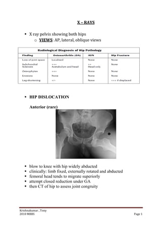
  • 1. Krishnakumar , Tony 2010 MBBS Page 1 X – RAYS  X ray pelvis showing both hips o VIEWS: AP, lateral, oblique views  HIP DISLOCATION Anterior (rare)  blow to knee with hip widely abducted  clinically: limb fixed, externally rotated and abducted  femoral head tends to migrate superiorly  attempt closed reduction under GA  then CT of hip to assess joint congruity
  • 2. Krishnakumar , Tony 2010 MBBS Page 2 Posterior  severe forces to knee with hip flexed and adducted (e.g. knee into dashboard in MVA)  clinically: limb shortened, internally rotated and adducted  femoral head tends to migrate inferiorly/medially  +/– fracture of posterior lip of acetabulum or intra-articular fracture  sciatic nerve injury common especially with associated acetabular fracture  assess knee, femoral shaft for other injuries/fractures  +/– fracture of posterior lip of acetabulum or intra-articular fracture  attempt closed reduction under GA +/– image intensifier  then CT to assess congruity and acetabular integrity  traction x 6 weeks, then ROM  ORIF if unstable, intra-articular fragments, or posterior wall fractures Central  associated with acetabular fracture After Total Hip Arthroplasty (THA)  occurs in 1-4% of primary THA and 16% in revision cases
  • 3. Krishnakumar , Tony 2010 MBBS Page 3  about 74% of THA dislocations are posterior, 16% anterior and 8% central  THA are unstable in the position of flexion and internal rotation Treatment  complete muscle relaxation is key – conscious sedation (IV fentanyl and versed) or spinal or GA  assistant applies downward pressure to pelvis  reduction for posterior dislocation – fully flex hip, abduct and externally rotate hip,  apply upward traction on femur  reduction for anterior dislocation – fully flex hip , adduct and internally rotate hip,  apply downward pressure on femur  Complications  post-traumatic arthritis due to cartilage injury or intra-articular loose body  femoral head injury including osteonecrosis + fracture; 100% if nothing12 hours before reduction  sciatic nerve palsy in 25% (10% permanent)  fracture of femoral shaft or neck  heterotopic ossification  coxa magna (occurs in up to 50% of children after a hip dislocation)  sciatic nerve palsy in 25% (10% permanent)  fracture of femoral shaft or neck  knee injury (posterior cruciate ligament (PCL) tear with dashboard injury) DISLOCATED KNEE (anterior)
  • 4. Krishnakumar , Tony 2010 MBBS Page 4  bad high energy injury  associated injuries • popliteal artery intimal tear or disruption 35-50% • capsular, ligamentous and common peroneal nerve injury Investigations  angiogram Treatment  closed reduction, above knee cylinder cast x 4 weeks  alternately, external fixation especially if vascular repair • surgical repair of all ligaments if high demand patient SUPRACONDYLAR FRACTURE  usually in children  fall on outstretched hand  type of fracture is based on the distal segment .extension type common
  • 5. Krishnakumar , Tony 2010 MBBS Page 5  Treatment  children o closed reduction +/– percutaneous pinning in O.R. with fluoroscopy o cast in flexion x 3 weeks  adult o undisplaced fracture, may be treated in cast o displaced fracture, ORIF since closed reduction usually inadequate  Complications  stiffnes most common RADIAL HEAD FRACTURE  mechanism: fall on outstretched hand (FOOSH)  clinically: progressive pain due to hemarthrosis with loss of ROM and pain on lateral side of elbow  aggravated by forearm pronation or supination  careful, may not be seen radiographically  look for “sail sign” of anterior fat pad or the prescence of a posterior fat pad on x-ray to detect occult  radial head fractures Mason Classification Type 1: undisplaced segmental fracture, usually normal ROM Type 2: displaced segmental fracture, ROM compromised Type 3: comminuted fracture Type 4: Type 3 with posterior dislocation Treatment  Type 1: elbow slab, sling 3-5 days, early ROM  Type 2: ORIF radial head  Type 3/4: excision of radial head +/– prosthesis OLECRANON FRACTURE  fall on point of elbow with avulsion by triceps or fall on outstretched arm
  • 6. Krishnakumar , Tony 2010 MBBS Page 6  active extension absent  gross displacement can not be reduced closed because of pull of triceps Treatment  undisplaced: above elbow cast 2 weeks, early ROM  displaced: ORIF, above elbow slab x 1 week, early ROM ELBOW DISLOCATION  usually young people in sporting events or high speed MVA  > 90% are posterior or posterior-lateral  fall on outstretched hand  rule out concurrent radial head or coracoid process fractures Treatment of Posterior Dislocation  closed reduction: traction then flexion  ❏ above elbow backslab with elbow 90 degrees and wrist pronated  ❏ open reduction if unstable or loose body (unusual) Complications  stiffness  intra-articular loose body • usually from joint surface cartilage • not obvious on x-ray
  • 7. Krishnakumar , Tony 2010 MBBS Page 7 • occasionally medial epicondyle is pulled into joint, especially in children  heterotopic ossification (bone formation)  • prevented by indomethacin immediately following surgery RADIAL HEAD FRACTURE  mechanism: fall on outstretched hand (FOOSH)  clinically: progressive pain due to hemarthrosis with loss of ROM and pain on lateral side of elbow  aggravated by forearm pronation or supination  careful, may not be seen radiographically  look for “sail sign” of anterior fat pad or the prescence of a posterior fat pad on x-ray to detect occult  radial head fractures  Mason Classification  Type 1: undisplaced segmental fracture, usually normal ROM  Type 2: displaced segmental fracture, ROM compromised  Type 3: comminuted fracture  Type 4: Type 3 with posterior dislocation Treatment  ❏ Type 1: elbow slab, sling 3-5 days, early ROM  ❏ Type 2: ORIF radial head  ❏ Type 3/4: excision of radial head +/– prosthesis
  • 8. Krishnakumar , Tony 2010 MBBS Page 8 GALEAZZI FRACTURE  fracture of distal radius  dislocation of distal radio-ulnar joint (DRUJ) at wrist  treatment: immobilize in supination to reduce DRUJ, ORIF MONTEGGIA FRACTURE  fracture of ulna with associated dislocation of radial head  treatment: ORIF is recommended- open reduction of the ulna is usually followed by  indirect reduction of the radius
  • 9. Krishnakumar , Tony 2010 MBBS Page 9 COLLES' FRACTURE Etiology  most common wrist fracture  fall on outstretched hand (FOOSH)  most common in osteoporotic bone  Diagnosis clinical o swelling, ecchymosis, tenderness o “dinner fork” deformity (Figure 14) o assess neurovascular status (carpal tunnel syndrome) X-ray: distal fragment is
  • 10. Krishnakumar , Tony 2010 MBBS Page 10  dorsally displaced with dorsal comminution  dorsally tilted fragment with apex of fracture volar  supinated  radially deviated  shortened (radial styloid normally 1cm distal to ulna) +/– fracture of ulnar styloid Treatment  nondisplaced o short arm cast applied to wrist under gentle traction o neutral wrist position  displaced  anesthesia - hematoma block commonly used  disimpaction - axial traction with increasing force over 2 minutes  (pull on thumb and ring finger, with countertraction at the elbow)  reduce by pulling hand into o slight flexion o full pronation o full ulnar deviation  maintain reduction with direct pressure to fracture site, apply well moulded dorsal-radial slab (splint)  post-reduction x-ray (AP/lateral), goal to correct dorsal angulation and regain radial length  check arm after 24 hours for swelling, neurovascular status  circular cast after 1-2 weeks; check cast at 1, 2, 6 weeks; cast off after 6 weeks, physiotherapy  (ROM, grip strength)  if inadequate reduction at any time o try closed reduction under GA o ORIF
  • 11. Krishnakumar , Tony 2010 MBBS Page 11 ANTERIOR SHOULDER DISLOCATION  over 90% of all shoulder dislocations, usually traumatic  may be of two general types: • involuntary: traumatic, unidirectional, Bankart lesion, responds to surgery • voluntary: atraumatic, multidirectional, bilateral, rehab, surgery is last resort occurs when abducted arm is externally rotated or hyperextended recurrence rate depends on age of first dislocation Ossification around elbow Capitulum ………………………… 1 year Radial head………………………… 3 year Internal epicondyle……………... 5 year (last to fuse – 16 years ) Trochlea……………………………... 7 year Olecranon…………………………… 1year External epicondyle……………… 11 year
  • 12. Krishnakumar , Tony 2010 MBBS Page 12 • at age 20: 80%; at age 21-40: 60-70%; at age 40-60: 40-60%; at age > 60: < 10% associated with Hill-Sachs and Bankart lesion • indentation of humeral head after impaction on glenoid rim SHOULDER . . . CONT.  avulsion of capsule when shoulder dislocates  associated bony avulsion called "Bony Bankart Lesion"  occurs in 85% of all anterior dislocations  axillary nerve and musculocutaneous nerve at risk  some associated injuries more common in elderly  vascular injury and fracture of greater tuberosity Physical Examination  humeral head can be palpated anteriorly  arm held in slight abduction and external rotation  ❏ loss of internal rotation with anterioinferior humeral head  axillary nerve may be damaged, therefore check sensation and contraction over lateral deltoid;  for musculocutaneous nerve check sensation of lateral forearm and contraction of biceps  apprehension test: for recurrent shoulder instability  with patient supine, gently abduct and externally rotate patient’s arm to a position where it may  easily dislocate; if shoulder is dislocatable, patient will have a look of apprehension on face X-Rays  humeral head anterior (to Mercedes Benz sign) in trans- scapular view  axillary view is diagnostic  AP view may show Hill-Sachs lesion if recurrent  rule out associated humeral neck fracture Treatment  intravenous sedation and muscle relaxation  gentle longitudinal traction and countertraction  +/– alternating internal and external rotation
  • 13. Krishnakumar , Tony 2010 MBBS Page 13  Hippocratic Method - foot used in axilla for countertraction (not recommended - risk of nerve damage)  Stimsons’s method - patient prone with arm hanging over edge of table, weight hung on wrist  (typically 5 lbs for 15-20 mins)  X-Ray to verify reduction and check neurologic status ❏ sling x 3 weeks with movement of elbow, wrist, fingers  • rehabilitation aimed at strengthening dynamic stabilizers and avoiding the unstable position (i.e. external rotation and abduction)  recurrent instability and dislocations may require surgery COLD ORTHOPEDICSBONE TUMOURS ❏ primary bone tumours are rare after 3rd decade ❏ metastases to bone are relatively common after 3rd decade BENIGN BONE TUMOURS 1. Osteoid Osteoma ❏ age 10-25 years ❏ small, round radiolucent nidus (< 1 cm) surrounded by dense bone
  • 14. Krishnakumar , Tony 2010 MBBS Page 14 ❏ tibia and femur; diaphyseal ❏ produces severe intermittent pain, mostly at night ❏ characteristically relieved by ASA 2. Osteochondroma ❏ metaphysis of long bone ❏ cartilage-capped bony spur on surface of bone (“mushroom” on x- ray) ❏ may be multiple (hereditary form) - higher risk of malignant change ❏ generally not painful unless impinging on neurovascular structure ❏ malignant degeneration occurs in 1-2 % 3. Enchondroma ❏ age 20-40 years ❏ 50% occur in the small tubular bones of the hand and foot; others in femur, humerus, ribs ❏ benign cartilage growth, develops in medullary cavity ❏ single/multiple enlarged rarefied areas in tubular bones ❏ lytic lesion with specks of calcification on x-ray 4. Cystic Lesions ❏ includes unicameral bone cyst, aneurysmal bone cyst, fibrous cortical defect ❏ children and young adults ❏ local pain, pathological fracture or accidental detection
  • 15. Krishnakumar , Tony 2010 MBBS Page 15 ❏ translucent area on metaphyseal side of growth plate ❏ cortex thinned/expanded; well defined lesion ❏ treatment of unicameral bone cyst with steroid injections +/– bone graft Treatment ❏ in general, curettage +/– bone graft BENIGN AGGRESSIVE BONE TUMOURS 1. Giant Cell Tumours ❏ 80% occur > 20 years, average 35 years ❏ distal femur, proximal tibia, distal radius ❏ pain and swelling ❏ cortex appears thinned, expanded; well demarcated sclerotic margin ❏ 1/3 benign, 1/3 invasive, 1/3 metastasize ❏ 30% reccur within 2 years of surgery Soap bubble appearance
  • 16. Krishnakumar , Tony 2010 MBBS Page 16 2. Osteoblastoma ❏ aggressive tumour forming osteoid ❏ lesions > 2 cm in size and grow rapidly ❏ painful ❏ most frequent in spine and long bones (humerus, femur, tibia) Treatment ❏ controversial, should do metastatic work up ❏ wide local excision +/– bone graft 1. Osteosarcoma ❏ bimodal age distribution • ages 10-20 (60%) • > 50 with history of Paget's disease ❏ invasive, variable histology; frequent metastases ❏ predilection for distal femur (45%), tibia (20%) and proximal humerus (15%) ❏ history of trauma common ❏ painful, tender, poorly defined swelling ❏ x-ray shows Codman's Triangle: characteristic periosteal elevation and spicule formation representing tumour extension into periosteum with calcification
  • 17. Krishnakumar , Tony 2010 MBBS Page 17 ❏ treatment with complete resection (limb salvage, rarely amputation) adjuvant chemo, radiotherapy 2. Chondrosarcoma ❏ primary: previous normal bone, patient over 40; expands into cortex to give pain, pathological fracture, flecks of calcification ❏ secondary: malignant degeneration of preexisting cartilage tumour such as enchondroma or osteochondroma ❏ occurs in pelvis, femur, ribs, shoulder ❏ x-ray shows large exostosis with calcification in cap ❏ highly resistant to chemotherapy, treat with aggressive surgical resection 2. Ewing's Sarcoma ❏ thought to be undifferentiated member of a family of neural tumours distinct form neuroblastoma ❏ most occur between 5 - 20 years old ❏ florid periosteal reaction in diaphysis of long bone; ages 10-20 ❏ present with mild fever, anemia, leukocytosis and elevated ESR ❏ moth-eaten appearance with periosteal "onion-skinning" ❏ metastases frequent ❏ treatment: chemotherapy, resection, radiation