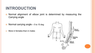
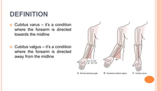
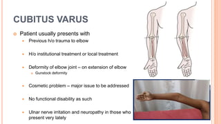
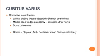
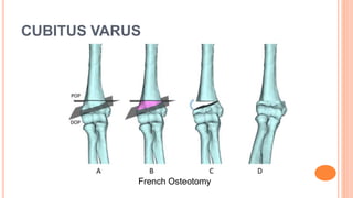
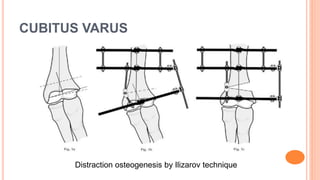
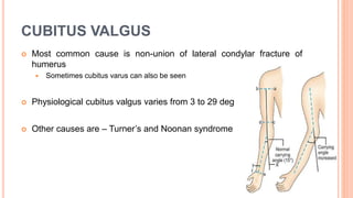
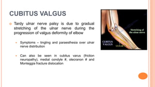
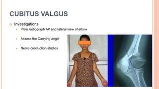
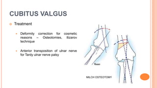
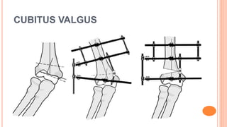
CUBITUS VARUS AND
CUBITUS VALGUS
DR. BIPUL BORTHAKUR
PROFESSOR, DEPT OF ORTHOPAEDICS, SMCH
INTRODUCTION
 Normal alignment of elbow joint is determined by measuring the
Carrying angle
 Normal carrying angle – 5 to 15 deg
 More in females than in males
DEFINITION
 Cubitus varus – it’s a condition
where the forearm is directed
towards the midline
 Cubitus valgus – it’s a condition
where the forearm is directed
away from the midline
CUBITUS VARUS
 Synonyms – Bow elbow or Gunstock deformity
 Most common cause is Malunion of supracondylar fracture of
humerus
 Rarely, osteonecrosis with or without growth disturbance
 Causes of malunion
 Malreduction or loss of proper reduction when treated conservatively
 Treatment by local bone setters
 Severely comminuted fracture
CUBITUS VARUS
 Patient usually presents with
 Previous h/o trauma to elbow
 H/o institutional treatment or local treatment
 Deformity of elbow joint – on extension of elbow
 Gunstock deformity
 Cosmetic problem – major issue to be addressed
 No functional disability as such
 Ulnar nerve irritation and neuropathy in those who
present very lately
CUBITUS VARUS
 Investigations:
 Plain radiograph of elbow AP and lateral view
 Assess the Carrying angle
CUBITUS VARUS
 Treatment – cubitus varus has no tendency for spontaneous
correction by remodelling. Hence has to corrected surgically
 Mostly done for cosmetic reasons
 Options available are:
 Hemiepiphysiodesis and growth alteration – in patients with medial growth
arrest and progressive deformity
 Corrective osteotomies – Commonly done
 Distraction osteogenesis by Ilizarov technique
CUBITUS VARUS
 Corrective osteotomies
 Lateral closing wedge osteotomy (French osteotomy)
 Medial open wedge osteotomy – stretches ulnar nerve
 Dome osteotomy
 Others – Step cut, Arch, Pentalateral and Oblique osteotomy
CUBITUS VARUS
French Osteotomy
CUBITUS VARUS
Distraction osteogenesis by Ilizarov technique
CUBITUS VALGUS
 Most common cause is non-union of lateral condylar fracture of
humerus
 Sometimes cubitus varus can also be seen
 Physiological cubitus valgus varies from 3 to 29 deg
 Other causes are – Turner’s and Noonan syndrome
CUBITUS VALGUS
 H/o previous trauma to elbow along with
fracture lateral condyle of humerus
 Patient presents with external deformity of
elbow joint
 Usually asymptomatic till patient develops
TARDY ULNAR NERVE PALSY
CUBITUS VALGUS
 Tardy ulnar nerve palsy is due to gradual
stretching of the ulnar nerve during the
progression of valgus deformity of elbow
 Symptoms – tingling and paraesthesia over ulnar
nerve distribution
 Can also be seen in cubitus varus (friction
neuropathy), medial condyle #, olecranon # and
Monteggia fracture dislocation
CUBITUS VALGUS
 Investigations
 Plain radiograph AP and lateral view of elbow
 Assess the Carrying angle
 Nerve conduction studies
CUBITUS VALGUS
 Treatment
 Deformity correction for cosmetic
reasons – Osteotomies, Ilizarov
technique
 Anterior transposition of ulnar nerve
for Tardy ulnar nerve palsy
MILCH OSTEOTOMY
CUBITUS VALGUS
THANK YOU

Cubitus varus and valgus

  • 1.
    CUBITUS VARUS AND CUBITUSVALGUS DR. BIPUL BORTHAKUR PROFESSOR, DEPT OF ORTHOPAEDICS, SMCH
  • 2.
    INTRODUCTION  Normal alignmentof elbow joint is determined by measuring the Carrying angle  Normal carrying angle – 5 to 15 deg  More in females than in males
  • 3.
    DEFINITION  Cubitus varus– it’s a condition where the forearm is directed towards the midline  Cubitus valgus – it’s a condition where the forearm is directed away from the midline
  • 4.
    CUBITUS VARUS  Synonyms– Bow elbow or Gunstock deformity  Most common cause is Malunion of supracondylar fracture of humerus  Rarely, osteonecrosis with or without growth disturbance  Causes of malunion  Malreduction or loss of proper reduction when treated conservatively  Treatment by local bone setters  Severely comminuted fracture
  • 5.
    CUBITUS VARUS  Patientusually presents with  Previous h/o trauma to elbow  H/o institutional treatment or local treatment  Deformity of elbow joint – on extension of elbow  Gunstock deformity  Cosmetic problem – major issue to be addressed  No functional disability as such  Ulnar nerve irritation and neuropathy in those who present very lately
  • 6.
    CUBITUS VARUS  Investigations: Plain radiograph of elbow AP and lateral view  Assess the Carrying angle
  • 7.
    CUBITUS VARUS  Treatment– cubitus varus has no tendency for spontaneous correction by remodelling. Hence has to corrected surgically  Mostly done for cosmetic reasons  Options available are:  Hemiepiphysiodesis and growth alteration – in patients with medial growth arrest and progressive deformity  Corrective osteotomies – Commonly done  Distraction osteogenesis by Ilizarov technique
  • 8.
    CUBITUS VARUS  Correctiveosteotomies  Lateral closing wedge osteotomy (French osteotomy)  Medial open wedge osteotomy – stretches ulnar nerve  Dome osteotomy  Others – Step cut, Arch, Pentalateral and Oblique osteotomy
  • 9.
  • 10.
  • 11.
    CUBITUS VALGUS  Mostcommon cause is non-union of lateral condylar fracture of humerus  Sometimes cubitus varus can also be seen  Physiological cubitus valgus varies from 3 to 29 deg  Other causes are – Turner’s and Noonan syndrome
  • 12.
    CUBITUS VALGUS  H/oprevious trauma to elbow along with fracture lateral condyle of humerus  Patient presents with external deformity of elbow joint  Usually asymptomatic till patient develops TARDY ULNAR NERVE PALSY
  • 13.
    CUBITUS VALGUS  Tardyulnar nerve palsy is due to gradual stretching of the ulnar nerve during the progression of valgus deformity of elbow  Symptoms – tingling and paraesthesia over ulnar nerve distribution  Can also be seen in cubitus varus (friction neuropathy), medial condyle #, olecranon # and Monteggia fracture dislocation
  • 14.
    CUBITUS VALGUS  Investigations Plain radiograph AP and lateral view of elbow  Assess the Carrying angle  Nerve conduction studies
  • 15.
    CUBITUS VALGUS  Treatment Deformity correction for cosmetic reasons – Osteotomies, Ilizarov technique  Anterior transposition of ulnar nerve for Tardy ulnar nerve palsy MILCH OSTEOTOMY
  • 16.
  • 17.