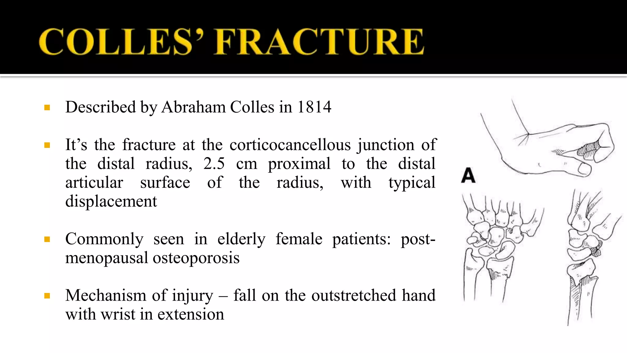
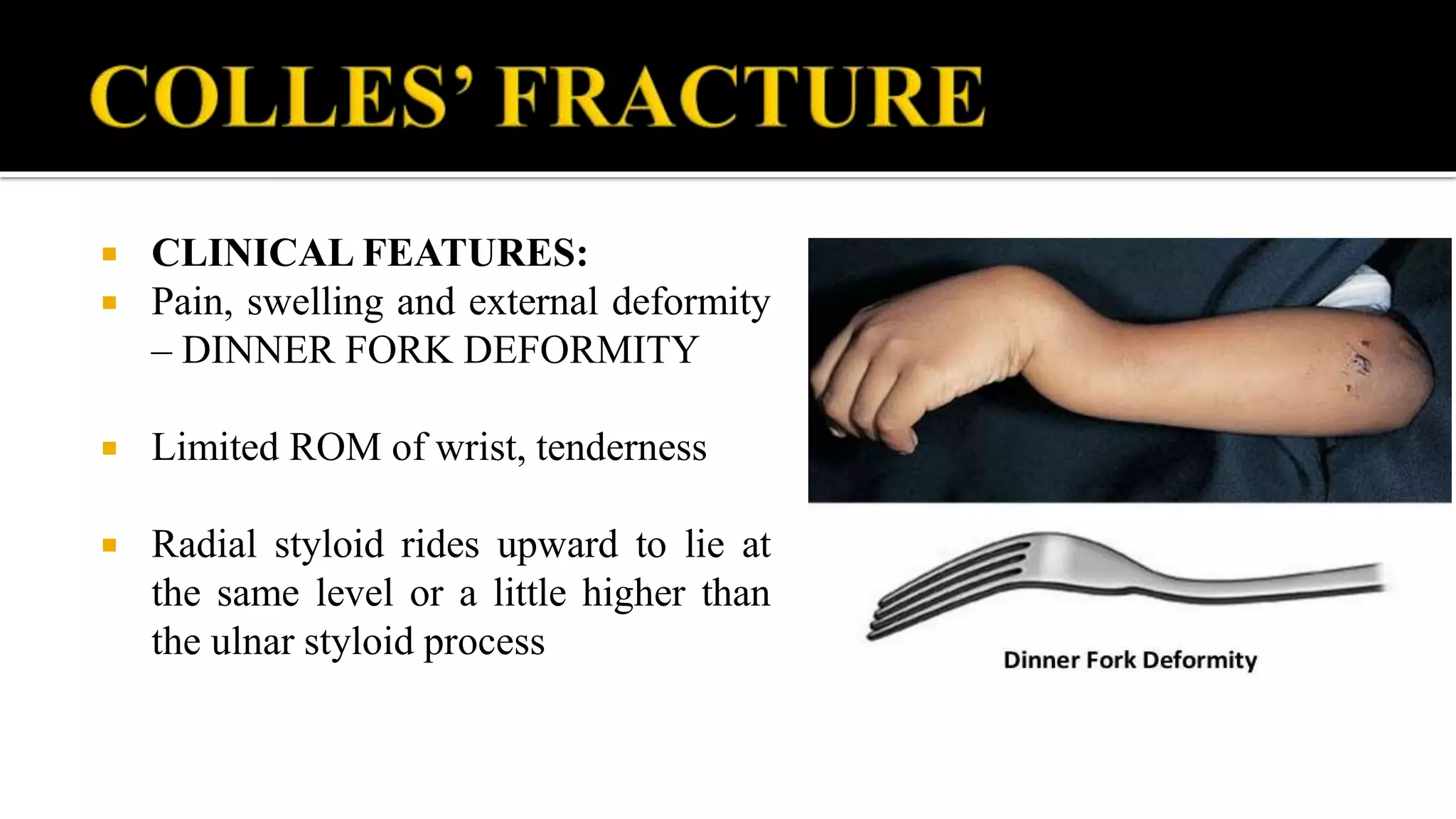
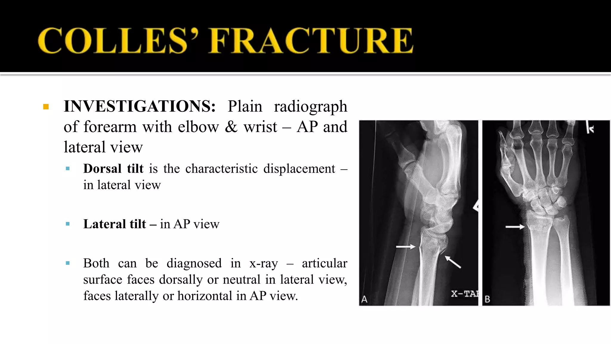
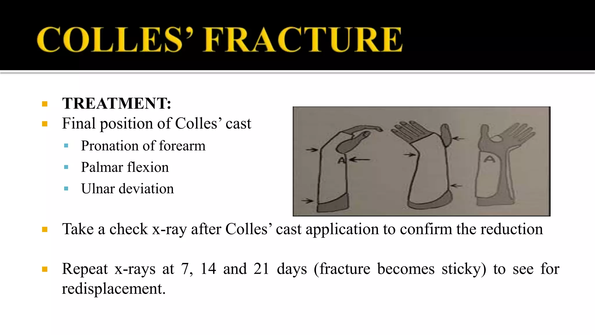
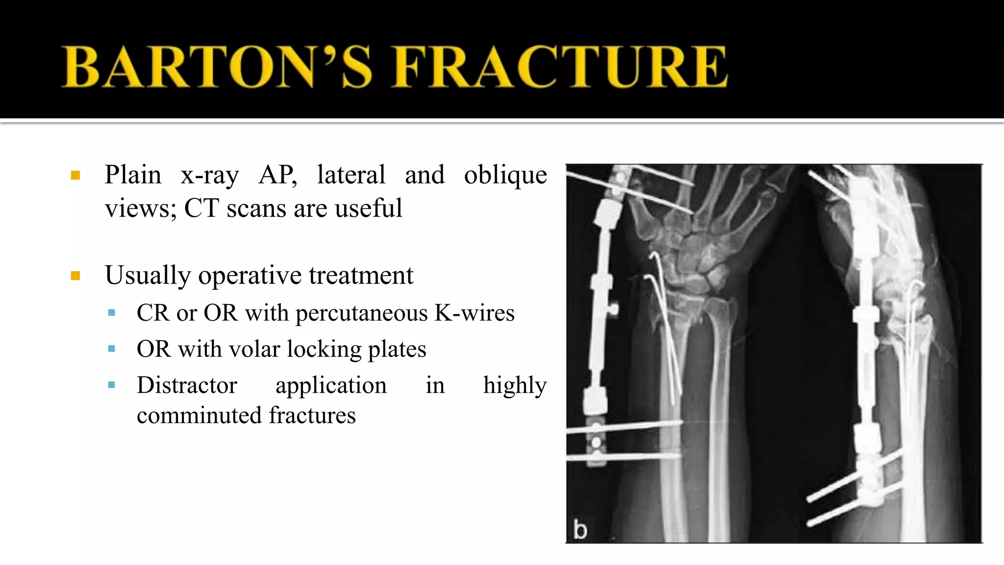
DR. BIPUL BORTHAKUR
PROFESSOR, DEPT OF ORTHOPAEDICS, SMCH
 Colle’s fracture
 Smith fracture
 Barton’s fracture
 Chauffeur’s fracture
 Described by Abraham Colles in 1814
 It’s the fracture at the corticocancellous junction of
the distal radius, 2.5 cm proximal to the distal
articular surface of the radius, with typical
displacement
 Commonly seen in elderly female patients: post-
menopausal osteoporosis
 Mechanism of injury – fall on the outstretched hand
with wrist in extension
 Displacements in Colles’ fracture
 Shifts – Dorsal and lateral
 Tilts – Dorsal and lateral
 Impaction or proximal shift
 Supination
 Associated injuries:
 Ulnar styloid fracture
 Rupture of ulnar collateral ligament
 Rupture TFCC (triangular fibrocartilage complex)
 Rupture of the interosseous radio-ulnar ligament – DRUJ subluxation
 CLINICAL FEATURES:
 Pain, swelling and external deformity
– DINNER FORK DEFORMITY
 Limited ROM of wrist, tenderness
 Radial styloid rides upward to lie at
the same level or a little higher than
the ulnar styloid process
 INVESTIGATIONS: Plain radiograph
of forearm with elbow & wrist – AP and
lateral view
 Dorsal tilt is the characteristic displacement –
in lateral view
 Lateral tilt – in AP view
 Both can be diagnosed in x-ray – articular
surface faces dorsally or neutral in lateral view,
faces laterally or horizontal in AP view.
 TREATMENT:
 Undisplaced fracture – below elbow cast immobilization without any
manipulation
 With typical displacement – closed manipulation and below elbow slab or
cast immobilization for 4 to 6 weeks.
 Traction and counter-traction: disimpaction
 Correction of dorsal tilt – palmar flexion
 Correction of radial tilt – ulnar deviation
 TREATMENT:
 Final position of Colles’ cast
 Pronation of forearm
 Palmar flexion
 Ulnar deviation
 Take a check x-ray after Colles’ cast application to confirm the reduction
 Repeat x-rays at 7, 14 and 21 days (fracture becomes sticky) to see for
redisplacement.
 TREATMENT:
 Patient advised to keep the limb elevated
 Shoulder, elbow and finger joint movements have to be emphasized
 Remove the cast immediately if signs of compartment syndrome develops
 Fracture heals by 6 weeks, following which physiotherapy has to be started
 Reverse Colles’ fracture – ventral tilt and shift
 Fall on the outstretched hand with wrist in flexion
 Volar tilt / shift instead of dorsal tilt / shift
 More unstable than Colles’ fracture
 Garden-spade deformity
 Plain x-ray of wrist confirms the diagnosis
 Treatment – closed manipulation + B/E slab
or cast for 6weeks
 Traction – supination – extension of the wrist
 Surgical fixation if fracture displaces again –
volar locking plates
 High energy injury
 Intra-articular fracture of distal radius
 Either with volar or dorsal displacement
 Highly unstable fracture
 Demands restoration of articular congruity, alignment and length.
 Plain x-ray AP, lateral and oblique
views; CT scans are useful
 Usually operative treatment
 CR or OR with percutaneous K-wires
 OR with volar locking plates
 Distractor application in highly
comminuted fractures
 It is an intra-articular fracture of radial
styloid process.
 It results due to fall on outstretched
hand.
 It can be managed conservatively.
 Early:
 Nerve injury – median nerve compression
 Complex regional pain syndrome (CRPS) or Sudeck’s osteodystrophy
 Ulnar corner pain and instability
 Associated injuries of the carpus
 Redisplacement
 Late:
 Malunion – due to improper reduction, improper fixation and redisplacement
 Delayed union and non-union – rare unless infected or severe bone loss with instability
 Tendon rupture – EPL, FPL
 Carpal instability – due to ligament injury
 Secondary osteoarthritis – in intra-articular fractures
Distal radius fracture

Distal radius fracture

  • 1.
    DR. BIPUL BORTHAKUR PROFESSOR,DEPT OF ORTHOPAEDICS, SMCH
  • 3.
     Colle’s fracture Smith fracture  Barton’s fracture  Chauffeur’s fracture
  • 4.
     Described byAbraham Colles in 1814  It’s the fracture at the corticocancellous junction of the distal radius, 2.5 cm proximal to the distal articular surface of the radius, with typical displacement  Commonly seen in elderly female patients: post- menopausal osteoporosis  Mechanism of injury – fall on the outstretched hand with wrist in extension
  • 5.
     Displacements inColles’ fracture  Shifts – Dorsal and lateral  Tilts – Dorsal and lateral  Impaction or proximal shift  Supination  Associated injuries:  Ulnar styloid fracture  Rupture of ulnar collateral ligament  Rupture TFCC (triangular fibrocartilage complex)  Rupture of the interosseous radio-ulnar ligament – DRUJ subluxation
  • 6.
     CLINICAL FEATURES: Pain, swelling and external deformity – DINNER FORK DEFORMITY  Limited ROM of wrist, tenderness  Radial styloid rides upward to lie at the same level or a little higher than the ulnar styloid process
  • 7.
     INVESTIGATIONS: Plainradiograph of forearm with elbow & wrist – AP and lateral view  Dorsal tilt is the characteristic displacement – in lateral view  Lateral tilt – in AP view  Both can be diagnosed in x-ray – articular surface faces dorsally or neutral in lateral view, faces laterally or horizontal in AP view.
  • 8.
     TREATMENT:  Undisplacedfracture – below elbow cast immobilization without any manipulation  With typical displacement – closed manipulation and below elbow slab or cast immobilization for 4 to 6 weeks.  Traction and counter-traction: disimpaction  Correction of dorsal tilt – palmar flexion  Correction of radial tilt – ulnar deviation
  • 9.
     TREATMENT:  Finalposition of Colles’ cast  Pronation of forearm  Palmar flexion  Ulnar deviation  Take a check x-ray after Colles’ cast application to confirm the reduction  Repeat x-rays at 7, 14 and 21 days (fracture becomes sticky) to see for redisplacement.
  • 10.
     TREATMENT:  Patientadvised to keep the limb elevated  Shoulder, elbow and finger joint movements have to be emphasized  Remove the cast immediately if signs of compartment syndrome develops  Fracture heals by 6 weeks, following which physiotherapy has to be started
  • 11.
     Reverse Colles’fracture – ventral tilt and shift  Fall on the outstretched hand with wrist in flexion  Volar tilt / shift instead of dorsal tilt / shift  More unstable than Colles’ fracture  Garden-spade deformity
  • 12.
     Plain x-rayof wrist confirms the diagnosis  Treatment – closed manipulation + B/E slab or cast for 6weeks  Traction – supination – extension of the wrist  Surgical fixation if fracture displaces again – volar locking plates
  • 13.
     High energyinjury  Intra-articular fracture of distal radius  Either with volar or dorsal displacement  Highly unstable fracture  Demands restoration of articular congruity, alignment and length.
  • 14.
     Plain x-rayAP, lateral and oblique views; CT scans are useful  Usually operative treatment  CR or OR with percutaneous K-wires  OR with volar locking plates  Distractor application in highly comminuted fractures
  • 15.
     It isan intra-articular fracture of radial styloid process.  It results due to fall on outstretched hand.  It can be managed conservatively.
  • 16.
     Early:  Nerveinjury – median nerve compression  Complex regional pain syndrome (CRPS) or Sudeck’s osteodystrophy  Ulnar corner pain and instability  Associated injuries of the carpus  Redisplacement
  • 17.
     Late:  Malunion– due to improper reduction, improper fixation and redisplacement  Delayed union and non-union – rare unless infected or severe bone loss with instability  Tendon rupture – EPL, FPL  Carpal instability – due to ligament injury  Secondary osteoarthritis – in intra-articular fractures