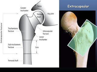
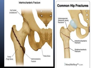
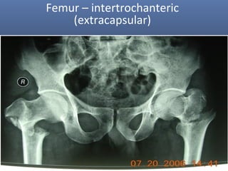
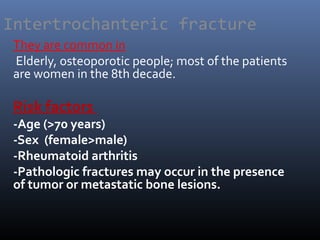
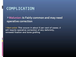
Colles fracture is the fracture at the distal end of radius, at its
cortico cancellous junction(about 2cm from the distal articular
surface).
It is not just the fracture of distal radius but the fracture
dislocation of the inferior radio-ulnar joint.
Colles fracture is the fracture at the distal end of radius, at its
cortico cancellous junction(about 2cm from the distal articular
surface).
It is not just the fracture of distal radius but the fracture
dislocation of the inferior radio-ulnar joint.
1) Subtrochanteric Fracture
Subtrochanteric typically defined as area from lesser trochanter to 5cm distal fractures with an associated intertrochanteric component may be called peritrochanteric fracture.
*Unique Aspect
Blood loss is greater than with femoral neck or trochanteric fractures – covered with anastomosing branches of the medial and lateral circumflex femoral arteries branch of profunda femoris trunk.
2) Femoral Shaft Fracture
Femoral shaft fracture is defined as a fracture of the diaphysis occurring between 5 cm distal to the lesser trochanter and 5 cm proximal to the adductor tubercle
The femoral shaft is padded with large muscles.
- reduction can be difficult as muscle contraction displaces the fracture
- healing potential is improved by having this well-vascularized
*Age
-usually a fracture of young adults and results from a high energy injury
-elderly patients should be considered ‘pathological’ until proved otherwise
-children under 4 years the suspected possibility of physical abuse
*FRACTURES ASSOCIATED WITH VASCULAR INJURY
Warning signs of an associated vascular injury are
(1) excessive bleeding or haematoma formation; and
(2) paraesthesia, pallor or pulselessness in the leg and foot.
~Warm ischemia in 2-3H
~If > 6H – salvage not possible
*‘FLOATING KNEE’
Ipsilateral fractures of the femur and tibia may leave the knee joint ‘floating’
3) Distal Femoral Fracture
Defined as fractures from articular surface to 5cm above metaphyseal flare
*clinical feature
The knee is swollen because of a haemarthrosis – this can be severe enough to cause blistering later
Movement is too painful to be attempted
The tibial pulses should always be checked to ensure the popliteal artery was not injured in the fracture.
Reference: Apley's System of Orthopaedic and Fracture (9th edition)
Prix Galien International 2024 Forum ProgramLevi Shapiro
June 20, 2024, Prix Galien International and Jerusalem Ethics Forum in ROME. Detailed agenda including panels:
- ADVANCES IN CARDIOLOGY: A NEW PARADIGM IS COMING
- WOMEN’S HEALTH: FERTILITY PRESERVATION
- WHAT’S NEW IN THE TREATMENT OF INFECTIOUS,
ONCOLOGICAL AND INFLAMMATORY SKIN DISEASES?
- ARTIFICIAL INTELLIGENCE AND ETHICS
- GENE THERAPY
- BEYOND BORDERS: GLOBAL INITIATIVES FOR DEMOCRATIZING LIFE SCIENCE TECHNOLOGIES AND PROMOTING ACCESS TO HEALTHCARE
- ETHICAL CHALLENGES IN LIFE SCIENCES
- Prix Galien International Awards Ceremony
Knee anatomy and clinical tests 2024.pdfvimalpl1234
This includes all relevant anatomy and clinical tests compiled from standard textbooks, Campbell,netter etc..It is comprehensive and best suited for orthopaedicians and orthopaedic residents.
Tom Selleck Health: A Comprehensive Look at the Iconic Actor’s Wellness Journeygreendigital
Tom Selleck, an enduring figure in Hollywood. has captivated audiences for decades with his rugged charm, iconic moustache. and memorable roles in television and film. From his breakout role as Thomas Magnum in Magnum P.I. to his current portrayal of Frank Reagan in Blue Bloods. Selleck's career has spanned over 50 years. But beyond his professional achievements. fans have often been curious about Tom Selleck Health. especially as he has aged in the public eye.
Follow us on: Pinterest
Introduction
Many have been interested in Tom Selleck health. not only because of his enduring presence on screen but also because of the challenges. and lifestyle choices he has faced and made over the years. This article delves into the various aspects of Tom Selleck health. exploring his fitness regimen, diet, mental health. and the challenges he has encountered as he ages. We'll look at how he maintains his well-being. the health issues he has faced, and his approach to ageing .
Early Life and Career
Childhood and Athletic Beginnings
Tom Selleck was born on January 29, 1945, in Detroit, Michigan, and grew up in Sherman Oaks, California. From an early age, he was involved in sports, particularly basketball. which played a significant role in his physical development. His athletic pursuits continued into college. where he attended the University of Southern California (USC) on a basketball scholarship. This early involvement in sports laid a strong foundation for his physical health and disciplined lifestyle.
Transition to Acting
Selleck's transition from an athlete to an actor came with its physical demands. His first significant role in "Magnum P.I." required him to perform various stunts and maintain a fit appearance. This role, which he played from 1980 to 1988. necessitated a rigorous fitness routine to meet the show's demands. setting the stage for his long-term commitment to health and wellness.
Fitness Regimen
Workout Routine
Tom Selleck health and fitness regimen has evolved. adapting to his changing roles and age. During his "Magnum, P.I." days. Selleck's workouts were intense and focused on building and maintaining muscle mass. His routine included weightlifting, cardiovascular exercises. and specific training for the stunts he performed on the show.
Selleck adjusted his fitness routine as he aged to suit his body's needs. Today, his workouts focus on maintaining flexibility, strength, and cardiovascular health. He incorporates low-impact exercises such as swimming, walking, and light weightlifting. This balanced approach helps him stay fit without putting undue strain on his joints and muscles.
Importance of Flexibility and Mobility
In recent years, Selleck has emphasized the importance of flexibility and mobility in his fitness regimen. Understanding the natural decline in muscle mass and joint flexibility with age. he includes stretching and yoga in his routine. These practices help prevent injuries, improve posture, and maintain mobilit
micro teaching on communication m.sc nursing.pdfAnurag Sharma
Microteaching is a unique model of practice teaching. It is a viable instrument for the. desired change in the teaching behavior or the behavior potential which, in specified types of real. classroom situations, tends to facilitate the achievement of specified types of objectives.
HOT NEW PRODUCT! BIG SALES FAST SHIPPING NOW FROM CHINA!! EU KU DB BK substit...GL Anaacs
Contact us if you are interested:
Email / Skype : kefaya1771@gmail.com
Threema: PXHY5PDH
New BATCH Ku !!! MUCH IN DEMAND FAST SALE EVERY BATCH HAPPY GOOD EFFECT BIG BATCH !
Contact me on Threema or skype to start big business!!
Hot-sale products:
NEW HOT EUTYLONE WHITE CRYSTAL!!
5cl-adba precursor (semi finished )
5cl-adba raw materials
ADBB precursor (semi finished )
ADBB raw materials
APVP powder
5fadb/4f-adb
Jwh018 / Jwh210
Eutylone crystal
Protonitazene (hydrochloride) CAS: 119276-01-6
Flubrotizolam CAS: 57801-95-3
Metonitazene CAS: 14680-51-4
Payment terms: Western Union,MoneyGram,Bitcoin or USDT.
Deliver Time: Usually 7-15days
Shipping method: FedEx, TNT, DHL,UPS etc.Our deliveries are 100% safe, fast, reliable and discreet.
Samples will be sent for your evaluation!If you are interested in, please contact me, let's talk details.
We specializes in exporting high quality Research chemical, medical intermediate, Pharmaceutical chemicals and so on. Products are exported to USA, Canada, France, Korea, Japan,Russia, Southeast Asia and other countries.
NVBDCP.pptx Nation vector borne disease control programSapna Thakur
NVBDCP was launched in 2003-2004 . Vector-Borne Disease: Disease that results from an infection transmitted to humans and other animals by blood-feeding arthropods, such as mosquitoes, ticks, and fleas. Examples of vector-borne diseases include Dengue fever, West Nile Virus, Lyme disease, and malaria.
Lung Cancer: Artificial Intelligence, Synergetics, Complex System Analysis, S...Oleg Kshivets
RESULTS: Overall life span (LS) was 2252.1±1742.5 days and cumulative 5-year survival (5YS) reached 73.2%, 10 years – 64.8%, 20 years – 42.5%. 513 LCP lived more than 5 years (LS=3124.6±1525.6 days), 148 LCP – more than 10 years (LS=5054.4±1504.1 days).199 LCP died because of LC (LS=562.7±374.5 days). 5YS of LCP after bi/lobectomies was significantly superior in comparison with LCP after pneumonectomies (78.1% vs.63.7%, P=0.00001 by log-rank test). AT significantly improved 5YS (66.3% vs. 34.8%) (P=0.00000 by log-rank test) only for LCP with N1-2. Cox modeling displayed that 5YS of LCP significantly depended on: phase transition (PT) early-invasive LC in terms of synergetics, PT N0—N12, cell ratio factors (ratio between cancer cells- CC and blood cells subpopulations), G1-3, histology, glucose, AT, blood cell circuit, prothrombin index, heparin tolerance, recalcification time (P=0.000-0.038). Neural networks, genetic algorithm selection and bootstrap simulation revealed relationships between 5YS and PT early-invasive LC (rank=1), PT N0—N12 (rank=2), thrombocytes/CC (3), erythrocytes/CC (4), eosinophils/CC (5), healthy cells/CC (6), lymphocytes/CC (7), segmented neutrophils/CC (8), stick neutrophils/CC (9), monocytes/CC (10); leucocytes/CC (11). Correct prediction of 5YS was 100% by neural networks computing (area under ROC curve=1.0; error=0.0).
CONCLUSIONS: 5YS of LCP after radical procedures significantly depended on: 1) PT early-invasive cancer; 2) PT N0--N12; 3) cell ratio factors; 4) blood cell circuit; 5) biochemical factors; 6) hemostasis system; 7) AT; 8) LC characteristics; 9) LC cell dynamics; 10) surgery type: lobectomy/pneumonectomy; 11) anthropometric data. Optimal diagnosis and treatment strategies for LC are: 1) screening and early detection of LC; 2) availability of experienced thoracic surgeons because of complexity of radical procedures; 3) aggressive en block surgery and adequate lymph node dissection for completeness; 4) precise prediction; 5) adjuvant chemoimmunoradiotherapy for LCP with unfavorable prognosis.
Title: Sense of Taste
Presenter: Dr. Faiza, Assistant Professor of Physiology
Qualifications:
MBBS (Best Graduate, AIMC Lahore)
FCPS Physiology
ICMT, CHPE, DHPE (STMU)
MPH (GC University, Faisalabad)
MBA (Virtual University of Pakistan)
Learning Objectives:
Describe the structure and function of taste buds.
Describe the relationship between the taste threshold and taste index of common substances.
Explain the chemical basis and signal transduction of taste perception for each type of primary taste sensation.
Recognize different abnormalities of taste perception and their causes.
Key Topics:
Significance of Taste Sensation:
Differentiation between pleasant and harmful food
Influence on behavior
Selection of food based on metabolic needs
Receptors of Taste:
Taste buds on the tongue
Influence of sense of smell, texture of food, and pain stimulation (e.g., by pepper)
Primary and Secondary Taste Sensations:
Primary taste sensations: Sweet, Sour, Salty, Bitter, Umami
Chemical basis and signal transduction mechanisms for each taste
Taste Threshold and Index:
Taste threshold values for Sweet (sucrose), Salty (NaCl), Sour (HCl), and Bitter (Quinine)
Taste index relationship: Inversely proportional to taste threshold
Taste Blindness:
Inability to taste certain substances, particularly thiourea compounds
Example: Phenylthiocarbamide
Structure and Function of Taste Buds:
Composition: Epithelial cells, Sustentacular/Supporting cells, Taste cells, Basal cells
Features: Taste pores, Taste hairs/microvilli, and Taste nerve fibers
Location of Taste Buds:
Found in papillae of the tongue (Fungiform, Circumvallate, Foliate)
Also present on the palate, tonsillar pillars, epiglottis, and proximal esophagus
Mechanism of Taste Stimulation:
Interaction of taste substances with receptors on microvilli
Signal transduction pathways for Umami, Sweet, Bitter, Sour, and Salty tastes
Taste Sensitivity and Adaptation:
Decrease in sensitivity with age
Rapid adaptation of taste sensation
Role of Saliva in Taste:
Dissolution of tastants to reach receptors
Washing away the stimulus
Taste Preferences and Aversions:
Mechanisms behind taste preference and aversion
Influence of receptors and neural pathways
Impact of Sensory Nerve Damage:
Degeneration of taste buds if the sensory nerve fiber is cut
Abnormalities of Taste Detection:
Conditions: Ageusia, Hypogeusia, Dysgeusia (parageusia)
Causes: Nerve damage, neurological disorders, infections, poor oral hygiene, adverse drug effects, deficiencies, aging, tobacco use, altered neurotransmitter levels
Neurotransmitters and Taste Threshold:
Effects of serotonin (5-HT) and norepinephrine (NE) on taste sensitivity
Supertasters:
25% of the population with heightened sensitivity to taste, especially bitterness
Increased number of fungiform papillae
Pulmonary Thromboembolism - etilogy, types, medical- Surgical and nursing man...VarunMahajani
Disruption of blood supply to lung alveoli due to blockage of one or more pulmonary blood vessels is called as Pulmonary thromboembolism. In this presentation we will discuss its causes, types and its management in depth.
The prostate is an exocrine gland of the male mammalian reproductive system
It is a walnut-sized gland that forms part of the male reproductive system and is located in front of the rectum and just below the urinary bladder
Function is to store and secrete a clear, slightly alkaline fluid that constitutes 10-30% of the volume of the seminal fluid that along with the spermatozoa, constitutes semen
A healthy human prostate measures (4cm-vertical, by 3cm-horizontal, 2cm ant-post ).
It surrounds the urethra just below the urinary bladder. It has anterior, median, posterior and two lateral lobes
It’s work is regulated by androgens which are responsible for male sex characteristics
Generalised disease of the prostate due to hormonal derangement which leads to non malignant enlargement of the gland (increase in the number of epithelial cells and stromal tissue)to cause compression of the urethra leading to symptoms (LUTS
- Video recording of this lecture in English language: https://youtu.be/lK81BzxMqdo
- Video recording of this lecture in Arabic language: https://youtu.be/Ve4P0COk9OI
- Link to download the book free: https://nephrotube.blogspot.com/p/nephrotube-nephrology-books.html
- Link to NephroTube website: www.NephroTube.com
- Link to NephroTube social media accounts: https://nephrotube.blogspot.com/p/join-nephrotube-on-social-media.html
Hemodialysis: Chapter 3, Dialysis Water Unit - Dr.Gawad
Intertrochanteric fracture
1.
2. Intertrochanteric fracture
Definition :
These fractures are ‘ extracapsular ’ and occur in the
wide metaphyseal region between the two
trochanters of the femur.
Why such fractures tend to unite without difficulty
and seldom cause avascular necrosis?
Because the blood supply to the fracture is adequate
6. Intertrochanteric fracture
They are common in
Elderly, osteoporotic people; most of the patients
are women in the 8th decade.
Risk factors
-Age (>70 years)
-Sex (female>male)
-Rheumatoid arthritis
-Pathologic fractures may occur in the presence
of tumor or metastatic bone lesions.
7. Intertrochanteric fracture
Mechanism of injury
The fracture is caused either by a fall
directly onto the greater trochanter or by
an indirect twisting injury.
The crack runs up between the lesser and
greater trochanter and the proximal
fragment tends to displace in varus.
10. Intertrochanteric fracture
X-ray
Undisplaced, stable fractures
may show no more than a
thin crack along the
intertrochanteric line;
indeed,there is often
doubt as to whether
the bone is fractured
and the diagnosis may
have to be confirmed by MRI.
11. Intertrochanteric fracture
Treatment
Intertrochanteric fractures are almost always
treated by early internal fixation
not because they fail to unite with conservative
treatment but
(a) to obtain the best possible position and
(b) to get the patient up and walking as soon as
possible and there by reduce the complications
associated with prolonged lying down
12. Intertrochanteric fracture
Treatment
Fracture reduction at
surgery is performed
on a fracture
table that provides slight
traction and internal
rotation;the position is checked by
x-ray and the fracture is fixed with an angled device
preferably a sliding screw in conjunction with a plate or
intramedullary nail.
13. Intertrochanteric fracture
Treatment
Positioning the screw is
important if it is to be
Prevented from cutting
out of the osteoporotic
bone.
It should pass up the
femoral neck to end
within the centre
of the femoral head,
with the tip resting
about 5 mm
from the subchondral
bone plate.
14. Intertrochanteric fracture
Treatment
Non-operative treatment may be appropriate for a
small group who are too ill to undergo anaesthesia;
traction in bed until there is sufficient reduction of
pain to allow mobilization can yield reasonable results
but much depends on the quality of nursing care and
physical therapy.
15. Intertrochanteric fracture
PRIMARY PROSTHETIC REPLACEMENT
Peritrochanteric fractures in the presence of severe
arthritis of the hip, especially if the hip is stiff
Pathologic fractures in which the bone stock
preclude internal fixation
Unstable, severely comminuted fractures in the
very elderly, whose bone is so osteoporotic that
internal fixation, even with cement augmentation,
is expected to fail
17. Intertrochanteric fracture
Complications
LATE
1-Failed fixation Screws may cut out of the osteoporotic bone
if reduction is poor or if the fixation device is incorrectly
positioned
2-Malunion Varus and external rotation deformities are
common. Fortunately they are seldom severe and rarely
interfere with function.
3-Non-union Intertrochanteric fractures seldom fail to unite
19. Subtrochanteric Fracture
They are common in
In elderly patient with osteoporosis,
osteomalacia, paget’s disease or secondary
deposit
Blood loss is greater than with femoral neck
or trochanteric fracture
20. Subtrochanteric Fracture
29.18 Subtrochanteric fractures of the femur –
warning signs on the x-ray X-ray findings that should
caution the surgeon: (a) comminution, with extension into
the piriform fossa; (b) displacement of a medial fragment
including the lesser trochanter and, (c) lytic lesions in the
femur.
)a( )b( )c(
21. Subtrochanteric Fracture
Subtrochanteric fractures have several features
which make them interesting (and challenging to
treat):
1. Blood loss is greater than with femoral neck or
trochanteric fractures – the region is covered with
anastomosing branches of the medial and lateral
circumflex femoral arteries which come off the
profunda femoris trunk
22. Subtrochanteric Fracture
2. There may be subtle extensions of the fracture
into the intertrochanteric region, which may
influence the manner in which internal fixation
can be performed.
3. The proximal part is abducted and externally
rotated by the gluteal muscles, and flexed by the
psoas. The shaft of the femur has to be brought
into a position to match the proximal part or else
a malunion is created by internal fixation
24. Subtrochanteric Fracture
X-ray
The fracture is through or below the lesser trochanter.
It may be transverse, oblique or spiral, and is frequently
comminuted. The upper fragment is flexed
and appears deceptively short; the shaft is adducted
and is displaced proximally
25. Subtrochanteric Fracture
Open reduction and internal fixation is the treatment
of hoice
Two main types of implant are used
For fracture fixation:
(a ) an intramedullary nail with aproximal interlocking
screw.
(b) a 95 degree hip screw-and-plate device.
27. Subtrochanteric Fracture
Treatment
Traction may help to reduce blood loss and pain. It is
an interim measure until the patient, especially if elderly
and with multiple medical problems, is stabilized
and prepared for surgery
28. Malunion :Is Fairly common and may need
operative correction
Non-union This occurs in about 5 per cent of cases; it
will require operative correction of any deformity,
renewed fixation and bone grafting