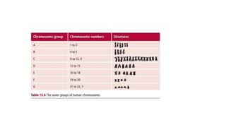
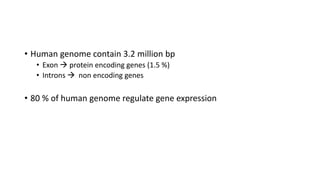
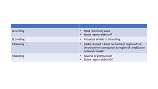
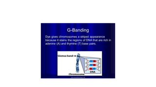
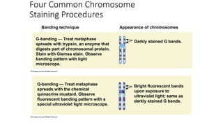
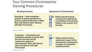
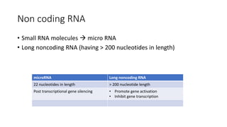
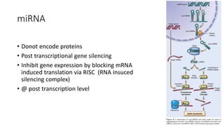
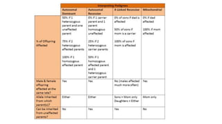
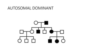
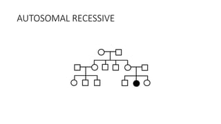
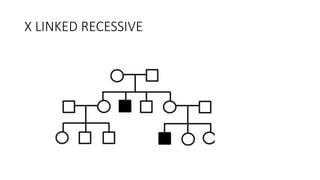
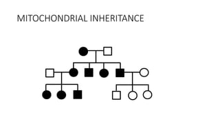
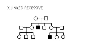
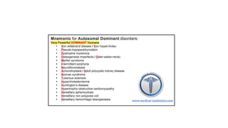
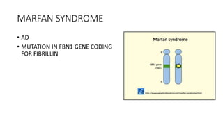
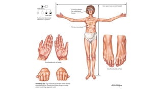
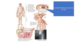
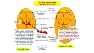
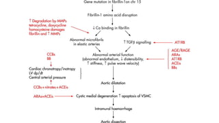
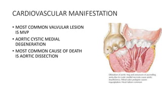
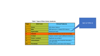
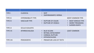
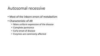
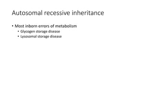
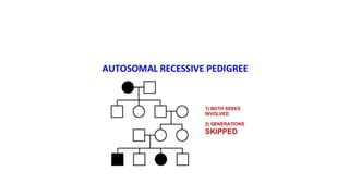
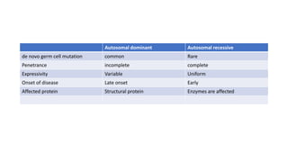
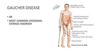
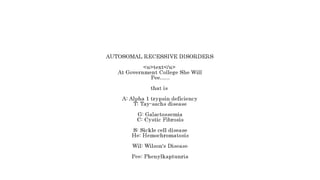
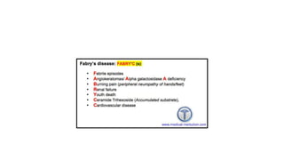
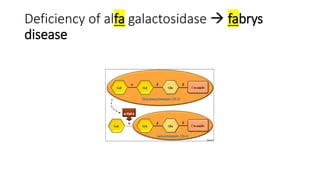
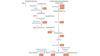
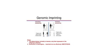
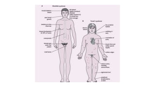
genetics
• Genome: The sum total of genetic information of an individual which
is encoded in structure of DNA
• Genomics: Is the study of genome
Karyotyping
• For karyotyping lymphocytes are used
• Lymphocytes complete cell division earlier than other cells
• Chromocytes appear early (with in 3 days of incubation in culture medium)
• Human genome contain 3.2 million bp
• Exon  protein encoding genes (1.5 %)
• Introns  non encoding genes
• 80 % of human genome regulate gene expression
G banding • Most commonly used
• Stains regions rich in AT
Q banding • Pattern is similar to G banding
C banding • Darkly stained C band centromeric region of the
chromosome corresponds to region of constitutive
heterochromatin
R banding • Reverse of geimsa stain
• Stains regions rich in GC
Four Common Chromosome
Staining Procedures
Banding technique Appearance of chromosomes
G-banding — Treat metaphase
spreads with trypsin, an enzyme that
digests part of chromosomal protein.
Stain with Giemsa stain. Observe
banding pattern with light
microscope.
Darkly stained G bands.
Q-banding — Treat metaphase
spreads with the chemical
quinacrine mustard. Observe
fluorescent banding pattern with a
special ultraviolet light microscope.
Bright fluorescent bands
upon exposure to
ultraviolet light; same as
darkly stained G bands.
Four Common Chromosome
Staining Procedures
R-banding — Heat metaphase
spreads at high temperatures to
achieve partial denaturation of DNA.
Stain with Giemsa stain. Observe
with light microscope.
Darkly stained R bands
correspond to light bands
in G-banded chromosomes.
Pattern is the reverse of G-
banding.
Banding technique Appearance of chromosomes
C-banding — Chemically treat
metaphase spreads to extract DNA
from the arms but not the
centromeric regions of
chromosomes. Stain with Giemsa
stain and observe with light
microscope.
Darkly stained C band
centromeric region of the
chromosome corresponds
to region of constitutive
heterochromatin.
METACENTRIC
• 2 EQUALS ARMS
WITH
CENTROMERE IN
THE CENTRE
SUBMETACENTRIC
• CENTROMERE IS
AWAY SLIGHTLY
FROM CENTRE
• X
CHROMOSOME
ACROCENTRIC
• CENTROMERE IS
TOWARDS ONE
END
• Y
CHROMOSOME
TELOCENTRIC
• NOT SEEN IN
HUMANS
Polymorphism
• two individuals share > 99.5% of their DNA sequences
• DNA sequence variation b/w 2 individuals 0.5 % (15 million bp)
• DNA sequence variation b/w 2 individual genetic polymorphism
Types of polymorphism
Single nucleotide polymorphism Copy number variations Repeat length polymoprhism
• MC type of polymorphism
• Single nucleotide per 1000 bp
• More than 6 million SNP
• genetic variation consisting of
different numbers of large
contiguous stretches of DNA
from 1000 base pairs to millions
of base pairs
• Short repetitive sequence of
DNA variation
• Basis of phenotypic diversity Microsatellite Minisatellite
• Present both in exons & introns
• Less than 1 % in coding regions
• 50 % of coding regions 2-6 bp repeat
size
15-70 bp reoeat
size
< 1 kilobase 1-3 kilobase
Non coding RNA
• Small RNA molecules  micro RNA
• Long noncoding RNA (having > 200 nucleotides in length)
microRNA Long noncoding RNA
22 nucleotides in length > 200 nucleotide length
Post transcriptional gene silencing • Promote gene activation
• Inhibit gene transcription
miRNA
• Donot encode proteins
• Post transcriptional gene silencing
• Inhibit gene expression by blocking mRNA
induced translation via RISC (RNA insuced
silencing complex)
• @ post transcription level
siRNA
• Exogenous miRNA
• Useful laboratory tool
• To study gene function  knock down technology
• Study for therapeutic agents
Lnc RNA
• XIST
• A type of Lnc RNA
• itself escapes X inactivation, but forms a repressive “cloak” on the X
chromosome from which it is transcribed, resulting in gene silencing.
• Emerging studies are highlighting the roles of lncRNAs in various human
diseases, from atherosclerosis to cancer
Epigenetic Changes
• modulation of gene or protein expression in the absence of
alterations in DNA sequence (i.e., mutation) or structure of the
encoding gene
• Useful in regulation of
• Tissue specific gene expression
• Genomic imprinting
• X chromosome inactivation
• Fragile x syndrome
Mutations
Point mutation
d/t one nucleotide is replaced by another
Frame shift mutation
d/t addition or removal of nucleotide  frameshift
Mis-sense Nonsense Silent
Yield
different type
of proteins
Mutation lead to
termination
In sickle cell
anemia
Thalassemia
Pleiotropism
• Single mutant gene produces multiple end effects
• Sickle cell anemia  multiple effects like sickling RBC / splenic infarction
Genetic heterogeneity
• Multiple gene  single end effect
• DM
Gene disorders
Single gene disorders Multifactorial inheritance Chromosomal disorders
Classic mendelian
inheritance
Non menedelian
inheritance
• Complex polygenic inheritance
 2 or more mutant genes +
environmental factors
 DM
 Cleft lip & palate
 Coronary heart disease
 HTn
 Gout
Structural Numerical
• Euploidy
• Aneuploidy
• Deletion
• Translocation
• Ring
chromosome
• Isochromosome
• Inversion
• AR
• AD
• XR
• XD
• Trinucleotide
repeat
mutation
• Mitochondrial
inheritance
• Genomic
imprinting
• Gonadal
mosaicism
PEDIGREE ANALYSIS
AUTOSOMAL DOMINANT
AUTOSOMAL RECESSIVE
X LINKED RECESSIVE
MITOCHONDRIAL INHERITANCE
X LINKED RECESSIVE
SINGLE GENE DISORDERS
• SINGLE GENE OR MENDELIAN INHERITANCE
• CODOMINANT INHERITANCE
• PENETRANCE
• VARIABLE EXPRESSIVITY
• PLEIOTROPISM
• SINGLE MUTANT GENE PRODUCING MULTIPLE END EFFECTS
AUTOSOMAL DOMINANT
AUTOSOMAL DOMINANT MOST COMMON
PATTERN OF INHERITANCE
AUTOSOMAL DOMINANT
• ALL GENERATIONS ARE
AFFECTED
• DONOT SHOW GENDER BASIS
AUTOSOMAL DOMINANT
• VARIABLE EXPRESSIVITY
• IMCOMPLETE PENETRANCE
• Denovo germ cell mutation
• LOSS OF FUNCTION TYPE MUTATION IS MORE COMMON
DIFFERENT EFFECT
CAUSED BY SAME
MUTANT GENE
WITH MUTATED GENE BUT
PHENOTYPICALLY NORMAL
MARFAN SYNDROME
• AD
• MUTATION IN FBN1 GENE CODING
FOR FIBRILLIN
SUPEROTEMPORAL DISLOCATION
OF LENS
CARDIOVASCULAR MANIFESTATION
• MOST COMMON VALVULAR LESION
IS MVP
• AORTIC CYSTIC MEDIAL
DEGENERATION
• MOST COMMON CAUSE OF DEATH
IS AORTIC DISSECTION
HYPER EXTENSIBILITY
ARACHNODACTYLY
REVISED GHENT CRITERIA
• FAMILY HISTORY
• CARDINAL CLINICAL SIGNS
• FIBRILLIN MUTATION
EHLERS DANLOS SYNDROME
• ALL PATTERN OF MENDELIAN
INHERITANCE ARE SEEN BUT
MOST COMMON IS AD
• DEFECTIVE COLLGEN
SYNTHESIS
• MC AFFECTED COLLAGEN
TYPE 3
EHLERS DANLOS SYNDROME
HYPEREXTENSIBLE SKIN
RUBBER MAN
SYNDROME
ABNORMAL WOUND HEALING IN EHLER
DANLOS  CIGARETTE PAPER LIKE SCAR
• EHLERS DANLOS SYNDROME HAS ALL 3 PATTERNS OF INHERITANCE
MC IS TYPE III
TYPE I CLASSICAL • MVP
• DIAPHRAGMATIC HERNIA
TYPE III HYPERMOBILITY TYPE MOST COMMON TYPE
TYPE IV VASCULAR TYPE • RUPTURE OF COLON
• RUPTURE OF VESSELS
• MOST SERIOUS TYPE
• WORST PROGNOSIS
• SUDDEN DEATH
TYPE VI OCULO SCOLIOTIC
TYPE VII ATHRROCHALASIA • BLUE SCLERA
• RETINAL DETACHMENT
• CORNEAL RUPTURE
• KYPHOSCOLIOSIS
LEAST COMMON
TYPE VIII PERIODONTIC • PREMATURE LOSS OF TEETH
Autosomal recessive
Autosomal recessive
• Most of the inborn errors of metabolism
• Characteristic of AR
• More uniform expression of the disease
• Complete pentrance
• Early onset of disease
• Enzymes are commonly affected
Autosomal recessive inheritance
• Most inborn errors of metabolism
• Glycogen storage disease
• Lysosomal storage disease
Autosomal dominant Autosomal recessive
de novo germ cell mutation common Rare
Penetrance incomplete complete
Expressivity Variable Uniform
Onset of disease Late onset Early
Affected protein Structural protein Enzymes are affected
• All lysososmal storage disease are AR
• Except for hunters & fabrys disease (which are x linked recessive )
GAUCHER DISEASE
• AR
• MOST COMMON LYSOSOMAL
STORAGE DISORDER
CRUMPLED TISSUE PAPER APPEARANCE
ELONGATED
LYSOSOMES CONTAING
LIPIDS IN BILAYER
STACCKS
Fabrys disease
• Angiokeratomas & neuropathic pain
• d/t deficiency of alfa galctosidase
Angiokeratoma
Deficiency of alfa galactosidase  fabrys
disease
Maltese cross d/t lipid inclusions
X LINKED DOMINANT
• RARE PATTERN
• AFFECTED MALE WILL TRANSMIT THE DISEASE TO 100 % OF THEIR
DAUGHTERS
• AFFECTED FEMALE WILL TRANSMIT THE DISEASE TO 50 % OF THEIR
SONS & DAUGHTERS
X LINKED DOMINANT
Genomic imprinting
• o Human inherits two copies of each gene, i.e. two alleles, from homologous maternal and
paternal chromosomes.
• o There is no functional difference between the genes derived from mother or the father.
• o But, with respect to some genes, there are functional differences between the paternal gene
and maternal gene.
• o These differences result from an epigenetic process, called genomic imprinting.
• o In most cases, genomic imprinting selectively inactivates either the maternal or paternal allel.
• 1. Maternal genomic imprinting
• • Selective inactivation of maternal allel
• • Example is Angelman syndrome
• 2. Paternal genomic imprinting
• • Selective inactivation ofpatemal allel.
• • Example is Prader-willi syndrome.
Genomic imprinting
• Epigenetic process resulting in
differential inactivation of either
maternal or paternal alleles of certain
genes
• Heritable chemical modification
• Methylation of DNA or acetylation of
histones
• Decreases genetic expression (with out
affecting primary DNA sequence
EPIGENETICS)
• Occurs in sperm or ova before
fertilisation
Trinucleotide repeats
Not diseased but increased
susceptibility to get diseased
in next generation
premuttion (d/t genetic
anticipation)
Genetic anticipation
• In trinucleotide repeats
• Increase in severity in successive
generation
• Symptoms appear in an earlier age
• Premutation
Fragile x chromosome
Macrorochidism in post pubertal period
• Long face
• Large mandible
• Large everted ears
• High arched palate
• MVP
• Hyperextensible joints
• Inversion refers to a
rearrangement that involves two
breaks within a single chromosome
with reincorporation of the
inverted, intervening segment.
• It can be of the following two
types:
• a. An inversion involving only one
arm of the chromosome is known as
paracentric.
• b. If the breaks are on opposite sides
of the centromere, it is known as
pericentric
Mitochondrial inheritance
PATERNAL MITOCHONDRIA
LEFT OUTSIDE THE OVUM
ONLY MATERNAL
MITOCHONDRIA IS
TRANSFERRED TO ZYGOTE
MITOCHONDRIAL INHERITANCE
INFECTED MOTHER TRANSMITS
INFECTION TO ALL HER CHILDREN
NO TRANSMISSION FROM
INFECTED MALE
Heteroplasmy in mitochondrial inheritance
Mitochondrially transmitted disease
• LHON
• MELAS
• Mitochondrial encephalopathy lactic acidosis & stroke like syndrome
Chromosomal anomalies
Cat eye syndrome
 trisomy
chromosome 22
Cat eye syndrome
• Partial tetrasomy or trisomy of chromosome 22
• Coloboma iris
Klinefelter syndrome
Testes on biopsy
• Grossly atrophic
• on biopsy hyalanisation of seminiferous tubule
• Adenomatous clumping of sertoli cells
• Azoospermia
• Rx
• Testosterone at 11 – 12 years of age
Advanced paternal age
• Neurofibromatosis
• Marfan syndrome
• Klinefelter syndrome
Advanced maternal age
• Down syndrome
• XXX syndrome
• Klinefelter syndrome
Turner syndrome
• Mosaic with with XY pattern is associated with
gonadoblastoma & virilisation
• Single X chromosome is maternal in origin in 75 % of patients
Webbed neck  lymphatic dilation
• No of genomes in human gene  1,00,000
Genetics pathology revision notes

Genetics pathology revision notes

  • 1.
  • 2.
    • Genome: Thesum total of genetic information of an individual which is encoded in structure of DNA • Genomics: Is the study of genome
  • 3.
  • 4.
    • For karyotypinglymphocytes are used • Lymphocytes complete cell division earlier than other cells • Chromocytes appear early (with in 3 days of incubation in culture medium)
  • 8.
    • Human genomecontain 3.2 million bp • Exon  protein encoding genes (1.5 %) • Introns  non encoding genes • 80 % of human genome regulate gene expression
  • 9.
    G banding •Most commonly used • Stains regions rich in AT Q banding • Pattern is similar to G banding C banding • Darkly stained C band centromeric region of the chromosome corresponds to region of constitutive heterochromatin R banding • Reverse of geimsa stain • Stains regions rich in GC
  • 12.
    Four Common Chromosome StainingProcedures Banding technique Appearance of chromosomes G-banding — Treat metaphase spreads with trypsin, an enzyme that digests part of chromosomal protein. Stain with Giemsa stain. Observe banding pattern with light microscope. Darkly stained G bands. Q-banding — Treat metaphase spreads with the chemical quinacrine mustard. Observe fluorescent banding pattern with a special ultraviolet light microscope. Bright fluorescent bands upon exposure to ultraviolet light; same as darkly stained G bands.
  • 13.
    Four Common Chromosome StainingProcedures R-banding — Heat metaphase spreads at high temperatures to achieve partial denaturation of DNA. Stain with Giemsa stain. Observe with light microscope. Darkly stained R bands correspond to light bands in G-banded chromosomes. Pattern is the reverse of G- banding. Banding technique Appearance of chromosomes C-banding — Chemically treat metaphase spreads to extract DNA from the arms but not the centromeric regions of chromosomes. Stain with Giemsa stain and observe with light microscope. Darkly stained C band centromeric region of the chromosome corresponds to region of constitutive heterochromatin.
  • 17.
    METACENTRIC • 2 EQUALSARMS WITH CENTROMERE IN THE CENTRE SUBMETACENTRIC • CENTROMERE IS AWAY SLIGHTLY FROM CENTRE • X CHROMOSOME ACROCENTRIC • CENTROMERE IS TOWARDS ONE END • Y CHROMOSOME TELOCENTRIC • NOT SEEN IN HUMANS
  • 18.
  • 19.
    • two individualsshare > 99.5% of their DNA sequences • DNA sequence variation b/w 2 individuals 0.5 % (15 million bp) • DNA sequence variation b/w 2 individual genetic polymorphism
  • 20.
    Types of polymorphism Singlenucleotide polymorphism Copy number variations Repeat length polymoprhism • MC type of polymorphism • Single nucleotide per 1000 bp • More than 6 million SNP • genetic variation consisting of different numbers of large contiguous stretches of DNA from 1000 base pairs to millions of base pairs • Short repetitive sequence of DNA variation • Basis of phenotypic diversity Microsatellite Minisatellite • Present both in exons & introns • Less than 1 % in coding regions • 50 % of coding regions 2-6 bp repeat size 15-70 bp reoeat size < 1 kilobase 1-3 kilobase
  • 21.
    Non coding RNA •Small RNA molecules  micro RNA • Long noncoding RNA (having > 200 nucleotides in length) microRNA Long noncoding RNA 22 nucleotides in length > 200 nucleotide length Post transcriptional gene silencing • Promote gene activation • Inhibit gene transcription
  • 22.
    miRNA • Donot encodeproteins • Post transcriptional gene silencing • Inhibit gene expression by blocking mRNA induced translation via RISC (RNA insuced silencing complex) • @ post transcription level
  • 23.
    siRNA • Exogenous miRNA •Useful laboratory tool • To study gene function  knock down technology • Study for therapeutic agents
  • 24.
    Lnc RNA • XIST •A type of Lnc RNA • itself escapes X inactivation, but forms a repressive “cloak” on the X chromosome from which it is transcribed, resulting in gene silencing. • Emerging studies are highlighting the roles of lncRNAs in various human diseases, from atherosclerosis to cancer
  • 25.
    Epigenetic Changes • modulationof gene or protein expression in the absence of alterations in DNA sequence (i.e., mutation) or structure of the encoding gene • Useful in regulation of • Tissue specific gene expression • Genomic imprinting • X chromosome inactivation • Fragile x syndrome
  • 26.
  • 27.
    Point mutation d/t onenucleotide is replaced by another Frame shift mutation d/t addition or removal of nucleotide  frameshift Mis-sense Nonsense Silent Yield different type of proteins Mutation lead to termination In sickle cell anemia Thalassemia
  • 28.
    Pleiotropism • Single mutantgene produces multiple end effects • Sickle cell anemia  multiple effects like sickling RBC / splenic infarction
  • 29.
    Genetic heterogeneity • Multiplegene  single end effect • DM
  • 30.
  • 31.
    Single gene disordersMultifactorial inheritance Chromosomal disorders Classic mendelian inheritance Non menedelian inheritance • Complex polygenic inheritance  2 or more mutant genes + environmental factors  DM  Cleft lip & palate  Coronary heart disease  HTn  Gout Structural Numerical • Euploidy • Aneuploidy • Deletion • Translocation • Ring chromosome • Isochromosome • Inversion • AR • AD • XR • XD • Trinucleotide repeat mutation • Mitochondrial inheritance • Genomic imprinting • Gonadal mosaicism
  • 33.
  • 37.
  • 38.
  • 39.
  • 40.
  • 41.
  • 42.
  • 43.
    • SINGLE GENEOR MENDELIAN INHERITANCE • CODOMINANT INHERITANCE • PENETRANCE • VARIABLE EXPRESSIVITY • PLEIOTROPISM • SINGLE MUTANT GENE PRODUCING MULTIPLE END EFFECTS
  • 44.
    AUTOSOMAL DOMINANT AUTOSOMAL DOMINANTMOST COMMON PATTERN OF INHERITANCE
  • 45.
    AUTOSOMAL DOMINANT • ALLGENERATIONS ARE AFFECTED • DONOT SHOW GENDER BASIS
  • 46.
    AUTOSOMAL DOMINANT • VARIABLEEXPRESSIVITY • IMCOMPLETE PENETRANCE • Denovo germ cell mutation • LOSS OF FUNCTION TYPE MUTATION IS MORE COMMON
  • 47.
    DIFFERENT EFFECT CAUSED BYSAME MUTANT GENE WITH MUTATED GENE BUT PHENOTYPICALLY NORMAL
  • 49.
    MARFAN SYNDROME • AD •MUTATION IN FBN1 GENE CODING FOR FIBRILLIN
  • 51.
  • 54.
    CARDIOVASCULAR MANIFESTATION • MOSTCOMMON VALVULAR LESION IS MVP • AORTIC CYSTIC MEDIAL DEGENERATION • MOST COMMON CAUSE OF DEATH IS AORTIC DISSECTION
  • 56.
  • 58.
  • 60.
    REVISED GHENT CRITERIA •FAMILY HISTORY • CARDINAL CLINICAL SIGNS • FIBRILLIN MUTATION
  • 61.
    EHLERS DANLOS SYNDROME •ALL PATTERN OF MENDELIAN INHERITANCE ARE SEEN BUT MOST COMMON IS AD • DEFECTIVE COLLGEN SYNTHESIS • MC AFFECTED COLLAGEN TYPE 3
  • 62.
    EHLERS DANLOS SYNDROME HYPEREXTENSIBLESKIN RUBBER MAN SYNDROME
  • 63.
    ABNORMAL WOUND HEALINGIN EHLER DANLOS  CIGARETTE PAPER LIKE SCAR
  • 64.
    • EHLERS DANLOSSYNDROME HAS ALL 3 PATTERNS OF INHERITANCE
  • 65.
  • 66.
    TYPE I CLASSICAL• MVP • DIAPHRAGMATIC HERNIA TYPE III HYPERMOBILITY TYPE MOST COMMON TYPE TYPE IV VASCULAR TYPE • RUPTURE OF COLON • RUPTURE OF VESSELS • MOST SERIOUS TYPE • WORST PROGNOSIS • SUDDEN DEATH TYPE VI OCULO SCOLIOTIC TYPE VII ATHRROCHALASIA • BLUE SCLERA • RETINAL DETACHMENT • CORNEAL RUPTURE • KYPHOSCOLIOSIS LEAST COMMON TYPE VIII PERIODONTIC • PREMATURE LOSS OF TEETH
  • 67.
  • 68.
    Autosomal recessive • Mostof the inborn errors of metabolism • Characteristic of AR • More uniform expression of the disease • Complete pentrance • Early onset of disease • Enzymes are commonly affected
  • 69.
    Autosomal recessive inheritance •Most inborn errors of metabolism • Glycogen storage disease • Lysosomal storage disease
  • 71.
    Autosomal dominant Autosomalrecessive de novo germ cell mutation common Rare Penetrance incomplete complete Expressivity Variable Uniform Onset of disease Late onset Early Affected protein Structural protein Enzymes are affected
  • 72.
    • All lysososmalstorage disease are AR • Except for hunters & fabrys disease (which are x linked recessive )
  • 74.
    GAUCHER DISEASE • AR •MOST COMMON LYSOSOMAL STORAGE DISORDER
  • 75.
    CRUMPLED TISSUE PAPERAPPEARANCE ELONGATED LYSOSOMES CONTAING LIPIDS IN BILAYER STACCKS
  • 78.
    Fabrys disease • Angiokeratomas& neuropathic pain • d/t deficiency of alfa galctosidase Angiokeratoma
  • 81.
    Deficiency of alfagalactosidase  fabrys disease
  • 82.
    Maltese cross d/tlipid inclusions
  • 85.
    X LINKED DOMINANT •RARE PATTERN • AFFECTED MALE WILL TRANSMIT THE DISEASE TO 100 % OF THEIR DAUGHTERS • AFFECTED FEMALE WILL TRANSMIT THE DISEASE TO 50 % OF THEIR SONS & DAUGHTERS
  • 86.
  • 88.
    Genomic imprinting • oHuman inherits two copies of each gene, i.e. two alleles, from homologous maternal and paternal chromosomes. • o There is no functional difference between the genes derived from mother or the father. • o But, with respect to some genes, there are functional differences between the paternal gene and maternal gene. • o These differences result from an epigenetic process, called genomic imprinting. • o In most cases, genomic imprinting selectively inactivates either the maternal or paternal allel. • 1. Maternal genomic imprinting • • Selective inactivation of maternal allel • • Example is Angelman syndrome • 2. Paternal genomic imprinting • • Selective inactivation ofpatemal allel. • • Example is Prader-willi syndrome.
  • 89.
    Genomic imprinting • Epigeneticprocess resulting in differential inactivation of either maternal or paternal alleles of certain genes • Heritable chemical modification • Methylation of DNA or acetylation of histones • Decreases genetic expression (with out affecting primary DNA sequence EPIGENETICS) • Occurs in sperm or ova before fertilisation
  • 94.
  • 95.
    Not diseased butincreased susceptibility to get diseased in next generation premuttion (d/t genetic anticipation)
  • 96.
    Genetic anticipation • Intrinucleotide repeats • Increase in severity in successive generation • Symptoms appear in an earlier age • Premutation
  • 97.
  • 101.
    Macrorochidism in postpubertal period • Long face • Large mandible • Large everted ears • High arched palate • MVP • Hyperextensible joints
  • 102.
    • Inversion refersto a rearrangement that involves two breaks within a single chromosome with reincorporation of the inverted, intervening segment. • It can be of the following two types: • a. An inversion involving only one arm of the chromosome is known as paracentric. • b. If the breaks are on opposite sides of the centromere, it is known as pericentric
  • 104.
  • 105.
    PATERNAL MITOCHONDRIA LEFT OUTSIDETHE OVUM ONLY MATERNAL MITOCHONDRIA IS TRANSFERRED TO ZYGOTE
  • 106.
    MITOCHONDRIAL INHERITANCE INFECTED MOTHERTRANSMITS INFECTION TO ALL HER CHILDREN NO TRANSMISSION FROM INFECTED MALE
  • 108.
  • 109.
    Mitochondrially transmitted disease •LHON • MELAS • Mitochondrial encephalopathy lactic acidosis & stroke like syndrome
  • 110.
  • 111.
    Cat eye syndrome trisomy chromosome 22
  • 112.
    Cat eye syndrome •Partial tetrasomy or trisomy of chromosome 22 • Coloboma iris
  • 113.
  • 119.
    Testes on biopsy •Grossly atrophic • on biopsy hyalanisation of seminiferous tubule • Adenomatous clumping of sertoli cells • Azoospermia
  • 120.
    • Rx • Testosteroneat 11 – 12 years of age
  • 121.
    Advanced paternal age •Neurofibromatosis • Marfan syndrome • Klinefelter syndrome Advanced maternal age • Down syndrome • XXX syndrome • Klinefelter syndrome
  • 122.
  • 123.
    • Mosaic withwith XY pattern is associated with gonadoblastoma & virilisation
  • 124.
    • Single Xchromosome is maternal in origin in 75 % of patients
  • 125.
    Webbed neck lymphatic dilation
  • 132.
    • No ofgenomes in human gene  1,00,000