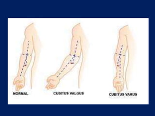
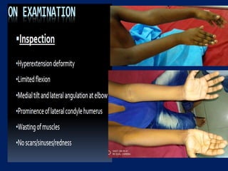
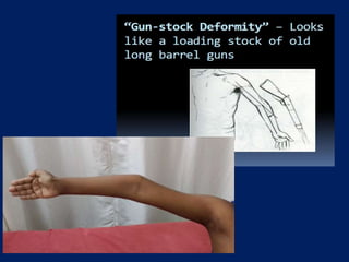
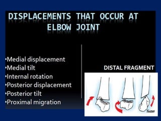
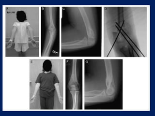
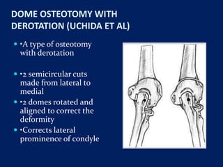
CUBITUS VARUS DEFORMITY
DR. RAMACHANDRA REDDY
(DNB RESIDENT)
Under the guidance of
Prof.A.DEVADOSS
Dr. SATHISH DEVADOSS
Dr. JAYAKUMAR
DEVADOSS MULTISPECIALITY HOSPITALS
 Cubitus varus or gunstock deformity as it is commonly
known is the most common complication of displaced
supracondylar fractures in children with an incidence
ranging from 3% to 57% .
 cubitus varus is a triplanar deformity with components
of varus, hyperextension and internal rotation.
 Forearm deviated inwards with respect to arm at elbow
with resulting lateral angulation in full extension.
 Reduction of physiological valgus
 8 ̊-15 ̊ ; Males : 10 ̊
 Females : 15 ̊- 20 ̊
 Normally forearm is aligned in valgus with respect to
arm in full extension with medial angulation.
 Decrease in valgus with neutral alignment (loss of
angulation) is called “Cubitus Rectus”.
 It is still a deformity as it deviates from the normal for
population.
CASUES
 MC cause is malunited supracondylar humerus
fracture.
 INFECTIVE: medial growth plate damage.
 VASCULAR: osteonecrosis of trochlea
 TRAUMATIC: lateral condyle fracture
 NEOPLASTIC: secondary to exostosis in distal, lateral
humerus
 CONGENITAL : epiphyseal dysplasia
factors for malunion are:
 1. Impacted / comminuted type I supracondylar
fractures
 2. Rotationally unstable type II fractures treated in a
cast with subsequent loss of reduction
 3. Poorly stabilized or reduced type III fractures or
delayed neglected fractures
 Smith has demonstrated that changes in the carrying
angle are a result of angular displacement or tilting of
the distal fragment, not translation or rotation.
 Problems arising from cubitus varus or valgus include
functional limitation, recurrent elbow fracture, and
cosmetic deformity.
 Functional problems are almost always related to
limitation of flexion, although tardy ulnar nerve palsy
and elbow instability
 The limitation in flexion is a result of the
hyperextension associated with varus malunion.
 The resultant cubitus varus deformity is a combined
deformity of varus, extension, and internal rotation to
various degrees.
 Most corrective osteotomies have focused on the
correction of varus and extension deformity. The
rotational deformity is well tolerated and best left
untreated because rotation of the distal fragment
makes the osteotomy unstable.
 Loss of fixation and persistent deformity are the most
common complications after corrective supracondylar
osteotomy.
 In an effort to limit these complications, a wide variety
of osteotomy and fixation techniques have been
described.
On palpation
 There is thickening and irregularity of supracondylar
ridges.
 Lateral condyle appear prominent due to rotation of
distal fragment.
 Decrease in carrying angle.
 Three point relationship do not make an equilateral
triangle.
GRADED BY SEVERITY :
 Grade I - loss of the physiological valgus angle;
 Grade II - 0 to 10 degrees of varus
 Grade III - 1 1 to 20 degrees
 Grade IV - more than 20 degrees
AP VIEW X RAY
 Baumann’s angle (or the humero capitellar angle)
radiographic measurement used to assess the normal
relationships of the distal humerus and is measured
on the AP projection of the elbow.
 Drawing a line parallel to the longitudinal axis of the
humeral shaft as well as a bisecting line parallel to the
lateral condylar physis creates Baumann’s angle.
 A normal angle is 70-75 degrees or within 5 degrees of
the contralateral elbow
LATERAL VIEW
 The anterior humeral line (AHL) is an important
radiographic landmark used to assess the alignment of
the distal humerus and is often used to evaluate the
anteriroposterior displacement of supracondylar
humerus fractures.
 This line is drawn on the lateral projection of the
elbow along the anterior cortex of the humerus and
should intersect the middle third of the capitellum in
most normal elbows.
TREATMENT
 Cubitus varus deformity has no tendency for
spontaneous correction but it always has to be
corrected.
 Treatment options include:
 (a) Observation with expectant remodelling
 (b) Hemi epiphysiodesis and growth alteration
 c) Corrective Osteotomy
Observation with expectant remodelling
 Not appropriate because although hyperextension
may remodel to some degree in a young child, in an
older child little remodelling occurs even in the joint’s
plane of motion.
 Hence, it is not recommended.
Hemi epiphysiodesis and growth alteration
 It is used to prevent cubitus varus deformity in a
patient with medial growth arrest and progressive
deformity, rather than correcting it.
 It has no role in a child with a normal physis.
CORRECTIVE OSTEOTOMY
 Osteotomy is the only way to correct a cubitus varus
deformity with a high probability of success.
 Options include:
 Medial open wedge osteotomy
 Lateral closing wedge osteotomy also known as
French osteotomy .
 Oblique osteotomy with derotation.
 Dome osteotomy .
 Step cut osteotomy
APPROACHES
 Three surgical approaches are described namely
MEDIAL, LATERAL AND POSTERIOR.
 Lateral approach is most frequently used as it provides
good exposure with less dissection.
 Complex osteotomies may require posterior approach
which offer more extensive exposure
Pre-requisites:
 1. Atleast 1 year following fracture (Bone remodeling
and tissue equilibrium)
 2. Patient demanding surgery
 3. Calculation of wedge to be removed→Normal side
Xray→
 Wedge angle = Varus + Normal physiological Valgus
Lateral closing wedge osteotmy
 Easiest, the safest, and the most stable osteotomy.
 Lateral closing wedge osteotomy with a medial hinge will correct the
varus deformity, with some minor correction of hyperextension
 Types
 Lateral closing wedge osteotomy (Voss et al)
 French osteotomy
 Modified french osteotomy
Different methods of fixation
 – Two screws and a wire attached between them
 – Plate fixation
 – Crossed Kirschner wires
 – Staples
FRENCH OSTEOTOMY
 French, in 1959, first described a lateral wedge
osteotomy held with screws and a figure-of-eight wire,
and this remains the most popular method of
correction.
 Lateral closed wedge osteotomy.
`
MODIFIED FRENCH OSTEOTOMY
 modifications of French’s osteotomy appears to fulfill
these criteria.
 The procedure is easy and,
 There is minimal dissection, and little possibility of
nerve damage.
 By operating with the arm in the extended position,
the adequacy of the correction can be seen during
operation and, if necessary, adjusted.
 The capacity for remodelling is reduced in the older
child undergoing osteotomy, and for this reason, the
medial “hinge” is an important feature of the
osteotomy.
 This hinge, with the screws and wire acting as a bone
suture, ensures that anatomical alignment is
maintained.
Post op management
 Postoperatively, the arm is maintained in the extended
position for two weeks.
Medial open wedge osteotomy
 King and Secor decsribed this osteotomy.
 a medial opening wedge osteotomy with external
fixation and with or without bone graft.
 advantage of this technique is that the alignment can
be manipulated after the wound is closed.
 Requires BG
 Gains length→ inherent instability
 May stretch the ulnar nerve- transferred anteriorly to
avoid this.
OBLIQUE OSTEOTOMY WITH DEROTATION
(AMSPACHER & MESSENBAUGH)
 Types
 Amspacher and Messenbaugh
 correct a two-plane deformity with one osteotomy.
 Dome osteotomy with derotation (Uchida)
 three-dimensional osteotomy Correction of medial
tilt, internal rotation & posterior tilt
Amspacher and Messenbaugh:
 Expose the elbow posteriorly
 Expose subperiosteally the supracondylar part of the
humerus
 Make an oblique osteotomy 3.8cm proximal to distal
end of humerus.
 Osteotomy directing it posteriorly above to anterior
below.
 Later tilt and rotate distal fragment until internal
rotation and cubitus varus is corrected.
 Fix with screw inserted across osteotomy site.
 Arm is immobilized in a long arm cast or splint until
union at 4-6 weeks.
Step Cut Osteotomy (DeRosa and Graziano)
 A standard posterior approach used.
 Incision extended proximally from distal 3rd upper arm
to a distance of 1 to 2 cm beyond the tip of the
olecranon distally.
 mobilize the ulnar nerve anteriorly.
 The triceps muscle was then split longitudinally.
 Circumferential subperiosteal dissection done.
 The osteotomy was performed by first making a
proximal, transverse cut perpendicular to the
anatomical axis of the humerus.
 Then, the angular correction cut was made based on
the degree of correction desired, as determined from
the preoperative planning template.
 cut was made in a proximal-medial to distal-
lateral direction.
 next cut, perpendicular to the angular correction cut
was made at its lateral margin, creating a step cut in
the distal humeral fragment.
 Once these steps were
completed, the proximal
and distal segments were
aligned and the clinical
carrying angle
reassessed.
 Internal fixation was
achieved by placing two
1.6mm k-wires through
the lateral epicondyle
and 1 k-wire through the
medial epicondyle.
DOME OSTEOTOMY WITH
DEROTATION (UCHIDA ET AL)
 •A type of osteotomy
with derotation
 •2 semicircular cuts
made from lateral to
medial
 •2 domes rotated and
aligned to correct the
deformity
 •Corrects lateral
prominence of condyle
COMPLICATIONS OF OSTEOTOMY
 1. Stiffness(myositis ossificans)
 2. Nerve injury(radial and ulnar nerve )
 3. Persistent deformity (under correction)
 4. Recurrent deformity
 5. Non-union
 6. Osteomyelitis
 7. unsatisfactory scar
 8.lateral prominence
Pseudo Cubitus Varus
 Lateral spur formation in lateral condyle humerus
fracture due to elevation of periosteum and new bone
formation leads to lateral bulge with normal carrying
angle.
Thank you all

Cubitus varus deformity

  • 1.
    CUBITUS VARUS DEFORMITY DR.RAMACHANDRA REDDY (DNB RESIDENT) Under the guidance of Prof.A.DEVADOSS Dr. SATHISH DEVADOSS Dr. JAYAKUMAR DEVADOSS MULTISPECIALITY HOSPITALS
  • 2.
     Cubitus varusor gunstock deformity as it is commonly known is the most common complication of displaced supracondylar fractures in children with an incidence ranging from 3% to 57% .  cubitus varus is a triplanar deformity with components of varus, hyperextension and internal rotation.
  • 3.
     Forearm deviatedinwards with respect to arm at elbow with resulting lateral angulation in full extension.  Reduction of physiological valgus  8 ̊-15 ̊ ; Males : 10 ̊  Females : 15 ̊- 20 ̊
  • 4.
     Normally forearmis aligned in valgus with respect to arm in full extension with medial angulation.  Decrease in valgus with neutral alignment (loss of angulation) is called “Cubitus Rectus”.  It is still a deformity as it deviates from the normal for population.
  • 6.
    CASUES  MC causeis malunited supracondylar humerus fracture.  INFECTIVE: medial growth plate damage.  VASCULAR: osteonecrosis of trochlea  TRAUMATIC: lateral condyle fracture  NEOPLASTIC: secondary to exostosis in distal, lateral humerus  CONGENITAL : epiphyseal dysplasia
  • 7.
    factors for malunionare:  1. Impacted / comminuted type I supracondylar fractures  2. Rotationally unstable type II fractures treated in a cast with subsequent loss of reduction  3. Poorly stabilized or reduced type III fractures or delayed neglected fractures
  • 8.
     Smith hasdemonstrated that changes in the carrying angle are a result of angular displacement or tilting of the distal fragment, not translation or rotation.  Problems arising from cubitus varus or valgus include functional limitation, recurrent elbow fracture, and cosmetic deformity.  Functional problems are almost always related to limitation of flexion, although tardy ulnar nerve palsy and elbow instability
  • 9.
     The limitationin flexion is a result of the hyperextension associated with varus malunion.  The resultant cubitus varus deformity is a combined deformity of varus, extension, and internal rotation to various degrees.  Most corrective osteotomies have focused on the correction of varus and extension deformity. The rotational deformity is well tolerated and best left untreated because rotation of the distal fragment makes the osteotomy unstable.
  • 10.
     Loss offixation and persistent deformity are the most common complications after corrective supracondylar osteotomy.  In an effort to limit these complications, a wide variety of osteotomy and fixation techniques have been described.
  • 13.
    On palpation  Thereis thickening and irregularity of supracondylar ridges.  Lateral condyle appear prominent due to rotation of distal fragment.  Decrease in carrying angle.  Three point relationship do not make an equilateral triangle.
  • 15.
    GRADED BY SEVERITY:  Grade I - loss of the physiological valgus angle;  Grade II - 0 to 10 degrees of varus  Grade III - 1 1 to 20 degrees  Grade IV - more than 20 degrees
  • 17.
    AP VIEW XRAY  Baumann’s angle (or the humero capitellar angle) radiographic measurement used to assess the normal relationships of the distal humerus and is measured on the AP projection of the elbow.  Drawing a line parallel to the longitudinal axis of the humeral shaft as well as a bisecting line parallel to the lateral condylar physis creates Baumann’s angle.  A normal angle is 70-75 degrees or within 5 degrees of the contralateral elbow
  • 20.
    LATERAL VIEW  Theanterior humeral line (AHL) is an important radiographic landmark used to assess the alignment of the distal humerus and is often used to evaluate the anteriroposterior displacement of supracondylar humerus fractures.  This line is drawn on the lateral projection of the elbow along the anterior cortex of the humerus and should intersect the middle third of the capitellum in most normal elbows.
  • 23.
    TREATMENT  Cubitus varusdeformity has no tendency for spontaneous correction but it always has to be corrected.  Treatment options include:  (a) Observation with expectant remodelling  (b) Hemi epiphysiodesis and growth alteration  c) Corrective Osteotomy
  • 24.
    Observation with expectantremodelling  Not appropriate because although hyperextension may remodel to some degree in a young child, in an older child little remodelling occurs even in the joint’s plane of motion.  Hence, it is not recommended.
  • 25.
    Hemi epiphysiodesis andgrowth alteration  It is used to prevent cubitus varus deformity in a patient with medial growth arrest and progressive deformity, rather than correcting it.  It has no role in a child with a normal physis.
  • 26.
    CORRECTIVE OSTEOTOMY  Osteotomyis the only way to correct a cubitus varus deformity with a high probability of success.  Options include:  Medial open wedge osteotomy  Lateral closing wedge osteotomy also known as French osteotomy .  Oblique osteotomy with derotation.  Dome osteotomy .  Step cut osteotomy
  • 27.
    APPROACHES  Three surgicalapproaches are described namely MEDIAL, LATERAL AND POSTERIOR.  Lateral approach is most frequently used as it provides good exposure with less dissection.  Complex osteotomies may require posterior approach which offer more extensive exposure
  • 28.
    Pre-requisites:  1. Atleast1 year following fracture (Bone remodeling and tissue equilibrium)  2. Patient demanding surgery  3. Calculation of wedge to be removed→Normal side Xray→  Wedge angle = Varus + Normal physiological Valgus
  • 29.
    Lateral closing wedgeosteotmy  Easiest, the safest, and the most stable osteotomy.  Lateral closing wedge osteotomy with a medial hinge will correct the varus deformity, with some minor correction of hyperextension  Types  Lateral closing wedge osteotomy (Voss et al)  French osteotomy  Modified french osteotomy Different methods of fixation  – Two screws and a wire attached between them  – Plate fixation  – Crossed Kirschner wires  – Staples
  • 30.
    FRENCH OSTEOTOMY  French,in 1959, first described a lateral wedge osteotomy held with screws and a figure-of-eight wire, and this remains the most popular method of correction.  Lateral closed wedge osteotomy.
  • 32.
  • 35.
    MODIFIED FRENCH OSTEOTOMY modifications of French’s osteotomy appears to fulfill these criteria.  The procedure is easy and,  There is minimal dissection, and little possibility of nerve damage.  By operating with the arm in the extended position, the adequacy of the correction can be seen during operation and, if necessary, adjusted.
  • 37.
     The capacityfor remodelling is reduced in the older child undergoing osteotomy, and for this reason, the medial “hinge” is an important feature of the osteotomy.  This hinge, with the screws and wire acting as a bone suture, ensures that anatomical alignment is maintained.
  • 38.
    Post op management Postoperatively, the arm is maintained in the extended position for two weeks.
  • 39.
    Medial open wedgeosteotomy  King and Secor decsribed this osteotomy.  a medial opening wedge osteotomy with external fixation and with or without bone graft.  advantage of this technique is that the alignment can be manipulated after the wound is closed.  Requires BG  Gains length→ inherent instability  May stretch the ulnar nerve- transferred anteriorly to avoid this.
  • 41.
    OBLIQUE OSTEOTOMY WITHDEROTATION (AMSPACHER & MESSENBAUGH)  Types  Amspacher and Messenbaugh  correct a two-plane deformity with one osteotomy.  Dome osteotomy with derotation (Uchida)  three-dimensional osteotomy Correction of medial tilt, internal rotation & posterior tilt
  • 42.
    Amspacher and Messenbaugh: Expose the elbow posteriorly  Expose subperiosteally the supracondylar part of the humerus  Make an oblique osteotomy 3.8cm proximal to distal end of humerus.  Osteotomy directing it posteriorly above to anterior below.  Later tilt and rotate distal fragment until internal rotation and cubitus varus is corrected.  Fix with screw inserted across osteotomy site.  Arm is immobilized in a long arm cast or splint until union at 4-6 weeks.
  • 44.
    Step Cut Osteotomy(DeRosa and Graziano)  A standard posterior approach used.  Incision extended proximally from distal 3rd upper arm to a distance of 1 to 2 cm beyond the tip of the olecranon distally.  mobilize the ulnar nerve anteriorly.  The triceps muscle was then split longitudinally.  Circumferential subperiosteal dissection done.
  • 45.
     The osteotomywas performed by first making a proximal, transverse cut perpendicular to the anatomical axis of the humerus.  Then, the angular correction cut was made based on the degree of correction desired, as determined from the preoperative planning template.  cut was made in a proximal-medial to distal- lateral direction.  next cut, perpendicular to the angular correction cut was made at its lateral margin, creating a step cut in the distal humeral fragment.
  • 47.
     Once thesesteps were completed, the proximal and distal segments were aligned and the clinical carrying angle reassessed.  Internal fixation was achieved by placing two 1.6mm k-wires through the lateral epicondyle and 1 k-wire through the medial epicondyle.
  • 50.
    DOME OSTEOTOMY WITH DEROTATION(UCHIDA ET AL)  •A type of osteotomy with derotation  •2 semicircular cuts made from lateral to medial  •2 domes rotated and aligned to correct the deformity  •Corrects lateral prominence of condyle
  • 51.
    COMPLICATIONS OF OSTEOTOMY 1. Stiffness(myositis ossificans)  2. Nerve injury(radial and ulnar nerve )  3. Persistent deformity (under correction)  4. Recurrent deformity  5. Non-union  6. Osteomyelitis  7. unsatisfactory scar  8.lateral prominence
  • 52.
    Pseudo Cubitus Varus Lateral spur formation in lateral condyle humerus fracture due to elevation of periosteum and new bone formation leads to lateral bulge with normal carrying angle.
  • 53.