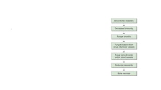
2. Mucormycosis
• Mucormycosis is an uncommon but aggressive opportunistic fungal
infection that afflicts patients with severe underlying
immunosuppression, uncontrolled hyperglycemia and/or
ketoacidosis, patients with iron overload resulting from frequent
blood transfusions or blood disorders and occasionally healthy
patients who are inoculated with fungal spores through traumatic
injuries.
• a/w high mortality
3. • third most common cause of invasive fungal infections following
Aspergillosis and Candidiasis with an increase of mucormycosis in the
adult population and in infants less than 1 year old
4. Zygomycosis or mucormycosis
• Fungi of class zygomycetes
• Rhizpous or mucor or absidia
• These fungi form aseptate or coenocytic hyphae.
• They reproduce sexually by zygospore formation
• They reproduce asexually by sporangiospore formation.
• Predisposing factors
• Leukemia/lymphoma/metabolic acidosis/DM
10. • Mucor are ubiquitous in nature and can be found on decaying
vegetation and in the soil. These fungi grow rapidly and release large
numbers of spores that can become airborne
11. • The morphology shows aseptate/pauci septate broad (3–25 μm),
ribbon-like, hyaline hyphae with irregular or right angle branching
12. Growth of the Fungus
Role of Iron
Acquisition of Iron by Using Siderophores
Hemoglobin as a Source of Iron
Evasion of Host Defenses
Resistance to AntifungalAgents
13. ketone reductase
• Rhizopus organisms have an enzyme, ketone reductase, which allows
them to thrive in high glucose, acidic conditions. Serum from healthy
individuals inhibits growth of Rhizopus, whereas serum from
individuals in diabetic ketoacidosis stimulates growth
15. • Specimen tissue biopsy
• Staining
• H& E best
• Aseptate hyphae irregular branching
of hyphae at OBTUSE angle
• Culture
• SDA @37*c
• Dense hairy colony with aseptate
hyphae sporangiophore and
sporangiospores
16.
17.
18. Risk factors
• Diabetes mellitus, particularly with
ketoacidosis
• Treatment with glucocorticoids
• Hematologic malignancies
• Hematopoietic cell transplantation
• Solid organ transplantation
• Treatment with deferoxamine
• Iron overload
• AIDS
• Malnutrition
• Injection drug use
These risk factors impair host
defenses and
permit growth and
dissemination of fungus
resulting in invasive disease
19. • The individuals at highest risk of developing invasive disease are those
who have decreased numbers (quantitative defects) of mononuclear
and polymorphonuclear phagocytes as in neutropenia or who have
diseases that impair the function (qualitative defects) of phagocytes
as in hyperglycemia/ acidosis or administration of glucocorticoid
23. Host Defenses
• intact skin/mucosal barrier and innate immunity
• mucus layer of sinus/lung has anti-microbial properties and poor in
nutrients
• nutrient immunity of mucosa and skin
• host keeps the essential nutrient iron firmly bound to the serum proteins to
restrict access to pathogens in a process called nutrient immunity and keeps
an iron poor environment as a defense strategy against fungal invasion
• Mononuclear cells
• Tissue macrophages phagocytose the inhaled spores and kill them
24.
25.
26. Altered Host Factors
• Neutropenia and immunosuppression
• hamper the host defenses and allow growth of the fungus
• Administration of corticosteroids impairs host’sability to prevent
germination of spores, whereas exposure to voriconazole makes the
organism highly virulent, possibly due to an epigenetic modification
27.
28. • exposure to voriconazole makes the organism highly virulent, possibly
due to an epigenetic modification
29.
30. • In healthy individuals, cilia transport these spores to the pharynx and
they are cleared through the gastrointestinal tract. In susceptible
individuals, infection usually begins in the nasal turbinates or the
alveoli
31.
32. • The spores enter the body by
• I. inhalation
• II. ingestion of contaminated food
• III. implantation in injured skin by trauma/burns/surgery, or
• IV. percutaneous route by contaminated needles or catheters
33.
34. • Inhaled or inoculated Mucorales spores elicit a robust inflammatory
response in healthy hosts
35. Rhinocerebral Mucormycosis
• Most common form
• diabetic ketoacidosis/uncontrolled diabetes is the most common risk
factor
• Acquired by inhalation of spores
36.
37. • The hallmarks of spread beyond the sinuses are tissue necrosis of the
palate resulting in palatal eschars, destruction of the turbinates,
perinasal swelling, and erythema and cyanosis of the facial skin
overlying the involved sinuses and/or orbit
43. Diagnosis of Rhino-orbital-cerebral infection
• Endoscopic evaluation of the sinuses should be performed to look for
tissue necrosis and to obtain specimens.
46. Diagnosis of Pulmonary infection
• radiographic evidence to support the diagnosis
• Chest radiographs or CT scans
• demonstrate focal consolidation, masses, pleural effusions, or multiple nodules
• halo sign (ground-glass attenuation surrounding a nodule) is characteristic of angioinvasive
fungi,
• reversed halo sign, a focal area of ground-glass attenuation surrounded by a ring of
consolidation
47. Gastrointestinal mucormycosis
Stomach is the most common site of involvement, followed by
colon and ileum, though any part of the gastro intestinal tract
may be involved. It may also involve liver, spleen, and pancreas
48.
49. • In premature neonates, it causes necrotizing enterocolitis whereas in
neutropenic patients, it forms mass lesions. Microscopically, it shows
angioinvasion leading to hemorrhage, gangrene, perforation, and
peritonitis
51. cutaneous mucormycosis
• cutaneous mucormycosis is almost always associated with trauma or
wounds. The entry of the fungi into the dermis can result from
seemingly innocuous insults, such as the entry site for an intravenous
catheter, spider bites, and insulin injection sites
52. • Majority of the patients are immunocompetent who have disruption
of skin barrier due to injury
• Patients have necrotic eschar, ulcer, or fasciitis
• It is locally invasive with involvement of subcutaneous tissue, adipose
tissue, muscle, and fascia
• Involvement of vessels may lead to hematogenous dissemination to
other organs and skin may be involved secondarily in disseminated
disease. Necrosis with suppurative inflammation and infarction may
be seen
53.
54. Disseminated Mucormycosis
• It involves two or more noncontiguous organs and lung is the most
common organ involved. Patients with iron overload and severe
immunosuppression develop disseminated disease
• The infection may spread from lung, GIT, or skin to other organs by
hematogenous dissemination. Microscopically, angioinvasion with
hemorrhagic infarcts and necrosis are seen
55. Uncommon Forms of Mucormycosis
• Intravenous drug use is the most common risk factor.
• The involvement may be osteomyelitis, pyelonephritis, endocarditis,
peritonitis, or focal involvement of any organ
56. Diagnosis
• identification of organisms in tissue by histopathology with culture
confirmation
• Serum tests, such as the 1,3-beta-D-glucan assay and the Aspergillus
galactomannan assay
• polymerase chain reaction (PCR)-based techniques on histologic
specimens
57.
58. Rx
• Intravenous (IV) amphotericin B (lipid formulation) is the drug of
choice for initial therapy
• Posaconazole or isavuconazole is used as step-down therapy for
patients who have responded to amphotericin B
59.
60. • Surgery — Aggressive surgical debridement of involved tissues should
be considered as soon as the diagnosis of any form of mucormycosis
is suspected. Surgical intervention with removal of necrotic tissue and
debulking infection has been associated with improved survival in
anecdotal clinical reviews of rhinocerebral and pulmonary infection
61. Infection in immunocompetent individual
causes
• skin injuries, burns, trauma
• natural calamities like tornadoes, tsunami, volcanic eruption
• combat-related injuries
• contaminated bandages, tongue depressors
• injection drug use
• prolonged hospital stay