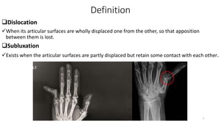
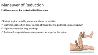
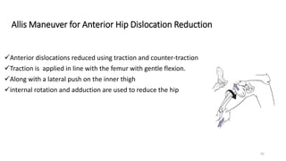
Principles of Dislocation Management
Moderator :Dr.Theodros
Assistant professor of Orthopedics and Traumatology
Presenter: Dr.Bol Kuol
(OSR1)
111
28thDec, 2023 1
Outlines
Introduction
Principles of dislocation Management of;
 GH joint
 Elbow joint
 Hip joint
 Knee joint
 Ankle joint
Summery
References
2
Types of Joints
Fibrous/Synarthrosis
No movement are permitted
E.g. suture,syndesmosis,gomphosis
Cartilaginous/Amphiarthrosis
 Little movements are permitted
E.g. rib cage, symphysis pubis
3
Synovial(Diarthrosis )
Movement is free
 Pivot
 Hinge
 Condylar
 Saddle
 Plane
 Ball and Socket
4
Definition
Dislocation
When its articular surfaces are wholly displaced one from the other, so that apposition
between them is lost.
Subluxation
Exists when the articular surfaces are partly displaced but retain some contact with each other.
5
Types of dislocation
Congenital
Acquired
1.Traumatic
2.Pathological e.g TB hip, Septic Arthritis
3.Paralytic e.g Poliomyelitis ,cerebral palsy ,etc.
4.Inflammatory disorders e.g. rheumatoid arthritis
NO joint is immune from dislocation
6
Most commonly occur in the following joints
 Shoulder
 Elbow
 Hip
 Knee
 Ankle
 Metacarpophalangeal joint
 Facet Joint dislocation in cervical spine
 Acromioclavicular joint dislocation
7
Complications
Acute
Injury to peripheral nerve and blood vessels
Chronic
Unreduced dislocation
Recurrent dislocation
Traumatic osteoarthritis
Joint stiffness
Avascular necrosis
Myositis ossificans
8
Shoulder(GH) Dislocation
 The glenohumeral joint is inherently unstable
 Stability provided by glenoid labrum, ligaments and muscles
 Most common dislocated joint
 Young/athletic patients(recurrence >90% if <25 year old )
Associated with
 labral tears(<40 year old )
 Rotator cuff tears(> 40 year old )
 Glenoid rim (‘’bony Bankart’’) fracture
 Hill-Sachs lesion
9
Anatomic Classification
 Based on direction of humeral head
Anterior Dislocation Most common
Posterior Dislocation often missed
Superior Dislocation extremely rare
Inferior Dislocation luxatio erecta: abducted arm cannot be lowered (rare)
 Recurrent Dislocation happen repeatedly
10
Anterior Dislocation
Most common and more than 90%
Mechanism
Direct force anteriorly impact to the posterior shoulder
 fall on the backward-stretching hand
forced abduction and external rotation of the shoulder
11
History
Trauma/fall
Pain
Inability to move arm
Neurovascular Examination
 Test axillary nerve function
P/E
‘’Flattened’ ’shoulder
Patient carries affected arm with other hand
 Empty glenoid fossa, compared with
contralateral side
Palpable dislocated head(anterior in axilla )
12
Imaging
Trauma series
 AP view
 LT view
 Axillary view
 Scapular Y view
overlapping shadow of the humeral head and glenoid fossa
Hill-Sachs lesion
Bony Bankart lesion
Humeral neck #
Tuberosity
13
Non Operative
Closed Reduction
Traction and Counter traction Method
Most commonly used
Axial traction is applied to arm
 counter traction is applied with sheet wrapped over shoulder
Kocher’s Maneuver
Traction
External rotation
Adduction
Internal rotation 14
Stimson Maneuver
Gravity method in prone position

Milch Maneuver
 Patient supine
steady downward traction applied at elbow
combined with slow
gradual external rotation and abduction of limb
15
Operative
Soft tissue interposition
Displaced greater tuberosity fracture > 5mm
Glenoid rim #> 5 mm in size
Glenoid labrum has been damaged or detached
16
Immobilization and Rehabilitation
 over 30 years of age, stiffness is more of a risk than recurrent dislocation so
movements are begun after 1 week.
under 30 years, recurrence is more of a risk and so the sling is retained for 3
weeks before mobilizing
Complications
Recurrent dislocation/instability (esp.in young/<25 y.o)
Neurovascular injury (axillary n, musculocutaneous n and axillary a)
Rotator cuff tear in older people
17
Posterior Shoulder Dislocation
 commonly missed
Rare than anterior shoulder dislocation
Mechanism
Direct Trauma
 Blow on the front of the shoulder
indirect trauma
 Electric shock
 convulsion
18
 History
Trauma/fall
Pain
Inability to move arm
Convulsion
 Neurovascular Examination
 Test axillary nerve function
 P/E
Shoulder internally rotated and
adduction
Palpable mass posterior to the
shoulder
Flattening of the anterior
shoulder
Tenderness
19
AP VIEW
Half Moon Sign Light bulb sign
20
Reduction technique
The arm is pulled and rotated laterally, while the head of the humerus is pushed forwards.
21
Immobilization and Rehabilitation
over 30 years of age, stiffness is more of a risk than recurrent dislocation so
movements are begun after 1 week.
under 30 years, recurrence is more of a risk and so the sling is retained for 3
weeks before mobilizing
22
Recurrent dislocation
Once a shoulder has been dislocated, this may happen repeatedly.
the capsule and labrum have usually been stripped from the margin of the
glenoid and the humeral head may be indented
In the vast majority of cases recurrence is anterior
23
History
Dislocation
the patient complains that the shoulder ‘slips out’
 when the arm is lifted into abduction and lateral rotation
Physical examination
The apprehension test is positive
 the shoulder is passively manipulated into abduction, extension and lateral rotation.
the patient tenses up and resists further movement
24
Imaging
AP x-ray
 MRI
25
Treatment
 Anterior dislocation some form of anterior capsular reconstruction is
usually successful
recurrent posterior dislocation
 is more difficult and may require soft-tissue reconstruction combined with a
bone operation to block abnormal movement at the back of the shoulder
26
Elbow Dislocation
The elbow is the second most commonly dislocated joint in adults.
 Acute dislocation of the elbow is almost always reducible by closed methods.
 most are stable after reduction.
 Open reduction may be required if fracture fragments in a fracture-dislocation block closed
reduction
Approximately 20% of dislocations are associated with fractures:
 “terrible triad”=elbow dx with radial head&coronoid fracture
Collateral ligaments &anterior capsule are typically torn
Mechanism
Usually a fall/RTA in elderly and young patient
27
History
Trauma/fall
Inability to move elbow
 Neurovascular Examination
P/E
Swelling
Deformity
Limited /no elbow ROM
Good neurovascular exam
28
Classification
By direction of forearm bones
Posterior-posterolateral(>80%)>posteromedial
Anterior (rare)
Medial
Lateral(rare)
Divergent(rare)
29
Radial Head Subluxation
Common in children age group(2-5 years)
Reduction
With thumb in antecubital space as a fulcrum
The forearm is supinated and flexed
30
Imaging
 XR
Elbow series
CT
To define associated fracture
31
Treatment
Acute-Closed reduction
Prone position
Only forearm hangs from the stretcher
Gentle downward traction
 Reduction of the olecranon with the opposite hand
32
Operative
ORIF(corocoid,radial head ,olecranon ),LCL repair,+/- MCL repair
Acute complex elbow dislocation
Persistent instability after reduction
Open reduction, capsular release, and dynamic hinged elbow fixator
Chronic dislocation
33
Immobilization
Stable
Splint in 90 degree of flexion for 7-10 day
Unstable
Splint for 2-3 week
Complications
Elbow stiffness and stability
Recurrent dislocation
Neurovascular injury (Median and ulnar nerves, brachial artery)
Secondary osteoarthritis
34
Hip Dislocation
The hip joint is inherently stable
 Orthopedic emergency risk of femoral head AVN increases with late/delayed reduction
Mechanism of Injury
High-energy trauma E.g. MVA, dashboard injury
 significant fall
 History
Trauma, severe pain, cannot move thigh/hip
35
Physical Examination
 Anterior Dislocation
Occurs with the hip in abduction and
external rotation
Characteristic position of affected limb.
 Hip flexed, thigh abducted and externally
rotated
 Posterior Dislocation
Most common (90%)
Axial load through flexed knee(dashboard
injury)
Femur adducted and internally rotated
 hip flexed
Associated with posterior wall acetabular
#,femoral head #,and sciatic nerve injury
36
Cont,,,,
Central Dislocation
This is the least common and difficult of all dislocation of the hip joint
Due to direct trauma to greater trochanter fracture of floor of acetabulum
Lateral force against adducted femur
Always fracture- dislocation
37
 XR:
AP pelvis, femur and knee series
CT: R/o fx or bony fragments/ loose bodies
post reduction
38
Treatment
Nonoperative
Emergent closed reduction within 6 hours for acute anterior and posterior
dislocations
Contraindicated in ipsilateral displaced or non –displaced femoral neck fracture
39
Operative
Open reduction
 removal of incarcerated fragments
 Irreducible dislocation
 Delayed presentation
ORIF for
 Acetabulum fracture
 Femora head and neck
40
Maneuver of Reduction
Allis maneuver for posterior Hip Dislocation
Patient supine on table, under anesthesia or sedation.
 Examiner applies firm distal traction at flexed knee to pull head into acetabulum
 slight rotary motion may also help
 Assistant fixes pelvis by pressing on anterior superior iliac spine
41
Allis Maneuver for Anterior Hip Dislocation Reduction
Anterior dislocations reduced using traction and counter-traction
Traction is applied in line with the femur with gentle flexion.
Along with a lateral push on the inner thigh
internal rotation and adduction are used to reduce the hip
42
Post-reduction Care
For simple dislocation
Within 6 hours a brief period of rest for several days to 2 weeks followed by
mobilization
After 6 hours since the rate of AVS is highest, it may be reasonable to delay full
weight bearing for 8 to 12 weeks
Most patients can achieve full weight bearing by 6 weeks
Dislocations with Associated Fractures
The rehabilitation for patients who had associated fractures fixed is determined by the
associated injury
 In the case of posterior wall acetabular fractures or femoral head fractures, active hip motion
may be deferred for approximately 6 weeks 43
Complications
Posttraumatic osteonecrosis (AVN) (reduced risk with early reduction)
5-40% incidence
Increased risk with increased time to reduction
 sciatic nerve injury (posterior dislocations)
8-20% incidence
femoral artery/nerve injury (anterior dislocations)
Post traumatic arthritis
Up to 20% for simple dislocation, markedly increased with complex dislocation
Recurrent dislocations
Less than 2 %
44
Precautions !!!!
1.Prohibit patients from crossing their
legs
2.Bending their hip a 90-degree angle
3.Twisting their hip inward
45
Knee Dislocation
Frank dislocation of Knee joint is a devastating injury with the potential for limb-threatening
complications
Many spontaneously reduce, must keep index of suspicion of suspicion for injury
Multiple ligaments and other soft tissue are disrupted
High incidence of associated fracture and neurovascular injury
46
Mechanism of Injury
High energy
From MVA,fall from a height or dashboard
Low energy
An athletic injury, routine walking
47
Presentation
Symptoms
History of trauma and deformity of the knee
Knee pain
Inability to bear weight
48
Physical Examination
Appearance
No obvious deformity 50% spontaneously reduce before arrival to ED
Signs of trauma(swelling, effusion, abrasions, ecchymosis)
+/- distal pulses/peroneal nerve function
‘’Dimple Sign’’-buttonholing of medical femoral condyle through the medical capsule
Vascular exam
To rule out vascular injury both before and after reduction
Serial examinations are mandatory
Palpate the dorsalis pedis&posterior tibial pulses
Neurologic exam
Sensory and motor function of peroneal and tibial nerve
49
Classification
Kennedy classification based on the direction of displacement of the tibia
Anterior (30-50%)
Most common
Due to hyperextension injury
Usually involves tear of PCL
The highest rate of peroneal nerve injury
Posterior( 30-40%)
2nd most common
Due to axial load to flexed knee(dashboard injury)
The highest rate of vascular injury (25 %)
Highest incidence of a complete tear of popliteal artery
50
Lateral(13%)
Due to a Varus or valgus force
Usually involves tears of both ACL&PCL
Medial (3%)
Varus or valgus force
Usually disrupted PLC and PCL
Rotational (4%)
Posterolateral is most common rotational dislocation
Usually irreducible
Buttonholing of femoral condyle through the capsule
51
Imaging
X-ray
Pre-reduction and post reduction (AP&Lt of the Knee)
 May be normal if spontaneous reduction
 Look for asymmetric or irregular joint space
 Look for avulsion Fxs(segond sign-lateral tibial condyle avulsion Fx)
CT Scan
Facture identified on post reduction x-ray
Findings
 Tibial eminence, tibial tubercle, and tibial plateau fractures may be seen
52
Cont,,,,,,,,,,,,,,,,,,,,,,,,,,,,,
MRI
Obtain MRI after acute reduction but prior to hardware placement
Required to evaluate soft tissue injury (ligaments, meniscus) and for surgical planning
53
Closed reduction technique
Anterior
 Traction and anterior translation of femur
Posterior
 Traction,extension,and anterior translation of the tibia
Medial/Lateral
 Traction and medial or lateral translation
Rotatory
 Axial limb traction and rotation in the opposite direction of deformity
54
External Fixation
Indications
 Vascular repair
 Open fracture-dislocation
 Compartment syndrome
 Polytrauma patient
Open reduction
Irreducible knee
Vascular injury
Open fracture-dislocation
Posterolateral dislocation
55
Early Ligamentous reconstruction (<3 week)
Acute reconstruction has been shown to lead to improved clinical and functional
outcomes
Complications
Vascular compromise 40-50% in anterior or posterior dislocations
Stiffness( arthrofibrosis ) most common complication (38 %)
Laxity and instability 37% of some instability,redislocation is uncommon
Peroneal nerve injury 25% occurrence of a peroneal nerve injury,50% recover partially
56
Subtalar Dislocation
Accounts for 1% of all dislocations
More common in young or middle-aged males
25% may be open especially in lateral dislocation
Mechanism
Typically result from a high energy Mechanism
 RTA
Fall from height
Athletics
57
Presentation
 Severe Pain
Gross deformity
Foot will be locked in supination with medial dislocation
 Known as ‘’ acquired clubfoot ‘’
Foot will be locked in supination with lateral dislocation
 Known as ‘’ acquired flatfoot’’
58
Anatomic Classification
Medical dislocation
Most common(65-80%)
Due to lateral malleolus acting as strong butrres,preventing dislocation
Foot become locked in supination
Reduction blocked by peroneal tendon and talonavicular capsule
Lateral dislocation
More likely to be open
Foot become locked in pronation
Associated with lateral process ,anterior calcaneus and fibula #
Reduction blocked by PT tendon ,FHL,FDL
59
Anterior dislocation
Rare
Posterior dislocation
Rare
Total dislocation(extruded talus )
Usually open
Talus is completely dislocated from ankle and subtalar and talovavicular joints
60
Imaging
X-ray of ankle(AP&Lt view )
CT scan of ankle
 Subtalar osteochondral fragment causing subluxation
61
Non operative
60-70 % cab be reduced by closed methods
 short leg cast non-weight bearing cast for 4-6 week
Traction counter traction
62
Operative reduction
Open dislocations
Failure of closed reduction
63
Post-Op care
If joint stable
 Place in short leg cast with non-weight bearing for 4-6 weeks
If joint remains unstable
 place temporary tranarticular pins or spanning external fixator
64
Complications
Stiffness
Most common complication
Post-traumatic arthritis
65
Typical deformities in dislocation
Shoulder- Abduction deformities
Elbow-Flexion deformities
HIP:
 Anterior :Flexion ,abduction and external rotation deformities .
 Posterior- Flexion ,adduction and internal rotation deformity
Knee- flexion deformity
Ankle- Varus deformity
66
Summery
Principles of dislocation Management
Aims Save life
Save limb
save the function
Assessment of condition of the patient for shock and other injuries
Assessment of local condition of injured limb (vascular injury, nerve)
Resuscitation, if needed
Safe transport, this help to minimize complication in injuries to spine
(All dislocations associated with fractures should be immobilized immediately)
67
Radiography of the part
Reduction of dislocation
Closed manipulation
Open
Immobilization
Re-radiography of the part
Early physiotherapy
For the preservation of the function of the limb
Rehabilitation
68
Ameseginalehu !
69
References
Apley &Solmon’s concise system of orthopaedic &truma -4th edition
Campbell’s Operative Orthopaedics-13th edition
Netter’s Concise Orthopedics anatomy -2nd edition
Othrobullets-Trauma -2021
Rockwood &Green’s fracture in Adults-8th edition
Internet
70

32,Principles of Dislocation Manangment.pptx

  • 1.
    Principles of DislocationManagement Moderator :Dr.Theodros Assistant professor of Orthopedics and Traumatology Presenter: Dr.Bol Kuol (OSR1) 111 28thDec, 2023 1
  • 2.
    Outlines Introduction Principles of dislocationManagement of;  GH joint  Elbow joint  Hip joint  Knee joint  Ankle joint Summery References 2
  • 3.
    Types of Joints Fibrous/Synarthrosis Nomovement are permitted E.g. suture,syndesmosis,gomphosis Cartilaginous/Amphiarthrosis  Little movements are permitted E.g. rib cage, symphysis pubis 3
  • 4.
    Synovial(Diarthrosis ) Movement isfree  Pivot  Hinge  Condylar  Saddle  Plane  Ball and Socket 4
  • 5.
    Definition Dislocation When its articularsurfaces are wholly displaced one from the other, so that apposition between them is lost. Subluxation Exists when the articular surfaces are partly displaced but retain some contact with each other. 5
  • 6.
    Types of dislocation Congenital Acquired 1.Traumatic 2.Pathologicale.g TB hip, Septic Arthritis 3.Paralytic e.g Poliomyelitis ,cerebral palsy ,etc. 4.Inflammatory disorders e.g. rheumatoid arthritis NO joint is immune from dislocation 6
  • 7.
    Most commonly occurin the following joints  Shoulder  Elbow  Hip  Knee  Ankle  Metacarpophalangeal joint  Facet Joint dislocation in cervical spine  Acromioclavicular joint dislocation 7
  • 8.
    Complications Acute Injury to peripheralnerve and blood vessels Chronic Unreduced dislocation Recurrent dislocation Traumatic osteoarthritis Joint stiffness Avascular necrosis Myositis ossificans 8
  • 9.
    Shoulder(GH) Dislocation  Theglenohumeral joint is inherently unstable  Stability provided by glenoid labrum, ligaments and muscles  Most common dislocated joint  Young/athletic patients(recurrence >90% if <25 year old ) Associated with  labral tears(<40 year old )  Rotator cuff tears(> 40 year old )  Glenoid rim (‘’bony Bankart’’) fracture  Hill-Sachs lesion 9
  • 10.
    Anatomic Classification  Basedon direction of humeral head Anterior Dislocation Most common Posterior Dislocation often missed Superior Dislocation extremely rare Inferior Dislocation luxatio erecta: abducted arm cannot be lowered (rare)  Recurrent Dislocation happen repeatedly 10
  • 11.
    Anterior Dislocation Most commonand more than 90% Mechanism Direct force anteriorly impact to the posterior shoulder  fall on the backward-stretching hand forced abduction and external rotation of the shoulder 11
  • 12.
    History Trauma/fall Pain Inability to movearm Neurovascular Examination  Test axillary nerve function P/E ‘’Flattened’ ’shoulder Patient carries affected arm with other hand  Empty glenoid fossa, compared with contralateral side Palpable dislocated head(anterior in axilla ) 12
  • 13.
    Imaging Trauma series  APview  LT view  Axillary view  Scapular Y view overlapping shadow of the humeral head and glenoid fossa Hill-Sachs lesion Bony Bankart lesion Humeral neck # Tuberosity 13
  • 14.
    Non Operative Closed Reduction Tractionand Counter traction Method Most commonly used Axial traction is applied to arm  counter traction is applied with sheet wrapped over shoulder Kocher’s Maneuver Traction External rotation Adduction Internal rotation 14
  • 15.
    Stimson Maneuver Gravity methodin prone position  Milch Maneuver  Patient supine steady downward traction applied at elbow combined with slow gradual external rotation and abduction of limb 15
  • 16.
    Operative Soft tissue interposition Displacedgreater tuberosity fracture > 5mm Glenoid rim #> 5 mm in size Glenoid labrum has been damaged or detached 16
  • 17.
    Immobilization and Rehabilitation over 30 years of age, stiffness is more of a risk than recurrent dislocation so movements are begun after 1 week. under 30 years, recurrence is more of a risk and so the sling is retained for 3 weeks before mobilizing Complications Recurrent dislocation/instability (esp.in young/<25 y.o) Neurovascular injury (axillary n, musculocutaneous n and axillary a) Rotator cuff tear in older people 17
  • 18.
    Posterior Shoulder Dislocation commonly missed Rare than anterior shoulder dislocation Mechanism Direct Trauma  Blow on the front of the shoulder indirect trauma  Electric shock  convulsion 18
  • 19.
     History Trauma/fall Pain Inability tomove arm Convulsion  Neurovascular Examination  Test axillary nerve function  P/E Shoulder internally rotated and adduction Palpable mass posterior to the shoulder Flattening of the anterior shoulder Tenderness 19
  • 20.
    AP VIEW Half MoonSign Light bulb sign 20
  • 21.
    Reduction technique The armis pulled and rotated laterally, while the head of the humerus is pushed forwards. 21
  • 22.
    Immobilization and Rehabilitation over30 years of age, stiffness is more of a risk than recurrent dislocation so movements are begun after 1 week. under 30 years, recurrence is more of a risk and so the sling is retained for 3 weeks before mobilizing 22
  • 23.
    Recurrent dislocation Once ashoulder has been dislocated, this may happen repeatedly. the capsule and labrum have usually been stripped from the margin of the glenoid and the humeral head may be indented In the vast majority of cases recurrence is anterior 23
  • 24.
    History Dislocation the patient complainsthat the shoulder ‘slips out’  when the arm is lifted into abduction and lateral rotation Physical examination The apprehension test is positive  the shoulder is passively manipulated into abduction, extension and lateral rotation. the patient tenses up and resists further movement 24
  • 25.
  • 26.
    Treatment  Anterior dislocationsome form of anterior capsular reconstruction is usually successful recurrent posterior dislocation  is more difficult and may require soft-tissue reconstruction combined with a bone operation to block abnormal movement at the back of the shoulder 26
  • 27.
    Elbow Dislocation The elbowis the second most commonly dislocated joint in adults.  Acute dislocation of the elbow is almost always reducible by closed methods.  most are stable after reduction.  Open reduction may be required if fracture fragments in a fracture-dislocation block closed reduction Approximately 20% of dislocations are associated with fractures:  “terrible triad”=elbow dx with radial head&coronoid fracture Collateral ligaments &anterior capsule are typically torn Mechanism Usually a fall/RTA in elderly and young patient 27
  • 28.
    History Trauma/fall Inability to moveelbow  Neurovascular Examination P/E Swelling Deformity Limited /no elbow ROM Good neurovascular exam 28
  • 29.
    Classification By direction offorearm bones Posterior-posterolateral(>80%)>posteromedial Anterior (rare) Medial Lateral(rare) Divergent(rare) 29
  • 30.
    Radial Head Subluxation Commonin children age group(2-5 years) Reduction With thumb in antecubital space as a fulcrum The forearm is supinated and flexed 30
  • 31.
    Imaging  XR Elbow series CT Todefine associated fracture 31
  • 32.
    Treatment Acute-Closed reduction Prone position Onlyforearm hangs from the stretcher Gentle downward traction  Reduction of the olecranon with the opposite hand 32
  • 33.
    Operative ORIF(corocoid,radial head ,olecranon),LCL repair,+/- MCL repair Acute complex elbow dislocation Persistent instability after reduction Open reduction, capsular release, and dynamic hinged elbow fixator Chronic dislocation 33
  • 34.
    Immobilization Stable Splint in 90degree of flexion for 7-10 day Unstable Splint for 2-3 week Complications Elbow stiffness and stability Recurrent dislocation Neurovascular injury (Median and ulnar nerves, brachial artery) Secondary osteoarthritis 34
  • 35.
    Hip Dislocation The hipjoint is inherently stable  Orthopedic emergency risk of femoral head AVN increases with late/delayed reduction Mechanism of Injury High-energy trauma E.g. MVA, dashboard injury  significant fall  History Trauma, severe pain, cannot move thigh/hip 35
  • 36.
    Physical Examination  AnteriorDislocation Occurs with the hip in abduction and external rotation Characteristic position of affected limb.  Hip flexed, thigh abducted and externally rotated  Posterior Dislocation Most common (90%) Axial load through flexed knee(dashboard injury) Femur adducted and internally rotated  hip flexed Associated with posterior wall acetabular #,femoral head #,and sciatic nerve injury 36
  • 37.
    Cont,,,, Central Dislocation This isthe least common and difficult of all dislocation of the hip joint Due to direct trauma to greater trochanter fracture of floor of acetabulum Lateral force against adducted femur Always fracture- dislocation 37
  • 38.
     XR: AP pelvis,femur and knee series CT: R/o fx or bony fragments/ loose bodies post reduction 38
  • 39.
    Treatment Nonoperative Emergent closed reductionwithin 6 hours for acute anterior and posterior dislocations Contraindicated in ipsilateral displaced or non –displaced femoral neck fracture 39
  • 40.
    Operative Open reduction  removalof incarcerated fragments  Irreducible dislocation  Delayed presentation ORIF for  Acetabulum fracture  Femora head and neck 40
  • 41.
    Maneuver of Reduction Allismaneuver for posterior Hip Dislocation Patient supine on table, under anesthesia or sedation.  Examiner applies firm distal traction at flexed knee to pull head into acetabulum  slight rotary motion may also help  Assistant fixes pelvis by pressing on anterior superior iliac spine 41
  • 42.
    Allis Maneuver forAnterior Hip Dislocation Reduction Anterior dislocations reduced using traction and counter-traction Traction is applied in line with the femur with gentle flexion. Along with a lateral push on the inner thigh internal rotation and adduction are used to reduce the hip 42
  • 43.
    Post-reduction Care For simpledislocation Within 6 hours a brief period of rest for several days to 2 weeks followed by mobilization After 6 hours since the rate of AVS is highest, it may be reasonable to delay full weight bearing for 8 to 12 weeks Most patients can achieve full weight bearing by 6 weeks Dislocations with Associated Fractures The rehabilitation for patients who had associated fractures fixed is determined by the associated injury  In the case of posterior wall acetabular fractures or femoral head fractures, active hip motion may be deferred for approximately 6 weeks 43
  • 44.
    Complications Posttraumatic osteonecrosis (AVN)(reduced risk with early reduction) 5-40% incidence Increased risk with increased time to reduction  sciatic nerve injury (posterior dislocations) 8-20% incidence femoral artery/nerve injury (anterior dislocations) Post traumatic arthritis Up to 20% for simple dislocation, markedly increased with complex dislocation Recurrent dislocations Less than 2 % 44
  • 45.
    Precautions !!!! 1.Prohibit patientsfrom crossing their legs 2.Bending their hip a 90-degree angle 3.Twisting their hip inward 45
  • 46.
    Knee Dislocation Frank dislocationof Knee joint is a devastating injury with the potential for limb-threatening complications Many spontaneously reduce, must keep index of suspicion of suspicion for injury Multiple ligaments and other soft tissue are disrupted High incidence of associated fracture and neurovascular injury 46
  • 47.
    Mechanism of Injury Highenergy From MVA,fall from a height or dashboard Low energy An athletic injury, routine walking 47
  • 48.
    Presentation Symptoms History of traumaand deformity of the knee Knee pain Inability to bear weight 48
  • 49.
    Physical Examination Appearance No obviousdeformity 50% spontaneously reduce before arrival to ED Signs of trauma(swelling, effusion, abrasions, ecchymosis) +/- distal pulses/peroneal nerve function ‘’Dimple Sign’’-buttonholing of medical femoral condyle through the medical capsule Vascular exam To rule out vascular injury both before and after reduction Serial examinations are mandatory Palpate the dorsalis pedis&posterior tibial pulses Neurologic exam Sensory and motor function of peroneal and tibial nerve 49
  • 50.
    Classification Kennedy classification basedon the direction of displacement of the tibia Anterior (30-50%) Most common Due to hyperextension injury Usually involves tear of PCL The highest rate of peroneal nerve injury Posterior( 30-40%) 2nd most common Due to axial load to flexed knee(dashboard injury) The highest rate of vascular injury (25 %) Highest incidence of a complete tear of popliteal artery 50
  • 51.
    Lateral(13%) Due to aVarus or valgus force Usually involves tears of both ACL&PCL Medial (3%) Varus or valgus force Usually disrupted PLC and PCL Rotational (4%) Posterolateral is most common rotational dislocation Usually irreducible Buttonholing of femoral condyle through the capsule 51
  • 52.
    Imaging X-ray Pre-reduction and postreduction (AP&Lt of the Knee)  May be normal if spontaneous reduction  Look for asymmetric or irregular joint space  Look for avulsion Fxs(segond sign-lateral tibial condyle avulsion Fx) CT Scan Facture identified on post reduction x-ray Findings  Tibial eminence, tibial tubercle, and tibial plateau fractures may be seen 52
  • 53.
    Cont,,,,,,,,,,,,,,,,,,,,,,,,,,,,, MRI Obtain MRI afteracute reduction but prior to hardware placement Required to evaluate soft tissue injury (ligaments, meniscus) and for surgical planning 53
  • 54.
    Closed reduction technique Anterior Traction and anterior translation of femur Posterior  Traction,extension,and anterior translation of the tibia Medial/Lateral  Traction and medial or lateral translation Rotatory  Axial limb traction and rotation in the opposite direction of deformity 54
  • 55.
    External Fixation Indications  Vascularrepair  Open fracture-dislocation  Compartment syndrome  Polytrauma patient Open reduction Irreducible knee Vascular injury Open fracture-dislocation Posterolateral dislocation 55
  • 56.
    Early Ligamentous reconstruction(<3 week) Acute reconstruction has been shown to lead to improved clinical and functional outcomes Complications Vascular compromise 40-50% in anterior or posterior dislocations Stiffness( arthrofibrosis ) most common complication (38 %) Laxity and instability 37% of some instability,redislocation is uncommon Peroneal nerve injury 25% occurrence of a peroneal nerve injury,50% recover partially 56
  • 57.
    Subtalar Dislocation Accounts for1% of all dislocations More common in young or middle-aged males 25% may be open especially in lateral dislocation Mechanism Typically result from a high energy Mechanism  RTA Fall from height Athletics 57
  • 58.
    Presentation  Severe Pain Grossdeformity Foot will be locked in supination with medial dislocation  Known as ‘’ acquired clubfoot ‘’ Foot will be locked in supination with lateral dislocation  Known as ‘’ acquired flatfoot’’ 58
  • 59.
    Anatomic Classification Medical dislocation Mostcommon(65-80%) Due to lateral malleolus acting as strong butrres,preventing dislocation Foot become locked in supination Reduction blocked by peroneal tendon and talonavicular capsule Lateral dislocation More likely to be open Foot become locked in pronation Associated with lateral process ,anterior calcaneus and fibula # Reduction blocked by PT tendon ,FHL,FDL 59
  • 60.
    Anterior dislocation Rare Posterior dislocation Rare Totaldislocation(extruded talus ) Usually open Talus is completely dislocated from ankle and subtalar and talovavicular joints 60
  • 61.
    Imaging X-ray of ankle(AP&Ltview ) CT scan of ankle  Subtalar osteochondral fragment causing subluxation 61
  • 62.
    Non operative 60-70 %cab be reduced by closed methods  short leg cast non-weight bearing cast for 4-6 week Traction counter traction 62
  • 63.
  • 64.
    Post-Op care If jointstable  Place in short leg cast with non-weight bearing for 4-6 weeks If joint remains unstable  place temporary tranarticular pins or spanning external fixator 64
  • 65.
  • 66.
    Typical deformities indislocation Shoulder- Abduction deformities Elbow-Flexion deformities HIP:  Anterior :Flexion ,abduction and external rotation deformities .  Posterior- Flexion ,adduction and internal rotation deformity Knee- flexion deformity Ankle- Varus deformity 66
  • 67.
    Summery Principles of dislocationManagement Aims Save life Save limb save the function Assessment of condition of the patient for shock and other injuries Assessment of local condition of injured limb (vascular injury, nerve) Resuscitation, if needed Safe transport, this help to minimize complication in injuries to spine (All dislocations associated with fractures should be immobilized immediately) 67
  • 68.
    Radiography of thepart Reduction of dislocation Closed manipulation Open Immobilization Re-radiography of the part Early physiotherapy For the preservation of the function of the limb Rehabilitation 68
  • 69.
  • 70.
    References Apley &Solmon’s concisesystem of orthopaedic &truma -4th edition Campbell’s Operative Orthopaedics-13th edition Netter’s Concise Orthopedics anatomy -2nd edition Othrobullets-Trauma -2021 Rockwood &Green’s fracture in Adults-8th edition Internet 70