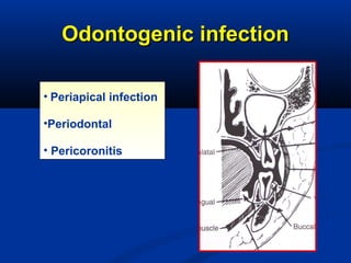
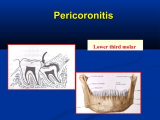
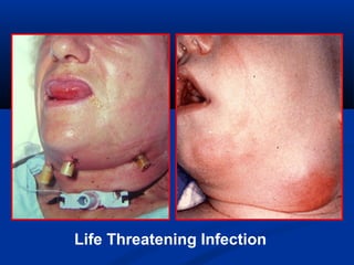
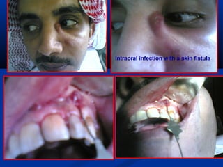
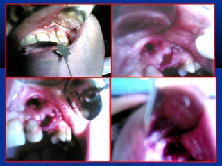
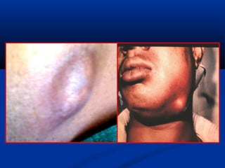
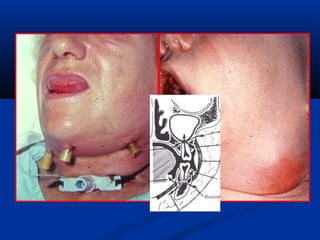
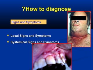
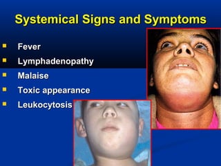
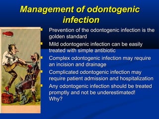
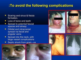
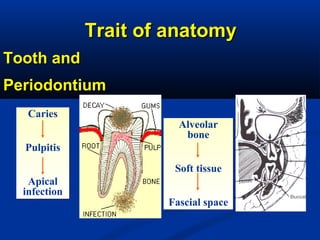
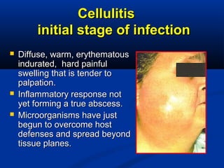
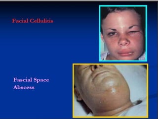
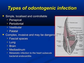
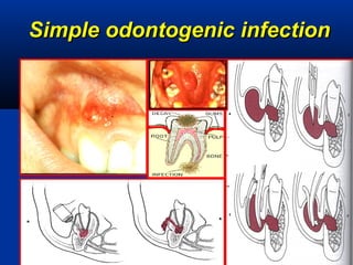
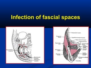
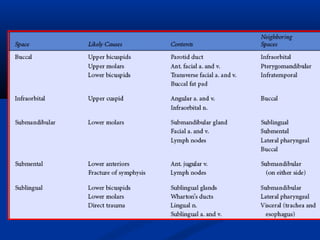
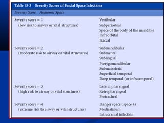
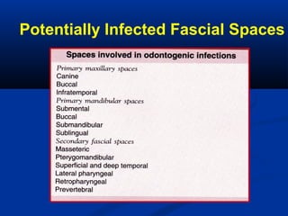
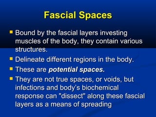
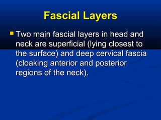
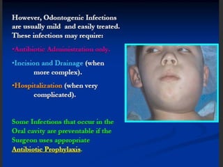
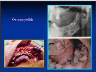
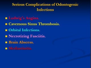
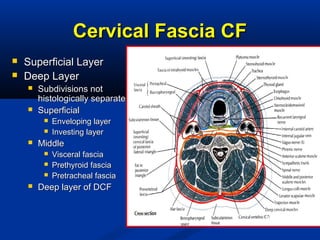
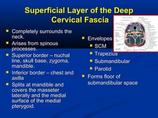
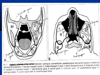
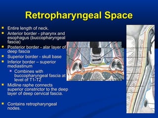
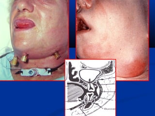
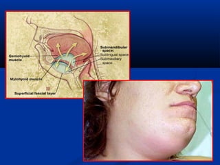
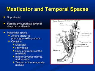
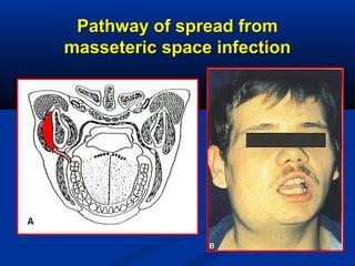
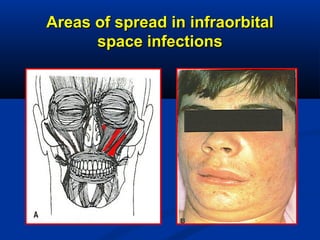
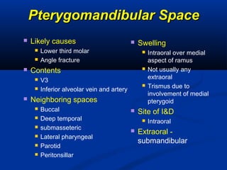
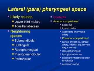
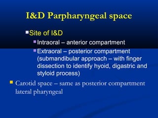
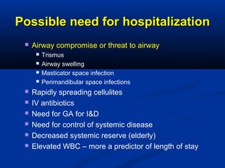
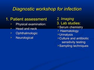
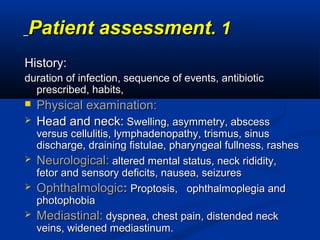
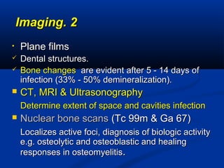
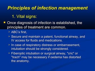
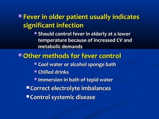
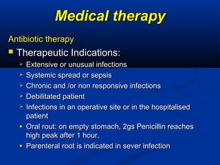
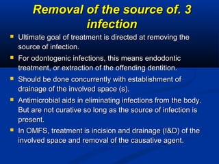
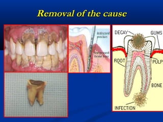
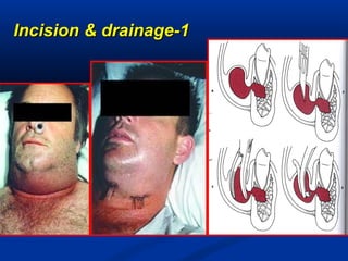
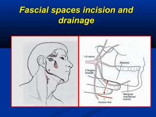
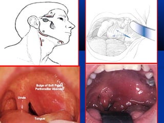
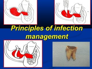
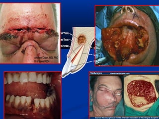
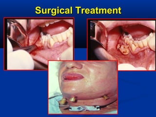
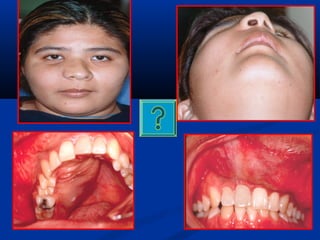
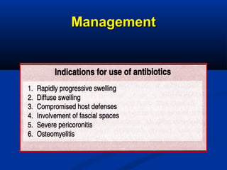
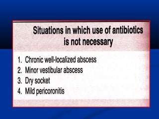
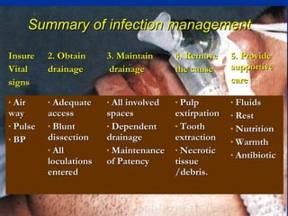
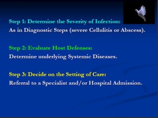
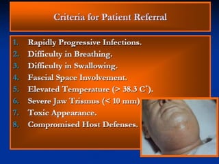
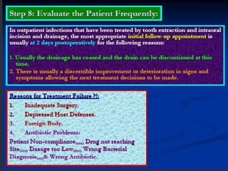
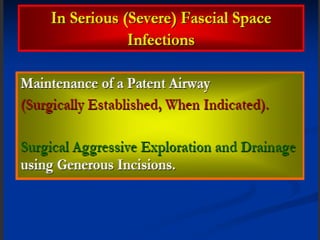
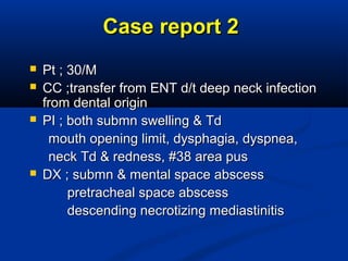
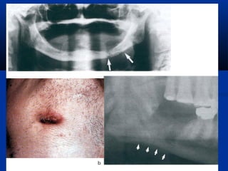
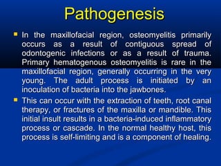
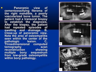
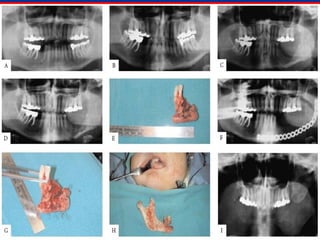
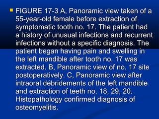
OdontogenicOdontogenic
Infection 1Infection 1
Dr. Adel I. AbdelhadyDr. Adel I. Abdelhady
BDS, MSc ( Tanta, Eg.), PhD (Egypt,USABDS, MSc ( Tanta, Eg.), PhD (Egypt,USA((
Ass. Prof. Oral and Maxillofacial surgeryAss. Prof. Oral and Maxillofacial surgery,,
Collage of DentistryCollage of Dentistry
King Faisal UniversityKing Faisal University
"Do not let sun sets on prisoned pus"
Odontogenic InfectionOdontogenic Infection
Infection due to
dental cause.
Oral flora is the
main source of OI:
Aerobic and
Anaerobic Gr-ve
and Gr. +ve cocci
and rods.
Oral flora is the main source ofOral flora is the main source of
odontogenic infectionodontogenic infection
bacteriabacteria PercentagePercentage % of Aerobic & Anaerobic% of Aerobic & Anaerobic
AerobicAerobic 7 %7 % Aerobic 25 %Aerobic 25 %
Anaerobic 75 %Anaerobic 75 %
Of the causative organismsOf the causative organisms
(number of patients was 404(.(number of patients was 404(.
AnaerobicAnaerobic 33 %33 %
MixedMixed 60 %60 %
Routes ofRoutes of
odontogenicodontogenic
infectionsinfections
▼▼
PulpalPulpal
oror
PeridontiumPeridontium
oror
SystemicSystemic
Infection Arising and SpreadingInfection Arising and Spreading
 Effectiveness of patientEffectiveness of patient
immune mechanismimmune mechanism
 Microbe---virulenceMicrobe---virulence
 QuantityQuantity
 Failure to drainFailure to drain
accumulation pusaccumulation pus
Balance ImbalanceBalance Imbalance
ScaleScale
The Anatomical Factors InfluencingThe Anatomical Factors Influencing
the direction of spread within thethe direction of spread within the
tissuestissues
 The site of the source of infection , upperThe site of the source of infection , upper
or lower jawor lower jaw
 The point at which the pus escapes fromThe point at which the pus escapes from
the bone to the soft tissues labiobucally orthe bone to the soft tissues labiobucally or
linguopalatallylinguopalatally
 The natural barriers to the spread of pus inThe natural barriers to the spread of pus in
the tissues as layer of fascia , muscle orthe tissues as layer of fascia , muscle or
jaw bonejaw bone
Sequence of odontogenicSequence of odontogenic
infectionsinfections
fascial spaces
Soft tissue &
cortical bone
Erosion of cancellous bone
Periapical through pulp necrosis
Periodontal through deep periodontal pocket
Alveolar
bone
Soft tissue
Fascial space
Alveolar
bone
Soft tissue
Fascial space
Trait of anatomyTrait of anatomy
Tooth andTooth and
PeriodontiumPeriodontium
Caries
Pulpitis
Apical
infection
Caries
Pulpitis
Apical
infection
Odontogenic infectionOdontogenic infection
• Periapical infection
•Periodontal
• Pericoronitis
• Periapical infection
•Periodontal
• Pericoronitis
PericoronitisPericoronitis
Lower third molarLower third molar
Periapical infectionPeriapical infection
Acute-chronic
Periapical
infection
Acute-chronic
Periapical
infection
Fistular
Cellulitis
Intraoral
soft tissue
abscess
Osteomyelitis
Septicemia
Deep fascial
space
infection
Ascending facial-
cerebral infection
Pathways of Periapical infectionPathways of Periapical infection
Changing directionsChanging directions
 Localization andLocalization and
recoveryrecovery
 Acute chronicAcute chronic
 Diffusion or spreadDiffusion or spread
Blood system---Septicemia
lymphoid system---
Lymphadenopathy
From submandibular
space infection
spread to chest region
Inflammation as a Sign of Odontogenic InfectionInflammation as a Sign of Odontogenic Infection
 Inflammation is an important pathologic process oftenInflammation is an important pathologic process often
encountered by dentists . As an indicator of disease, it hasencountered by dentists . As an indicator of disease, it has
been recognized for centuries. Almost 2,000 years ago,been recognized for centuries. Almost 2,000 years ago,
the Roman physicianthe Roman physician CelsusCelsus recognized the warmth,recognized the warmth,
redness, swelling, and pain associated with now is knownredness, swelling, and pain associated with now is known
asas "inflammation“."inflammation“.
 These events caused by a series of cellular and tissueThese events caused by a series of cellular and tissue
responses to some injurious agent. These responses areresponses to some injurious agent. These responses are
directed atdirected at

destroying the incitingdestroying the inciting ‫المحرض‬‫المحرض‬ agent or, rendering itagent or, rendering it
harmless, isolates the agent and prevents its spread toharmless, isolates the agent and prevents its spread to
other locations.other locations.
 All this activity may cause damage or destruction toAll this activity may cause damage or destruction to
normal tissue in the immediate area; the inflammatorynormal tissue in the immediate area; the inflammatory
process cleans up resulting debris and starts restoringprocess cleans up resulting debris and starts restoring
damaged tissuesdamaged tissues
Types of InflammationTypes of Inflammation
 There are two fundamentalThere are two fundamental
types of inflammation:types of inflammation:
acute and chronic.acute and chronic.
 A rapid onset, shortA rapid onset, short
duration, and profoundduration, and profound
signs and symptomssigns and symptoms
characterizecharacterize acuteacute
inflammation.inflammation.
 On the other hand, a slowOn the other hand, a slow
onset, long duration, andonset, long duration, and
less obvious signs andless obvious signs and
symptoms characterizesymptoms characterize
chronic inflammation.chronic inflammation.
 In addition to the two basicIn addition to the two basic
forms (acute and chronic),forms (acute and chronic),
there are two others thatthere are two others that
appear less commonly:appear less commonly:
subacute and granulomatoussubacute and granulomatous
chronic inflammation.chronic inflammation.
Subacute inflammationSubacute inflammation is anis an
ill-defined form that hasill-defined form that has
some clinical features ofsome clinical features of
acute and some of chronicacute and some of chronic
inflammation or predisposeinflammation or predispose
to chronic infection.to chronic infection.
 Granulomatous,Granulomatous, is ais a
special form of chronicspecial form of chronic
inflammation e.g.inflammation e.g.
tuberculosis.tuberculosis.

Life Threatening Infection
Intraoral infection with a skin fistula
Acute InflammationAcute Inflammation
 A series of responses of small bloodA series of responses of small blood
vessels and some blood and tissue cellsvessels and some blood and tissue cells
to "injurious" agents resulting into "injurious" agents resulting in
weakening, destruction, or isolation ofweakening, destruction, or isolation of
the agent.the agent.
 In the first minutes, small blood vesselsIn the first minutes, small blood vessels
(capillaries and venules) increase their(capillaries and venules) increase their
diameter (dilate) allowing more blood todiameter (dilate) allowing more blood to
flow into the area.flow into the area.
 This increased blood flow is fed byThis increased blood flow is fed by
dilation of supplying arterioles, a processdilation of supplying arterioles, a process
known as "active hyperemia" (hyper- =known as "active hyperemia" (hyper- =
increased; -emia = blood). Withincreased; -emia = blood). With
increased blood flow, increased numbersincreased blood flow, increased numbers
of blood cells enter the area too Fever,of blood cells enter the area too Fever,
leukocytosisleukocytosis, abscesses, and cellulitis, abscesses, and cellulitis
may be presentmay be present ..
MicrobiologyMicrobiology
 Odontogenic infections are multi-Odontogenic infections are multi-
microbial:microbial:
 Gram (+) cocci, aerobic and anaerobic:Gram (+) cocci, aerobic and anaerobic:
 Streptococci and their anaerobicStreptococci and their anaerobic
counterpart, peptostreptococcicounterpart, peptostreptococci
 Staphylococci, and their anaerobicStaphylococci, and their anaerobic
counterpart, peptococcicounterpart, peptococci
 Gram (+) rods:Gram (+) rods:
 Lactobacillus, diphtheroids, actinomycesLactobacillus, diphtheroids, actinomyces
 Gram (-) rods:Gram (-) rods:
 Fusobacterium, Bacteroids,Fusobacterium, Bacteroids,
Host FactorsHost Factors
Immunity against intraoral infection isImmunity against intraoral infection is
composed of three sets of mechanisms:composed of three sets of mechanisms:
 Humeral factorsHumeral factors
 Cellular factorsCellular factors
 Local factorsLocal factors
Decrease one of these mechanisms followedDecrease one of these mechanisms followed
by increases the potential for infection andby increases the potential for infection and
spread of infection may be supervene.spread of infection may be supervene.
Humoral FactorsHumoral Factors
 Circulating immunoglobulins, along withCirculating immunoglobulins, along with
complement, combine with microbes tocomplement, combine with microbes to
form opsonins that promote phagocytosisform opsonins that promote phagocytosis
by macrophages.by macrophages.
 IgA prevents colonization of microbes onIgA prevents colonization of microbes on
oral mucosal surfaces.oral mucosal surfaces.
 In the presence of infection, histamine,In the presence of infection, histamine,
serotonin, prostaglandins supportserotonin, prostaglandins support
inflammationinflammation →→ vasodilation and increasedvasodilation and increased
vascular permeability.vascular permeability.
Cellular factorsCellular factors
 Phagocytes engulf and kill microbes,Phagocytes engulf and kill microbes,
removing them, preventing replication.removing them, preventing replication.
 Lymphocytes produce lymphokines andLymphocytes produce lymphokines and
immunoglobulines (aids humoral).immunoglobulines (aids humoral).
 Lymphokines stimulate reproduction ofLymphokines stimulate reproduction of
other lymphocytes, and kills antigens.other lymphocytes, and kills antigens.
Local FactorsLocal Factors
 Specific factors leading to resistance:Specific factors leading to resistance:
 Abundant vascular supply allowingAbundant vascular supply allowing
humoral and cellular response.humoral and cellular response.
 Mechanical cleansing by salivary flow.Mechanical cleansing by salivary flow.
 Secretory IgA contained within saliva.Secretory IgA contained within saliva.
Host defence mechanismsHost defence mechanisms
LocalLocal
defencesdefences
HumoralHumoral
defencesdefences
CellularCellular
defencesdefences
• Intact anatomicIntact anatomic
barrierbarrier
• IndigenousIndigenous
bacteriabacteria
• ImmunoglobulinsImmunoglobulins
• ComplementComplement
• phagocytesphagocytes
• GranulocytesGranulocytes
• MonocytesMonocytes
• LymphocytesLymphocytes
Compromised host defencesCompromised host defences
UncontrolledUncontrolled
metabolic diseasesmetabolic diseases
SuppressingSuppressing
diseasesdiseases
Suppressing drugsSuppressing drugs
 UremiaUremia
 AlcoholismAlcoholism
 MalnutritionMalnutrition
 Severe diabetesSevere diabetes
 LeukaemiaLeukaemia
 LymphomaLymphoma
 MalignantMalignant
tumourstumours
 chemotherapeuticschemotherapeutics
 ImmunosuppressivesImmunosuppressives
Clinical FeaturesClinical Features
 Inflammation is tissue responseInflammation is tissue response
to injury or invasion byto injury or invasion by
microorganisms that involvesmicroorganisms that involves
vasodilation, capillaryvasodilation, capillary
permeability, mobilization ofpermeability, mobilization of
leukocytes, and phagocytosis.leukocytes, and phagocytosis.
 Cardinal signs of inflammationCardinal signs of inflammation::
 Red, hot, swelling, pain, with loss ofRed, hot, swelling, pain, with loss of
functionfunction
 Other findings:Other findings: regionalregional
lymphadenopathylymphadenopathy,, fever, elevated whitefever, elevated white
blood cell count, tachycardia,blood cell count, tachycardia,
tachypnea, dehydration, malaise.tachypnea, dehydration, malaise.

Oral tissue examinationOral tissue examination
 Examine quality and consistency:Examine quality and consistency:
 Soft to fluctuant (fluid filled) to hardSoft to fluctuant (fluid filled) to hard
(indurated)(indurated)
 Color and temperature determineColor and temperature determine
the presence and extent ofthe presence and extent of
infectioninfection
 Normal v abnormal tissueNormal v abnormal tissue
architecture:architecture:
 Distortion of mucobuccal foldDistortion of mucobuccal fold
 Soft palate symmetric with uvula inSoft palate symmetric with uvula in
midlinemidline (deviation → involvement of(deviation → involvement of
lateral pharyngeal space(lateral pharyngeal space(
 Nasolabial fold, circumorbital areasNasolabial fold, circumorbital areas
Examination, con’tExamination, con’t..
 Identify causative factors:Identify causative factors:
 Tooth, root tip, foreign body, etc.Tooth, root tip, foreign body, etc.
 Vital signs should be taken:Vital signs should be taken:
 TemperaturesTemperatures >> 101 to 102°F accompanied101 to 102°F accompanied
by an elevated heart rate indicate systemicby an elevated heart rate indicate systemic
involvement of the infection and increasedinvolvement of the infection and increased
urgency of treatment.urgency of treatment.
How to diagnoseHow to diagnose??
 Local Signs and SymptomsLocal Signs and Symptoms
 Systemical Signs and SymptomsSystemical Signs and Symptoms
Signs and Symptoms
Local Signs and SymptomsLocal Signs and Symptoms
 PainPain
 SwellingSwelling
 Surface erythemaSurface erythema
 Pus formationPus formation
 Limitation of motionLimitation of motion
LocallyLocally
Systemical Signs and SymptomsSystemical Signs and Symptoms
 FeverFever
 LymphadenopathyLymphadenopathy
 MalaiseMalaise
 Toxic appearanceToxic appearance
 LeukocytosisLeukocytosis
Management of odontogenicManagement of odontogenic
infectioninfection
 Prevention of the odontogenic infection is thePrevention of the odontogenic infection is the
golden standardgolden standard
 Mild odontogenic infection can be easilyMild odontogenic infection can be easily
treated with simple antibiotictreated with simple antibiotic
 Complex odontogenic infection may requireComplex odontogenic infection may require
an incision and drainagean incision and drainage
 Complicated odontogenic infection mayComplicated odontogenic infection may
require patient admission and hospitalizationrequire patient admission and hospitalization
 Any odontogenic infection should be treatedAny odontogenic infection should be treated
promptly and not be underestimated!promptly and not be underestimated!
Why?Why?
To avoid the following complicationsTo avoid the following complications::
 Scaring and sinus & fistulaScaring and sinus & fistula
formation.formation.
 Loss of bone and teethLoss of bone and teeth
 Spread to potential fascialSpread to potential fascial
spaces and airwayspaces and airway
 Orbital and intracranialOrbital and intracranial
spread via facial andspread via facial and
angular veinsangular veins
 Spread into the neck, withSpread into the neck, with
large vessel complicationslarge vessel complications
 Septic shock from gram –veSeptic shock from gram –ve
The routes by which theThe routes by which the
infection can spreadinfection can spread
 1-By direct continuity through the tissues1-By direct continuity through the tissues
 2-By the lymphatics to the regional nodes and2-By the lymphatics to the regional nodes and
eventually into the bloodstream, secondaryeventually into the bloodstream, secondary
abscess may develop.abscess may develop.
 3-By bloodstream ,local thrombophlebitis may3-By bloodstream ,local thrombophlebitis may
propagate along the veins, entering cranialpropagate along the veins, entering cranial
cavity via emissary vein to produce cavernouscavity via emissary vein to produce cavernous
sinus thrombophlebitis , organism or infectedsinus thrombophlebitis , organism or infected
emboli may be swept into blood stream leadingemboli may be swept into blood stream leading
to bacteraemia , septicemia or pyaemiato bacteraemia , septicemia or pyaemia
Site at Which Pus AccumulatesSite at Which Pus Accumulates
 Pus tends to accumulate in specific regions whichPus tends to accumulate in specific regions which
are referred to as tissue spaces, none of which areare referred to as tissue spaces, none of which are
actually spaces until pus has been formed.actually spaces until pus has been formed.
 Some of these potential spaces are compartmentsSome of these potential spaces are compartments
which contain structure such as SG,LN, BPF thesewhich contain structure such as SG,LN, BPF these
structures surrounded by loose connective tissue .structures surrounded by loose connective tissue .
 Pus destroys the loose connective tissue andPus destroys the loose connective tissue and
separate the anatomical boundaries of theseparate the anatomical boundaries of the
compartmentcompartment as it increase in volume, soas it increase in volume, so
creating an abscess cavity bounded bycreating an abscess cavity bounded by
fascia , muscle and bonefascia , muscle and bone
AnatomyAnatomy
Fascial space loose connective tissue
Among skin, maxillary and muscle
•Purulent--- spreading way
•Do not exist in healthy state
•Become filling during infection
Thank you & have a nice
day
Thank you & have a nice
day
Odontogenic Infection 2Odontogenic Infection 2
Dr. Adel I. AbdelhadyDr. Adel I. Abdelhady
BDS, MSc ( Tanta, Eg.(, PhD (Egypt,USABDS, MSc ( Tanta, Eg.(, PhD (Egypt,USA((
Ass. Prof. Oral and Maxillofacial surgeryAss. Prof. Oral and Maxillofacial surgery,,
Tanta UniversityTanta University
King Faisal UniversityKing Faisal University
"Do not let sun sets on prisoned pus"
Sequence of odontogenicSequence of odontogenic
infectionsinfections
fascial spaces
Soft tissue &
cortical bone
Erosion of cancellous bone
Periapical through pulp necrosis
Periodontal through deep periodontal pocket
Alveolar
bone
Soft tissue
Fascial space
Alveolar
bone
Soft tissue
Fascial space
Trait of anatomyTrait of anatomy
Tooth andTooth and
PeriodontiumPeriodontium
Caries
Pulpitis
Apical
infection
Caries
Pulpitis
Apical
infection
CellulitisCellulitis
initial stage of infectioninitial stage of infection
 Diffuse, warm, erythematousDiffuse, warm, erythematous
indurated, hard painfulindurated, hard painful
swelling that is tender toswelling that is tender to
palpation.palpation.
 Inflammatory response notInflammatory response not
yet forming a true abscess.yet forming a true abscess.
 Microorganisms have justMicroorganisms have just
begun to overcome hostbegun to overcome host
defenses and spread beyonddefenses and spread beyond
tissue planes.tissue planes.
True abscess formationTrue abscess formation
 As inflammatory responseAs inflammatory response
matures, may develop a focalmatures, may develop a focal
accumulation of pus.accumulation of pus.
 May have spontaneousMay have spontaneous
drainage intraorally ordrainage intraorally or
extraorally.extraorally.
 Abscess is a pocket of tissueAbscess is a pocket of tissue
containing necrotic tissue,containing necrotic tissue,
bacterial colonies,and deadbacterial colonies,and dead
white cells, the area may orwhite cells, the area may or
may not be fluctuant, the pat.may not be fluctuant, the pat.
Is often is a febrile, oftenIs often is a febrile, often
cused by anaerobic bacteria.cused by anaerobic bacteria.
Cellulitis Vs AbscessCellulitis Vs Abscess
Diffuse swelling
Localized swelling
Differences between cellulitis and abscessDifferences between cellulitis and abscess
CharacteristicsCharacteristics CellulitisCellulitis AbscessAbscess
DurationDuration AcuteAcute 3-5 days3-5 days Chronic 5 daysChronic 5 days
PainPain Sever andSever and
generalizedgeneralized
LocalizedLocalized
SizeSize LargeLarge SmallSmall
LocalizationLocalization Diffuse bordersDiffuse borders WellWell
circumscribedcircumscribed
PalpationPalpation Doughy to induratedDoughy to indurated Fluctuant, tenderFluctuant, tender
Presence of pusPresence of pus NoNo YesYes
Degree ofDegree of
seriousnessseriousness
GreaterGreater lessless
BacteriaBacteria AerobicAerobic AnaerobicAnaerobic
Types of odontogenic infectionTypes of odontogenic infection
 Simple, localised and controllableSimple, localised and controllable
 PeriapicalPeriapical
 PeriodontalPeriodontal
 VestibularVestibular
 PalatalPalatal
 Complex, invasive and may be dangerousComplex, invasive and may be dangerous
 Fascial spacesFascial spaces
 LungLung
 BrainBrain
 MediastinumMediastinum
 Metastatic infection to the heart subacuteMetastatic infection to the heart subacute
bacterial endocarditisbacterial endocarditis
Simple odontogenic infectionSimple odontogenic infection
Infection of fascial spacesInfection of fascial spaces
Potential fascial spacesPotential fascial spaces
Primary MandibularPrimary Mandibular
spacesspaces
Primary MaxillaryPrimary Maxillary
spacesspaces
Secondary fascialSecondary fascial
spacesspaces
SublingualSublingual
 BuccalBuccal
SubmandibularSubmandibular
SubmentalSubmental
CanineCanine
BuccalBuccal
InfratemporalInfratemporal
MassetericMasseteric
PterygomandibularPterygomandibular
Superficial and deepSuperficial and deep
temporaltemporal
Lateral pharyngealLateral pharyngeal
RetropharyngealRetropharyngeal
PrevertebralPrevertebral

Potentially Infected Fascial Spaces
Fascial SpacesFascial Spaces
 Bound by the fascial layers investingBound by the fascial layers investing
muscles of the body, they contain variousmuscles of the body, they contain various
structures.structures.
 Delineate different regions in the body.Delineate different regions in the body.
 These areThese are potential spaces.potential spaces.
 They are not true spaces, or voids, butThey are not true spaces, or voids, but
infections and body’s biochemicalinfections and body’s biochemical
response can "dissect" along these fascialresponse can "dissect" along these fascial
layers as a means of spreadinglayers as a means of spreading
Fascial LayersFascial Layers
 Two main fascial layers in head andTwo main fascial layers in head and
neck are superficial (lying closest toneck are superficial (lying closest to
the surface) and deep cervical fasciathe surface) and deep cervical fascia
(cloaking anterior and posterior(cloaking anterior and posterior
regions of the neck).regions of the neck).
Superficial Cervical FasciaSuperficial Cervical Fascia
 Continuation of deltopectoral fascia of ant.Continuation of deltopectoral fascia of ant.
chest wall, Camper’s fascia of abdomen.chest wall, Camper’s fascia of abdomen.
 Contains the intrinsic muscles of the faceContains the intrinsic muscles of the face
and neck innervated by CN VII.and neck innervated by CN VII.
 Most associated infections result fromMost associated infections result from
cellulitis, folliculitis, carbuncle, furuncle, orcellulitis, folliculitis, carbuncle, furuncle, or
trauma to overlying skin.trauma to overlying skin.
 Although there is potential for spread toAlthough there is potential for spread to
deeper layers, treatment is usually directdeeper layers, treatment is usually direct
incision over the fluctuance.incision over the fluctuance.
Deep Cervical FasciaDeep Cervical Fascia
 Contains muscles, viscera, andContains muscles, viscera, and
neurovascular bundles in fascial sheets.neurovascular bundles in fascial sheets.
 Acts as the lubricating system ofActs as the lubricating system of
musculoskeletal system.musculoskeletal system.
 The continuation and bony attachmentsThe continuation and bony attachments
form the planes and compartmentsform the planes and compartments
containing deeper structures.containing deeper structures.
Carotid SheathCarotid Sheath
 Contains, carotid artery, internal jugularContains, carotid artery, internal jugular
vein, and vagus nerve.vein, and vagus nerve.
 Ansa cervicalis, sympathetic trunk, andAnsa cervicalis, sympathetic trunk, and
lymphatics are adjacent, but not within.lymphatics are adjacent, but not within.
 Intrathoracic propagation may lead toIntrathoracic propagation may lead to
mediastinitis, empyema, and pericarditis.mediastinitis, empyema, and pericarditis.
Thank you & have a nice
day
Thank you & have a nice
day
Cervical Fascia CFCervical Fascia CF
 Superficial LayerSuperficial Layer
 Deep LayerDeep Layer
 Subdivisions notSubdivisions not
histologically separatehistologically separate
 SuperficialSuperficial
 Enveloping layerEnveloping layer
 Investing layerInvesting layer
 MiddleMiddle
 Visceral fasciaVisceral fascia
 Prethyroid fasciaPrethyroid fascia
 Pretracheal fasciaPretracheal fascia
 Deep layer of DCFDeep layer of DCF
Superficial fasciaSuperficial fascia
 Superior attachment –Superior attachment –
zygomatic processzygomatic process
 Inferior attachment –Inferior attachment –
thorax, axilla.thorax, axilla.
 Similar toSimilar to
subcutaneous tissuesubcutaneous tissue
 Ensheathes platysmaEnsheathes platysma
and muscles of facialand muscles of facial
expressionexpression
Superficial Layer of the DeepSuperficial Layer of the Deep
Cervical FasciaCervical Fascia
 Completely surrounds theCompletely surrounds the
neck.neck.
 Arises from spinousArises from spinous
processes.processes.
 Superior border – nuchalSuperior border – nuchal
line, skull base, zygoma,line, skull base, zygoma,
mandible.mandible.
 Inferior border – chest andInferior border – chest and
axillaaxilla
 Splits at mandible andSplits at mandible and
covers the massetercovers the masseter
laterally and the mediallaterally and the medial
surface of the medialsurface of the medial
pterygoid.pterygoid.
 EnvelopesEnvelopes
 SCMSCM
 TrapeziusTrapezius
 SubmandibularSubmandibular
 ParotidParotid
 Forms floor ofForms floor of
submandibular spacesubmandibular space
Deep Neck SpacesDeep Neck Spaces
 Described in relation to the hyoid.Described in relation to the hyoid.
 Entire length of neckEntire length of neck
 Superficial spaceSuperficial space
 RetropharyngealRetropharyngeal
 DangerDanger
 PrevertebralPrevertebral
 Vascular visceralVascular visceral
 SuprahyoidSuprahyoid
 SubmandibularSubmandibular
 PharyngomaxillaryPharyngomaxillary
(Parapharyngeal)(Parapharyngeal)
 ParotidParotid
 PeritonsillarPeritonsillar
 TemporalTemporal
 MasticatorMasticator
 InfrahyoidInfrahyoid
 Anterior visceralAnterior visceral
Superficial SpaceSuperficial Space
 Entire length of neckEntire length of neck
 Surrounds platysmaSurrounds platysma
 Contains areolar tissue,Contains areolar tissue,
nodes, nerves and vesselsnodes, nerves and vessels
 Subplatysmal FlapsSubplatysmal Flaps
 Involved with cellulitis andInvolved with cellulitis and
superficial abscessessuperficial abscesses
 Treat with incision alongTreat with incision along
Langer’s lines, drainageLanger’s lines, drainage
and antibioticsand antibiotics
Retropharyngeal SpaceRetropharyngeal Space
 Entire length of neck.Entire length of neck.
 Anterior border - pharynx andAnterior border - pharynx and
esophagus (buccopharyngealesophagus (buccopharyngeal
fascia)fascia)
 Posterior border - alar layer ofPosterior border - alar layer of
deep fasciadeep fascia
 Superior border - skull baseSuperior border - skull base
 Inferior border – superiorInferior border – superior
mediastinummediastinum
 Combines withCombines with
buccopharyngeal fascia atbuccopharyngeal fascia at
level of T1-T2level of T1-T2
 Midline raphe connectsMidline raphe connects
superior constrictor to the deepsuperior constrictor to the deep
layer of deep cervical fascia.layer of deep cervical fascia.
 Contains retropharyngealContains retropharyngeal
nodes.nodes.
Parotid SpaceParotid Space
 Superficial layer of deepSuperficial layer of deep
fasciafascia
 Dense septa fromDense septa from
capsule into glandcapsule into gland
 Direct communication toDirect communication to
parapharyngeal spaceparapharyngeal space
 ContainsContains
 External carotid arteryExternal carotid artery
 Posterior facial veinPosterior facial vein
 Facial nerveFacial nerve
 Lymph nodesLymph nodes
Odontogenic Infection 3Odontogenic Infection 3
Dr. Adel I. AbdelhadyDr. Adel I. Abdelhady
BDS, MSc ( Tanta, Eg.), PhD (Egypt,USABDS, MSc ( Tanta, Eg.), PhD (Egypt,USA))
Ass. Prof. Oral and Maxillofacial surgeryAss. Prof. Oral and Maxillofacial surgery,,
Tanta UniversityTanta University
King Faisal UniversityKing Faisal University
"Do not let sun sets on prisoned pus"
Fascial Layers of the Neck
 Two main fascial divisions exist, the superficial cervical
fascia and the deep cervical fascia.
 Superficial cervical fascia
 just deep to the dermis
 surrounds the muscles of fscial expression
 includes the superficial musculoaponeurotic system (SMAS)
 extends from the epicranium to the axillae and chest
 space deep to this layer contains fat, neurovascular bundles,
and lymphatics
Deep cervical fascia
 encloses the deep neck spaces
 3 layers, the superficial, middle, and deep
layers of the deep cervical fascia.
The superficial layer of the deep cervical fascia
 investing fascia that surrounds the neckencompasses the
sternocleidomastoid muscle, trapezius, muscles of
mastication, and submandibular and parotid glands,
limited superiorly by the nuchal ridge, mandible, zygoma,
mastoid, and hyoid bones
 Inferiorly, it is bounded by the clavicles, sternum,
scapula, hyoid, and acromion, contributes to the fascia
covering the digastric muscle and to the lateral aspect of
the carotid sheathIn its course from the hyoid bone to the
medial table of the ramus of the mandible, it envelops the
anterior belly of the digastric muscle and forms the floor
of the submandibular space
 Laterally, this fascia helps to define the parotid and
masticator spaces
The middle layer of the deep cervical
fascia
 2 divisions, muscular and visceral
 muscular division surrounds the strap muscles (ie,
sternohyoid, sternothyroid, thyrohyoid, omohyoid) and
the adventitia of the great vessels
 visceral division surrounds the constrictor muscles of the
pharynx and esophagus to create the buccopharyngeal
fascia and the anterior wall of the retropharyngeal space
 Both the muscular and visceral divisions contribute to the
formation of the carotid sheath
 also envelops the larynx, trachea, and thyroid gland
 attaches to the base of the skull superiorly and extends
inferiorly as low as the pericardium via the carotid sheath
The deep layer of the deep cervical
fascia
 2 divisions, prevertebral and alar
 prevertebral division adheres to the anterior aspect of the vertebral
body and extends laterally to the transverse processes of the
vertebrae.
 alar division lies between the prevertebral division and the visceral
division of the middle layer and defines the posterior border of the
retropharyngeal space
 surrounds the deep neck muscles and contributes to the carotid
sheath
 Posteriorly, the muscular division of the middle layer of the deep
cervical fascia fuses with the alar division of the deep layer of the
deep cervical fascia at the level of thoracic vertebrae 1-2 (T1-T2).
Sublingual spaceSublingual space 11
 Borders:Borders:
 Anterior – mandibleAnterior – mandible
 Posterior – submandibular spacePosterior – submandibular space
 Superior – oral mucosaSuperior – oral mucosa
 Inferior – mylohyoidInferior – mylohyoid
 Medial – tongue musclesMedial – tongue muscles
 Lateral – mandibleLateral – mandible
 Contains sublingual gland, lingual nerve, sublingual a &Contains sublingual gland, lingual nerve, sublingual a &
v Wharton's duct, hypoglossal nerve.v Wharton's duct, hypoglossal nerve.
 Infection would lead to dysphagia, pain, elevation ofInfection would lead to dysphagia, pain, elevation of
the floor of mouth and sup. displacement of tonguethe floor of mouth and sup. displacement of tongue..
Sublingual spaceSublingual space 22
Submandibular spaceSubmandibular space 11
 Borders:Borders:
 Anterior – anterior belly of digastricAnterior – anterior belly of digastric
 Posterior – posterior belly ofPosterior – posterior belly of
digastric/stylohyoid/digastric/stylohyoid/
stylopharyngeusstylopharyngeus
 Superior – mylohyoid/mandibleSuperior – mylohyoid/mandible
 Inferior – digastric tendon andInferior – digastric tendon and
hyoidhyoid
 Deep –Deep –
mylohyoid/mylohyoid/hyoglossus/styloglhyoglossus/stylogl
 Superficial –Superficial – platysma / faciaplatysma / facia
Pathways of spread of submandibularPathways of spread of submandibular
space infection from mandibular molarspace infection from mandibular molar
Submandibular SpaceSubmandibular Space
 Likely cause
 Lower molars
 Neighboring space
 Sublingual
 Lateral pharyngeal
 Submental
 Buccal
 Contents
 Submandibular gland
 Lymph nodes
 Hypoglossal nerve
 Nerve to mylohyoid
 Facial artery and vein
 S/S
 Extraoral below
inferior border
 May obliterate
inferior border
 Site for I&D
 Extraoral -
Submandibular
incision
Submandibular SpaceSubmandibular Space
Submental spaceSubmental space
 Borders:Borders:
 Sup.Sup. MylohyoidMylohyoid
 Inf.Inf. Platysma, Skin,Platysma, Skin,
 Ant.Ant. Lingual mandibleLingual mandible
 Post.Post. HyoidHyoid
 Med.Med. Common space, no medial wallCommon space, no medial wall
 Lat.Lat. Medial MandibleMedial Mandible
 Causes: from mand. incisor teeth or continuationCauses: from mand. incisor teeth or continuation
form submandibular space infection andform submandibular space infection and
SymphysisSymphysis fracturefracture
Ludwig’s anginaLudwig’s angina
 Hippocrates in 1836, a postmortem findings,Hippocrates in 1836, a postmortem findings,
Karl Friedrich WilhelmKarl Friedrich Wilhelm von Ludwigvon Ludwig
 A rapidly progressive gangrenous cellulitisA rapidly progressive gangrenous cellulitis
originating in submandibular gland.originating in submandibular gland.
 Inflammatory distention of the fascial planesInflammatory distention of the fascial planes
of the neck can lead to respiratory tractof the neck can lead to respiratory tract
obstruction and death.obstruction and death.
 It extends by continuity rather than lymphaticIt extends by continuity rather than lymphatic
spread.spread.
 Mortality rate exceeds 50% during the pre-Mortality rate exceeds 50% during the pre-
antibiotic era, attributed to overwhelmingantibiotic era, attributed to overwhelming
sepsis.sepsis.
Ludwig’s anginaLudwig’s angina
 Infection of 5 spaces;Infection of 5 spaces;
submental, and bilateralsubmental, and bilateral
submandibular andsubmandibular and
sublingual spaces.sublingual spaces.
 Foul serosanguinous fluid,Foul serosanguinous fluid,
no frank purulence. Fascia,no frank purulence. Fascia,
muscle, connective tissuemuscle, connective tissue
involvement, sparinginvolvement, sparing
glandsglands
Ludwig’s anginaLudwig’s angina
Signs and symptomsSigns and symptoms::
 Brauny oedema of theBrauny oedema of the
spaces.spaces.
 Paucity of pusPaucity of pus
(therefore not an(therefore not an
abscess).abscess).
 No lymphadenopathy.No lymphadenopathy.
 Minimal inflammation ofMinimal inflammation of
pharynx.pharynx.
Ludwig’s angina with bilateralLudwig’s angina with bilateral
involvement of sublingual andinvolvement of sublingual and
submandibular spacessubmandibular spaces
Ludwig’s anginaLudwig’s angina
Infection in multi-spaceInfection in multi-space
Ludwig’s anginaLudwig’s angina
Surgical interventionSurgical intervention
 DecompressionDecompression
sublingual andsublingual and
submandibular spaces.submandibular spaces.
Incision andIncision and
drainagedrainage
 DebridementDebridement
Masticator and Temporal SpacesMasticator and Temporal Spaces
 SuprahyoidSuprahyoid
 Formed by superficial layer ofFormed by superficial layer of
deep cervical fasciadeep cervical fascia
 Masticator spaceMasticator space
 Antero-lateral toAntero-lateral to
pharyngomaxillary space.pharyngomaxillary space.
 ContainsContains
 MasseterMasseter
 PterygoidsPterygoids
 Body and ramus of theBody and ramus of the
mandiblemandible
 Inferior alveolar nervesInferior alveolar nerves
and vesselsand vessels
 Tendon of the temporalisTendon of the temporalis
musclemuscle
Submasseteric spaceSubmasseteric space
 BordersBorders
 Anterior – buccal spaceAnterior – buccal space
 Posterior – parotid glandPosterior – parotid gland
 Superior – zygomatic archSuperior – zygomatic arch
 Inferior – inferior border of mandibleInferior – inferior border of mandible
 Superficial – masseterSuperficial – masseter
 Deep – ramusDeep – ramus
 Infection causes trismus.Infection causes trismus.
 Communicates with temporalCommunicates with temporal
fossafossa
Submasseteric spaceSubmasseteric space
 Likely causes
 Lower third molar
 Angle fracture
 Contents
 Masseteric artery
and vein
 Neighboring space
 Buccal
 Pterygomandibular
 Superficial temporal
 Parotid
 Swelling
 Extraoral over the
masseter/ascending
ramus
 Site of I&D
 Intraoral
 Extraoral –
submandibular
approach
Pathway of spread fromPathway of spread from
masseteric space infectionmasseteric space infection
Infection in masseteric spaceInfection in masseteric space
Superficial and Deep temporalSuperficial and Deep temporal
 Superficial Temporal spacSuperficial Temporal spac
 Anterior – superificalAnterior – superifical
temporalis fasciatemporalis fascia
 Posterior – superficialPosterior – superficial
temporalis fasciatemporalis fascia
 Superior – pericraniumSuperior – pericranium
 Inferior – masseteric spaceInferior – masseteric space
 Medial – temporalis muscleMedial – temporalis muscle
 Lateral – superficialLateral – superficial
temporalis fasciatemporalis fascia
Deep temporal
 Anterior – temporalis
muscle/infratemporal
space
 Posterior – temporalis
 Superior – temporalis
muscle attachment
 Lateral - temporalis
 Inferior –
infratemporal space
 Medial - squamous
temporal bone
 Likely cause
 Upper molars
 Extension from
submasseteric/pterygomandibular
/infratemporal spaces
 Neighboring spaces
 Pterygomandibular
 Submasseteric
 Infratemporal
 Contents
 Temporal arteries and veins
 Swelling
 Above zygomatic arch and
behind lateral orbital rim
 Almost always associated
with trismus
 Site of I&D
 Intraoral – incision at
superior aspect of ascending
ramus and dissect
posteriorly and superiorly on
temporalis into superficial
temporal space then
medially through temporalis
into deep temporal space
 Extraoral – incision parallel
to zygomatic branch of VII,
slightly superior to zygomatic
arch
Infratemporal spaceInfratemporal space
 The infratemporal fossa spaceThe infratemporal fossa space
forms the upper extremity offorms the upper extremity of
pterygomandibular spacepterygomandibular space
 Borders:Borders:
 Anterior – maxillary tuberosityAnterior – maxillary tuberosity
 Posterior – mandibular condylePosterior – mandibular condyle
 Superior – infratemporal crest ofSuperior – infratemporal crest of
sphenoid/deep temporal spacesphenoid/deep temporal space
 Inferior – lateral pterygoidInferior – lateral pterygoid
/pterygomandibular spac/pterygomandibular spac
 Medial – lateral pterygoidMedial – lateral pterygoid
plate/pterygopalatine foramenplate/pterygopalatine foramen
 Lateral – coronoidLateral – coronoid
process/temporalis tendonprocess/temporalis tendon
Infratemporal spaceInfratemporal space
 Contains maxillary artery, and pterygoidContains maxillary artery, and pterygoid
plexus of veins.plexus of veins.
 Communicates with submassetric andCommunicates with submassetric and
pterygo-mandibular spaces.pterygo-mandibular spaces.
 One of the potential spaces forOne of the potential spaces for
displacement of maxillary third molars.displacement of maxillary third molars.
Infratemporal space
 Likely cause
 Upper molars
 Extension from
neighboring sites
 Neighboring spaces
 Deep temporal
 Pterygomandibular
 Contents
 Internal maxillary artery
 Pterygoid plexus of
veins
 V3
 Swelling
 Not clinically seen –
behind tuberosity
 Trismus due to
involvement of
muscles of
mastication
 Site of I&D
 Intraoral – from
pterygomandibular
space
 Extraoral –
submandibular
approach
Buccal spaceBuccal space
 Borders:Borders:
 Sup.Sup. ZygomaZygoma
 Inf. deep fascia Inferior border of mandibleInf. deep fascia Inferior border of mandible
 AntromediallyAntromedially Buccinator ms.Buccinator ms.
 Posteromedially Masseter ms.Posteromedially Masseter ms.
 Lat.forward extension of deep fascia fromLat.forward extension of deep fascia from the capsule ofthe capsule of
parotid gland and platysma ms.parotid gland and platysma ms.
 Contains facial artery, vein, and nerve; Stenson’s duct,Contains facial artery, vein, and nerve; Stenson’s duct,
buccal fat pad.buccal fat pad.
 The buccal fat pad acts as an impediment for spread ofThe buccal fat pad acts as an impediment for spread of
infection from buccal to lateral pharyngeal space.infection from buccal to lateral pharyngeal space.
Pathway of spread for buccalPathway of spread for buccal
space infectionspace infection
Buccal space infectionBuccal space infection
Canine spaceCanine space
 If the canine root is short pus from periapical abscessIf the canine root is short pus from periapical abscess
will emerge below the origin of levator anguli oris inwill emerge below the origin of levator anguli oris in
buccal vestibule but if long pus will emerges betweenbuccal vestibule but if long pus will emerges between
levator labii superiors and levator labii superiors alaquelevator labii superiors and levator labii superiors alaque
nasinasi
 Borders:Borders:
 Sup.Sup. Origin of levator musclesOrigin of levator muscles
 Inf .Inf . Orbicularis orisOrbicularis oris
 Ant.Ant. Skin, subQSkin, subQ
 Post.Post. MaxillaMaxilla
 Med.Med. Levator labii alaquae nasiiLevator labii alaquae nasii
 Lat.Lat. Zygomaticus majorZygomaticus major
 Contains angular artery and vein, infraorbital foramen.Contains angular artery and vein, infraorbital foramen.
 These provide a path of communication to cavernousThese provide a path of communication to cavernous
sinus via ophthalmic vein, leading to cavernous sinusitissinus via ophthalmic vein, leading to cavernous sinusitis
and brain abscess.and brain abscess.
Areas of spread in infraorbitalAreas of spread in infraorbital
space infectionsspace infections
Pterygomandibular SpacePterygomandibular Space
Borders:Borders:
Anterior – pterygomandibularAnterior – pterygomandibular
raphe/buccal spaceraphe/buccal space
Posterior – parotidPosterior – parotid
Superior – lateral pteygoidSuperior – lateral pteygoid
Inferior – inferior border ofInferior – inferior border of
mandiblemandible
Lateral – ramusLateral – ramus
Medial – medial pterygoidMedial – medial pterygoid
Infection would causes trismus.Infection would causes trismus.
 Commonly would lead to para-Commonly would lead to para-
pharyngeal space involvement.pharyngeal space involvement.
Pterygomandibular SpacePterygomandibular Space
 Likely causes
 Lower third molar
 Angle fracture
 Contents
 V3
 Inferior alveolar vein and artery
 Neighboring spaces
 Buccal
 Deep temporal
 submasseteric
 Lateral pharyngeal
 Parotid
 Peritonsillar
 Swelling
 Intraoral over medial
aspect of ramus
 Not usually any
extraoral
 Trismus due to
involvement of medial
pterygoid
 Site of I&D
 Intraoral
 Extraoral -
submandibular
Lateral (Para) pharyngeal spaceLateral (Para) pharyngeal space
 Anterior – pterygomandibular
raphe, sublingual and
submandibular spaces
 Posterior –
retropharyngeal/carotid sheath
 Superior – skull base
 Inferior – hyoid bone
 Medial – superior and middle
constrictors and its coveringand its covering
buccopharyngeal fasciabuccopharyngeal fascia
 Lateral – medial
pterygoid/parotid capsule
 Note: Divided into anterior
(muscular) and posterior
(vascular) compartments, by the
stylohyoid process/ligaments
and muscles
Cone shapeCone shape
Lateral (para) pharyngeal spaceLateral (para) pharyngeal space
 Likely causes
 Lower third molars
 Tonsillar abscess
 Neighboring
spaces
 Submandibular
 Sublingual
 Retropharyngeal
 Pterygomandibular
 Peritonsillar
 Contents
 Anterior compartment
 Loose CT
 Lymph nodes
 Ascending pharyngeal
artery
 Posterior compartment
 carotid sheath (ie, carotid
artery, internal jugular vein,
vagus nerve)
 glossopharyngeal
 hypoglossal nerves
 superior sympathetic chain
lymphatics
 accessory nerve
S/S Of Parpharyngeal space
anterior compartment
 bulging of lateral pharyngeal wall
 deviation of uvula
 trismus
 swelling at angle indicates extension to inferior extent of
anterior compartmen
 Posterior compartment
 Posterolateral wall and posterior tonsillar pillar edema
 Minimal trismus
 Cranial nerve involvement (IX-XII)
 Horner’ syndrome (ptosis, miosis, anhidrosis) from
involvement of superior sympathetic chain
I&D Parpharyngeal space
Site of I&D
 Intraoral – anterior compartment
 Extraoral – posterior compartment
(submandibular approach – with finger
dissection to identify hyoid, digastric and
styloid process)
 Carotid space – same as posterior compartment
lateral pharyngeal
Pharyngomaxillary Space orPharyngomaxillary Space or
ParapharyngealParapharyngeal
 Communicates
with several deep
neck spaces.
 Parotid
 Masticator
 Peritonsillar
 Submandibular
 Retropharyngeal
Lateral (para) pharyngeal spaceLateral (para) pharyngeal space
 Infection manifests as:Infection manifests as:
 Trismus, DysphagiaTrismus, Dysphagia
 FeverFever
 Pharyngeal bulgePharyngeal bulge
 Induration at mandibular angleInduration at mandibular angle
 If the posterior compartment is involved:If the posterior compartment is involved:
 sepsissepsis
 dyspneadyspnea
 minimal trismusminimal trismus
 ? hearing loss due to blockade of Eustachian tube? hearing loss due to blockade of Eustachian tube
Retropharyngeal space
 Borders
 Anterior – superior and middle constrictor
muscles
 Posterior – alar fascia
 Superior – cranial base
 Inferior – fusion of alar and prevertebral
fascia (upper mediastinum - C6-T4)
 Medial – midline
 Lateral – lateral pharyngeal space/carotid
sheath
 Likely causes
 Extension from
lateral pharyngeal
 Neighboring space
 Lateral pharyngeal
 Prevertebral
 Mediastinum
 Contents
 Branches of cranial
nerves IX,X
 Pharyngeal vessels
 S/S
 Bulge in posterior wall of
pharynx
 Odynophagia/dysphagia
 Fever/leukocytosis/chills
 Sialorrhea/respiratory
distress
 Site of I&D
 Extraoral
 incision along anterior
border of SCM below hyoid
 muscle and carotid sheath
are retracted laterally
 finger inserted posterior to
inferior constrictor for blunt
dissection
 transoral
 for localized infections
 Patient who have infection of the lateralPatient who have infection of the lateral
pharyngeal space have serious potentialpharyngeal space have serious potential
problems. When the it is involved, theproblems. When the it is involved, the
Odontogenic infection is severe and may beOdontogenic infection is severe and may be
progressing at a rapid rate.progressing at a rapid rate.
 Another possible problem is the contents of theAnother possible problem is the contents of the
space, especially those of the posteriorspace, especially those of the posterior
compartment, as thrombosis of the internalcompartment, as thrombosis of the internal
jugular vein, erosion of the carotid artery or itsjugular vein, erosion of the carotid artery or its
branches and interference of cranial nerve IXbranches and interference of cranial nerve IX
through XII. The other serious complication arisethrough XII. The other serious complication arise
if the infection progress from the lateralif the infection progress from the lateral
pharyngeal to retropharyngeal space.pharyngeal to retropharyngeal space.
Retropharyngeal space infectionsRetropharyngeal space infections
 The retropharyngeal space lies behind the softThe retropharyngeal space lies behind the soft
tissue of the posterior aspect of the pharynx it istissue of the posterior aspect of the pharynx it is
bounded anteriorly by superior pharyngealbounded anteriorly by superior pharyngeal
constrictor muscle and posteriorly by the alarconstrictor muscle and posteriorly by the alar
layer of the prevertebral fascialayer of the prevertebral fascia
 The space begun at the base of skull andThe space begun at the base of skull and
extends inferiorly to the level of vertebra C7 orextends inferiorly to the level of vertebra C7 or
T1, where the alar fascia fuses anteriorly withT1, where the alar fascia fuses anteriorly with
the buccopharyngeal fascia.the buccopharyngeal fascia.
 Its danger when it is become infected theIts danger when it is become infected the
infection can extend inferiorly to posteriosuperiorinfection can extend inferiorly to posteriosuperior
mediastinummediastinum
 The final danger of retropharyngealThe final danger of retropharyngeal
space infection is progressivespace infection is progressive
involvement of the prevertebral space.involvement of the prevertebral space.
which is separated from retropharyngealwhich is separated from retropharyngeal
space by alar layer of prevertebral fasciaspace by alar layer of prevertebral fascia
if this fascia is perforated and the spaceif this fascia is perforated and the space
is involved .is involved .
 The prevertebral space extends from theThe prevertebral space extends from the
base of the skull to the diaphragmbase of the skull to the diaphragm
infection of this space can extends to theinfection of this space can extends to the
thorax and mediastinumthorax and mediastinum
 When the retropharyngeal or prevertebralWhen the retropharyngeal or prevertebral
spaces or both are involved as a result ofspaces or both are involved as a result of
odontogenic infection the patient is alwaysodontogenic infection the patient is always
seriously ill .The following potentialseriously ill .The following potential
complications:complications:
 1-Upper airway obstruction1-Upper airway obstruction
 2-Rupture of the retropharyngeal space2-Rupture of the retropharyngeal space
abscess and aspiration of pus to the lungabscess and aspiration of pus to the lung
and asphyxiationand asphyxiation
 3-Spread of infection into the mediastinum3-Spread of infection into the mediastinum
which results of severe infection in thewhich results of severe infection in the
thoraxthorax
Potential Pathways of Spread ofPotential Pathways of Spread of
Odontogenic InfectionsOdontogenic Infections
Cavernous sinus thrombosisCavernous sinus thrombosis
 Cavernous sinus contains; CN III, IV, VCavernous sinus contains; CN III, IV, V
(ophthalmic division) and VI, and internal carotid(ophthalmic division) and VI, and internal carotid
artery.artery.
 Valveless veins of head and neck result in aValveless veins of head and neck result in a
"venous lake" throughout the midface and skull"venous lake" throughout the midface and skull
base.base.
 This will result in retrograde flow dependent onThis will result in retrograde flow dependent on
pressure gradient;pressure gradient;
 Thus infection may spread from midface toThus infection may spread from midface to
cavernous sinus and other parts of brain viacavernous sinus and other parts of brain via
sup. and inf. ophthalmic veinssup. and inf. ophthalmic veins,, or emissaryor emissary
veins connecting pterygoidveins connecting pterygoid plexusplexus throughthrough
ovale and lacerum foramina to the cranial vault.ovale and lacerum foramina to the cranial vault.
Cavernous sinus thrombosisCavernous sinus thrombosis
 Earliest sign is vascularEarliest sign is vascular
congestion in periorbital,congestion in periorbital,
scleral and retinal veinsscleral and retinal veins
 Other signs include;Other signs include;
periorbital edemaperiorbital edema
proptosis ,dilated pupilsproptosis ,dilated pupils
abscent of corneal reflexabscent of corneal reflex
nausea, vomiting,nausea, vomiting,
diplopia, visualdiplopia, visual
impairment,impairment,
ophthalmoplegia,ophthalmoplegia,
photophobia,photophobia,
papilledema.papilledema.
Cavernous sinus thrombosisCavernous sinus thrombosis
 What are the pathways of odontogenicWhat are the pathways of odontogenic
infection to the cavernous sinus…..twoinfection to the cavernous sinus…..two
routes:routes:
 Anterior routeAnterior route: via angular and inferior: via angular and inferior
ophthalmic veinophthalmic vein
 Posterior route:Posterior route: via the transverse facialvia the transverse facial
vein and the pterygoid venous plexusvein and the pterygoid venous plexus
Principles of Management of
Odontogenic Infections
 Determine Severity of Infection
 Three major factors
 Anatomic location
 Rate of progression
 Airway compromise
2-Rate of Progression
 Onset of swelling
 Pain
 Trismus
 Airway compromise
Stages of odontogenic infections
 Days 1-3 - onset
 Soft
 Doughy
 Mildly tender
 Small, minimal
edema
 Aerobic bacteria
 Least severe
 Days 2-5 - cellulitis
 Hard
 Red
 hot
 ++ tender
 Diffuse and
spreading borders
 Mixed aerobic and
anaerobic
 Serosanguinous
fluid
 Day >5
 Cellulitis softens and abscess becomes apparent
 Compressible and shiny
 Fluctuant
 Tender
 Pus filled
 Moderate to severe
 Anaerobic
 Resolution
 After spontaneous or surgical drainage
 Swelling decreases
 May remain firm for weeks due to
inflammation and wound healing
Airway Compromise
 Most frequent cause of death is airway
compromise
 Complete obstruction requires
 Intubation
 Tracheostomy
 Cricothirotomy – emergency situations
 Partial airway obstruction
 Stridor
 Coarse breath sounds
 Drooling
 Accessory muscle use
 Trismus
 Ominous sign
 MIO of less than 20mm should be considered a
masticator space abscess until proven otherwise
 Need to observe the oropharynx and position of the
uvula to assess for swelling
 Pulse oximeter
 Below 94% is ominous sign
 Indicated insufficient oxygenation
 Radiographs
 Soft tissue radiographs of cervical airway
 CT scan (with contrast) – also identifies pus
Evaluate Host Defenses
 Immune system compromise
 Diabetes (WBC defect in phagocytosis and
chemotaxis and impaired vascular flow through small
vessels)
 Steroid therapy
 Organ transplant
 Malignancy
 Chemotherapy
 Chronic renal disease
 Malnutrition
 Alcoholism
 End stage AIDS (controversial – more defect of T
cells)
 Systemic Reserve
 Fever
 increases insensible fluid
loss and caloric
requirement
 ominous sign in elderly
patients (normally not
able to mount fever as
younger people)
 physiologic stress may
disrupt control of
systemic disease
 difficult glucose control
in diabetics
 Indications for hospital
admission
 Temperature > 38.3
 Increases fluid loss
 Dehydration
 Physical signs
 Dry skin
 Loss of turgor
 Chapped lips
 Dry mucous
membranes
 Elevated BUN
 Elevated urine
specific gravity
Possible need for hospitalizationPossible need for hospitalization
 Immunocompromised patient:Immunocompromised patient: diabetic,diabetic,
alcoholic, malnourishmentalcoholic, malnourishment
 Systemic involvement:Systemic involvement: fever >39 C, malaise,fever >39 C, malaise,
dehydrationdehydration
 Patient compliance:Patient compliance: patient is incapable of selfpatient is incapable of self
carecare
 Rapid spread:Rapid spread: Trismus, paresthesiaTrismus, paresthesia
 Need for parenteral antibioticsNeed for parenteral antibiotics
 Special features:Special features: resistant organisms,resistant organisms,
osteomyelitis, actinomycosisosteomyelitis, actinomycosis
Possible need for hospitalizationPossible need for hospitalization
 Airway compromise or threat to airway
 Trismus
 Airway swelling
 Masticator space infection
 Perimandibular space infections
 Rapidly spreading cellulites
 IV antibiotics
 Need for GA for I&D
 Need for control of systemic disease
 Decreased systemic reserve (elderly)
 Elevated WBC – more a predictor of length of stay
Diagnostic workshop for infectionDiagnostic workshop for infection
1. Patient assessment1. Patient assessment
 Physical examination:Physical examination:
 Head and neckHead and neck
 OphthalmologicOphthalmologic
 NeurologicalNeurological
2. Imaging2. Imaging
3. Lab studies:3. Lab studies:
Serum chemistrySerum chemistry
 HaematologyHaematology
UrinalysisUrinalysis
Culture and antibioticCulture and antibiotic
sensitivity testingsensitivity testing
Sampling techniquesSampling techniques
11..Patient assessmentPatient assessment
History:History:
duration of infection, sequence of events, antibioticduration of infection, sequence of events, antibiotic
prescribed, habits,prescribed, habits,
 Physical examination:Physical examination:
 Head and neck:Head and neck: Swelling, asymmetry, abscessSwelling, asymmetry, abscess
versus cellulitis, lymphadenopathy, trismus, sinusversus cellulitis, lymphadenopathy, trismus, sinus
discharge, draining fistulae, pharyngeal fullness, rashesdischarge, draining fistulae, pharyngeal fullness, rashes
 Neurological:Neurological: altered mental status, neck rididity,altered mental status, neck rididity,
fetor and sensory deficits, nausea, seizuresfetor and sensory deficits, nausea, seizures
 OphthalmologicOphthalmologic:: Proptosis, ophthalmoplegia andProptosis, ophthalmoplegia and
photophobiaphotophobia
 Mediastinal:Mediastinal: dyspnea, chest pain, distended neckdyspnea, chest pain, distended neck
veins, widened mediastinum.veins, widened mediastinum.
22..ImagingImaging
• Plane filmsPlane films
 Dental structures.Dental structures.
 Bone changesBone changes are evident after 5 - 14 days ofare evident after 5 - 14 days of
infection (33% - 50% demineralization).infection (33% - 50% demineralization).
 CT, MRI & UltrasonographyCT, MRI & Ultrasonography
Determine extent of space and cavities infectionDetermine extent of space and cavities infection
 Nuclear bone scansNuclear bone scans (Tc 99m & Ga 67)(Tc 99m & Ga 67)
Localizes active foci, diagnosis of biologic activityLocalizes active foci, diagnosis of biologic activity
e.g. osteolytic and osteoblastic and healinge.g. osteolytic and osteoblastic and healing
responses in osteomyelitisresponses in osteomyelitis..
33..Lab studiesLab studies
A. Serum chemistryA. Serum chemistry
 In Fever and dehydrationIn Fever and dehydration
 ↓↓Na++ and Cl- if ↑ sweatingNa++ and Cl- if ↑ sweating
 ↑↑ Na++ and Cl- if volume depletedNa++ and Cl- if volume depleted
 K and HCO3 remain unchangedK and HCO3 remain unchanged
 Bl. U/N may be ↑Bl. U/N may be ↑
 In septic shock → exaggeration of the above findingsIn septic shock → exaggeration of the above findings
 Evidence of acute renal failure:Evidence of acute renal failure:
 K, Cl and volume retentionK, Cl and volume retention
 Renal (metabolic) acidosisRenal (metabolic) acidosis
 ↓↓ HCO3-HCO3-
 Albumen may ↓ in osteomyelitis and necrotizing infection.Albumen may ↓ in osteomyelitis and necrotizing infection.
33..Lab studiesLab studies
B. HaematologyB. Haematology
 Leukocytosis >12, 000/ mm3Leukocytosis >12, 000/ mm3
 Normocytic, normochromic anaemiaNormocytic, normochromic anaemia
 Thrombocytosis (> 500,000/mm3Thrombocytosis (> 500,000/mm3
 ↑↑ ESR with most of the bacterial and fungalESR with most of the bacterial and fungal
infection but no ↑ in viral infection.infection but no ↑ in viral infection.
C. UrinalysisC. Urinalysis
 Proteinuria with extensive infectionProteinuria with extensive infection
 Oliguria and anaemia in septic shock.Oliguria and anaemia in septic shock.
33..Lab studiesLab studies
D. Sampling techniques:D. Sampling techniques:
 AspirationAspiration
 SwabbingSwabbing
 Tissue sample The specimen has to beTissue sample The specimen has to be
transferred directly to lab.transferred directly to lab.
 AspirateAspirate
 Dark, malodorous pusDark, malodorous pus,, is indicative of anaerobic infections.is indicative of anaerobic infections.
 White-yellow pusWhite-yellow pus,, implicates aerobic gram-positive cocciimplicates aerobic gram-positive cocci
 Dark-stained fluidDark-stained fluid,, is often produced by gram-negative entericis often produced by gram-negative enteric
bacteriabacteria
 Sulphur granules,Sulphur granules, in yellowish exudatesin yellowish exudates implicates actinomycesimplicates actinomyces
 Gas,Gas, with or without puswith or without pus, suggests clostridial or anaerobic, suggests clostridial or anaerobic
infections.infections.
OdontogenicOdontogenic
Infection 4Infection 4
Dr. Adel I. AbdelhadyDr. Adel I. Abdelhady
BDS, MSc ( Tanta, Eg.), PhD (Egypt,USABDS, MSc ( Tanta, Eg.), PhD (Egypt,USA))
Ass. Prof. Oral and Maxillofacial surgeryAss. Prof. Oral and Maxillofacial surgery,,
Collage of DentistryCollage of Dentistry
King Faisal UniversityKing Faisal University
"Do not let sun sets on prisoned pus"
POTENTIAL SPREAD
OF INFECTION
FROM LOWER
THIRD
MOLAR
SUPERIORLY
INFRATEMPORAL AND MASTICATOR SPACE
POSTERO INFERIORLY
PTERYGOMANDIBULAR
SPACE
INFERIORLY
SUBMANDIBULAR SPACE
LUDWIG’S ANGINA
ANTERIORLY,BUCCALY
BUCCAL SPACE
BUCCALY
MESSETRIC
SPACE
NOTE : DANGER SPACE IS THE SPACE BETWEEN
PREVERTIBRAL AND ALAR FASCIA
PTERYGOMANDIBULAR SPACE
PTERYGOID SPLEXUS
EMISSERY VEINS
CAVERNOUS SINUS
THROMBOSIS
LATERAL PHARYNGEAL SPACE
RETROPHARYNGEAL SPACE
MEDIASTINUM
CAROTID SHEATH
DANGER SPACE
33..Lab studiesLab studies
D. Sampling techniques:D. Sampling techniques:
 AspirationAspiration
 SwabbingSwabbing
 Tissue sample The specimen has to beTissue sample The specimen has to be
transferred directly to lab.transferred directly to lab.
 AspirateAspirate
 Dark, malodorous pusDark, malodorous pus,, is indicative of anaerobic infections.is indicative of anaerobic infections.
 White-yellow pusWhite-yellow pus,, implicates aerobic gram-positive cocciimplicates aerobic gram-positive cocci
 Dark-stained fluidDark-stained fluid,, is often produced by gram-negative entericis often produced by gram-negative enteric
bacteriabacteria
 Sulphur granules,Sulphur granules, in yellowish exudatesin yellowish exudates implicates actinomycesimplicates actinomyces
 Gas,Gas, with or without puswith or without pus, suggests clostridial or anaerobic, suggests clostridial or anaerobic
infections.infections.
33..Lab studiesLab studies
F. Culture and antibiotic sensitivity testingF. Culture and antibiotic sensitivity testing::
Even if there is no pus aspiratedEven if there is no pus aspirated forfor C&SC&S
 IndicationsIndications
 Rapidly spreading or extensive infectionRapidly spreading or extensive infection
 Infection in compromised patientInfection in compromised patient
 Infection not responding to antibioticsInfection not responding to antibiotics
 Recurrent infectionRecurrent infection
 OsteomyelitisOsteomyelitis
 Postoperative infectionPostoperative infection
 Infections with unusual features:Infections with unusual features:
 Tissue necrosisTissue necrosis
 Gas productionGas production
 Chronic or multiple fistulae or sinus tractsChronic or multiple fistulae or sinus tracts
 Hospital-acquired infections (NOSOCOMIAL)Hospital-acquired infections (NOSOCOMIAL)
Principles of infection managementPrinciples of infection management
 Once diagnosis of infection is established, theOnce diagnosis of infection is established, the
principles of treatment are common.principles of treatment are common.
 ABC’s first,ABC’s first,
 Secure and maintain a patent, functional airway, andSecure and maintain a patent, functional airway, and
IV access for fluids and medications.IV access for fluids and medications.
 In case of respiratory distress or embarrassment,In case of respiratory distress or embarrassment,
intubation should be strongly considered.intubation should be strongly considered.
 Fiberoptic intubation or surgical airway, "cric" orFiberoptic intubation or surgical airway, "cric" or
"trach" may be necessary if oedema has distorted"trach" may be necessary if oedema has distorted
the anatomy.the anatomy.
1. Vital signs:
Support medicallySupport medically
 HydrationHydration
 NutritionNutrition
 Control feverControl fever
 Fever below 39.4 isFever below 39.4 is
beneficialbeneficial
 Promotes phagocytosisPromotes phagocytosis
 Increases blood flow toIncreases blood flow to
areaarea
 Increases metabolicIncreases metabolic
raterate
 Enhances antibodyEnhances antibody
functionfunction
 Fever in older patient usually indicatesFever in older patient usually indicates
significant infectionsignificant infection
 Should control fever in elderly at a lowerShould control fever in elderly at a lower
temperature because of increased CV andtemperature because of increased CV and
metabolic demandsmetabolic demands
 Other methods for fever controlOther methods for fever control
 Cool water or alcohol sponge bathCool water or alcohol sponge bath
 Chilled drinksChilled drinks
 Immersion in bath of tepid waterImmersion in bath of tepid water
 Correct electrolyte imbalancesCorrect electrolyte imbalances
 Control systemic diseaseControl systemic disease
22..Medical therapyMedical therapy
Nutritional supportNutritional support
Daily requirements:Daily requirements:
 Adult male, 20-30 k Cal/kg body wt/d. (younger ↑Adult male, 20-30 k Cal/kg body wt/d. (younger ↑
require and elder ↓)require and elder ↓)
 Sepsis → ↑ caloric require. (13%/1 C° )Sepsis → ↑ caloric require. (13%/1 C° )
 Protein requirement for young adult is 0.45g/kg/d.Protein requirement for young adult is 0.45g/kg/d.
 Iron, Magnesium, and other trace elements must beIron, Magnesium, and other trace elements must be
monitored.monitored.
 Blood cultures: Indicated for all serous head and neckBlood cultures: Indicated for all serous head and neck
infections (2 culture bottle 5 cc bl. Each for aerobicinfections (2 culture bottle 5 cc bl. Each for aerobic
and anaerobic).and anaerobic).
Medical therapyMedical therapy
Antibiotic therapyAntibiotic therapy
 Therapeutic Indications:Therapeutic Indications:
 Extensive or unusual infectionsExtensive or unusual infections
 Systemic spread or sepsisSystemic spread or sepsis
 Chronic and /or non responsive infectionsChronic and /or non responsive infections
 Debilitated patientDebilitated patient
 Infections in an operative site or in the hospitalisedInfections in an operative site or in the hospitalised
patientpatient
● Oral rout: on empty stomach, 2gs Penicillin reachesOral rout: on empty stomach, 2gs Penicillin reaches
high peak after 1 hour.high peak after 1 hour.
● Parenteral root is indicated in sever infectionParenteral root is indicated in sever infection
Indications for antibiotic useIndications for antibiotic use
33..Removal of the source ofRemoval of the source of
infectioninfection
 Ultimate goal of treatment is directed at removing theUltimate goal of treatment is directed at removing the
source of infection.source of infection.
 For odontogenic infections, this means endodonticFor odontogenic infections, this means endodontic
treatment, or extraction of the offending dentition.treatment, or extraction of the offending dentition.
 Should be done concurrently with establishment ofShould be done concurrently with establishment of
drainage of the involved space (s).drainage of the involved space (s).
 Antimicrobial aids in eliminating infections from the body.Antimicrobial aids in eliminating infections from the body.
But are not curative so long as the source of infection isBut are not curative so long as the source of infection is
present.present.
 In OMFS, treatment is incision and drainage (I&D) of theIn OMFS, treatment is incision and drainage (I&D) of the
involved space and removal of the causative agent.involved space and removal of the causative agent.
Removal of the causeRemoval of the cause
44..Surgical treatmentSurgical treatment
 Sterile preparation and draping.Sterile preparation and draping.
 Aspiration of the swelling for investigation &Aspiration of the swelling for investigation &
samplingsampling
 Place 1-2 cm incision in a healthy skin orPlace 1-2 cm incision in a healthy skin or
mucosa not over most fluctuant areamucosa not over most fluctuant area
 Place skin incision in aesthetically acceptablePlace skin incision in aesthetically acceptable
area.area.
 blunt dissection with instrument and/or finger.blunt dissection with instrument and/or finger.
 Use shortest and most direct route to theUse shortest and most direct route to the
space.space.
 Secure drains, penrose or red rubberSecure drains, penrose or red rubber
catheters, avoid gauze drainscatheters, avoid gauze drains
Surgical drainage and incisionSurgical drainage and incision
 How to judge the pus formation?How to judge the pus formation?
 Purposes of surgical drainage and incisionPurposes of surgical drainage and incision
 Principles of surgical drainage and incisionPrinciples of surgical drainage and incision
How to judge the pus formationHow to judge the pus formation??
Three stagesThree stages
InoculationInoculation
CellulitisCellulitis
AbscessAbscess
 Duration--- >5 daysDuration--- >5 days
 Palpation---Palpation---
FluctuantFluctuant
 Appearance---Appearance---
ReddenedReddened
 Needle aspirationNeedle aspiration
 B-ultrasoundB-ultrasound
 CTCT
CharacteristicCharacteristic
Fluctuant examinationFluctuant examination
44..Surgical treatmentSurgical treatment
 Sterile preparation and draping.Sterile preparation and draping.
 Aspiration of the swelling for investigation &Aspiration of the swelling for investigation &
samplingsampling
 Place 1-2 cm incision in a healthy skin orPlace 1-2 cm incision in a healthy skin or
mucosa not over most fluctuant areamucosa not over most fluctuant area
 Place skin incision in aestheticallyPlace skin incision in aesthetically
acceptable area.acceptable area.
 blunt dissection with instrument and/orblunt dissection with instrument and/or
finger.finger.
 Use shortest and most direct route to theUse shortest and most direct route to the
space.space.
 Secure drains, penrose or red rubberSecure drains, penrose or red rubber
catheters, avoid gauze drainscatheters, avoid gauze drains
11--Incision & drainageIncision & drainage
Site of incision and drainageSite of incision and drainage
for FSfor FS
 Submandibular:Submandibular: Below inf mandible, inBelow inf mandible, in
submandibular trianglesubmandibular triangle
 Masticator:Masticator: E/O at Inferior border ofE/O at Inferior border of
mandible, I/O at pterygomandibular raphaemandible, I/O at pterygomandibular raphae
 Temporal:Temporal: Above zygomatic arch, I/O atAbove zygomatic arch, I/O at
raphaeraphae
 Infratemporal:Infratemporal: Above and lateral toAbove and lateral to
maxillary tuberositymaxillary tuberosity
 Buccal:Buccal: Inf. mandible, I/O buccal mucosaInf. mandible, I/O buccal mucosa
inf to Stenson’s duct.inf to Stenson’s duct.
 Lat Pharyngeal:Lat Pharyngeal: Angle of mandible, I/O atAngle of mandible, I/O at
raphae.raphae.
Principles of surgical drainage &Principles of surgical drainage &
incisionincision
 Place the incision in an estheticallyPlace the incision in an esthetically
acceptableacceptable
 Place the incision in a dependent positionPlace the incision in a dependent position
to encourage drainage by gravityto encourage drainage by gravity
 Dissect bluntly through deeper tissuesDissect bluntly through deeper tissues
and explore all pockets and portions ofand explore all pockets and portions of
the abscessthe abscess
 Place a drain and stabilize it with suturesPlace a drain and stabilize it with sutures
Principles of surgical drainage &Principles of surgical drainage &
incisionincision
Fascial spaces incision andFascial spaces incision and
drainagedrainage
Although no purulence is expressed, I&DAlthough no purulence is expressed, I&D
will alter the microenvironment whichwill alter the microenvironment which
promoted the infection.promoted the infection.
 This disruption of the balance amongstThis disruption of the balance amongst
different organism, together with adifferent organism, together with a
competent immune system and aid ofcompetent immune system and aid of
antimicrobials will lead to resolution of theantimicrobials will lead to resolution of the
infection.infection.
 If the infective source is removedIf the infective source is removed
simultaneously, the above manoeuvre issimultaneously, the above manoeuvre is
curative.curative.
Incision & drainage
Principles of infectionPrinciples of infection
managementmanagement
Establishment of DrainageEstablishment of Drainage
Drainage, con’tDrainage, con’t
Penrose drain in place to providePenrose drain in place to provide
drainage for vestibular abscessdrainage for vestibular abscess
 How the patientHow the patient
feels- Malaisefeels- Malaise
 PreviousPrevious
treatmenttreatment
 Self treatmentSelf treatment
 Past MedicalPast Medical
HistoryHistory
Severity of the InfectionSeverity of the Infection
 UncontrolledUncontrolled
metabolic diseasesmetabolic diseases
 AlcoholismAlcoholism
 MalnutritionMalnutrition
 DiabetesDiabetes
 Suppressing diseasesSuppressing diseases
 LeukemiaLeukemia
 LymphomaLymphoma
 Malignant TumorsMalignant Tumors
 Incision and drainageIncision and drainage
 Dependent siteDependent site
 Incision in healthyIncision in healthy
tissuetissue
 Adequate drainageAdequate drainage
 Exploration of allExploration of all
involved spacesinvolved spaces
 IrrigationIrrigation
Surgical TreatmentSurgical Treatment
Intraoral Aspiration
Surgical TreatmentSurgical Treatment
Purposes of surgical drainage &Purposes of surgical drainage &
incisionincision
 Get rid the body of toxic purulent materialGet rid the body of toxic purulent material
 Decompress the tissuesDecompress the tissues
 Allowing better perfusion of blood containingAllowing better perfusion of blood containing
antibiotics and defensive elementsantibiotics and defensive elements
 Increased oxygenation of the infected areaIncreased oxygenation of the infected area
AntibioticsAntibiotics
E. Antimicrobial susceptibility testingE. Antimicrobial susceptibility testing
 Based onBased on MICMIC ((Minimum InhibitoryMinimum Inhibitory
ConcentrationConcentration))
Therapeutic antibiotic dose is 3-4 times theTherapeutic antibiotic dose is 3-4 times the
MIC; and 8 times in compromised hosts.MIC; and 8 times in compromised hosts.
 Minimal Bactericidal Concentration (MBC):Minimal Bactericidal Concentration (MBC):
is the antimicrobial concentration that killsis the antimicrobial concentration that kills
99,9 % of bacteria.99,9 % of bacteria.
 Organisms are considered antibioticOrganisms are considered antibiotic
resistant if MBC> MIC by 32-fold.resistant if MBC> MIC by 32-fold.
22..Medical therapyMedical therapy::
Antibiotic therapyAntibiotic therapy a : Prophylactica : Prophylactic
 Principles of prophylactic antibiotic usePrinciples of prophylactic antibiotic use
CriteriaCriteria AdvantagesAdvantages DisadvantagesDisadvantages
 Significant risk ofSignificant risk of
infectioninfection
 Choose correctChoose correct
antibioticantibiotic
 High levelHigh level
 Timing (when)Timing (when)
 Shortest effectiveShortest effective
antibioticantibiotic
 ReducesReduces
incidence ofincidence of
infectioninfection
 Reduces healthReduces health
 care costscare costs
 Reduces totalReduces total
antibiotic useantibiotic use
 Allows fewerAllows fewer
resistant bacteriaresistant bacteria
 Alter hostAlter host
 flora?flora?
 Benefit?Benefit?
 Cost?Cost?
 Toxicity?Toxicity?
 Pharmacokinetics:Pharmacokinetics: What the bodyWhat the body
does to a drugdoes to a drug
 Pharmacodynamics:Pharmacodynamics: What the drugWhat the drug
does to a bodydoes to a body
 It can’t hurt and it might help shouldIt can’t hurt and it might help should
not be the reason you are prescribingnot be the reason you are prescribing
antibioticsantibiotics
Principles of antibioticPrinciples of antibiotic
administrationadministration
 Proper doseProper dose
 Proper time intervalProper time interval
 Proper route of administration (oral,Proper route of administration (oral,
parenteral)parenteral)
 Combination antibiotic therapy to obtainCombination antibiotic therapy to obtain
potentiation, to delay development of drugpotentiation, to delay development of drug
resistance, to broaden the spectrum of anti-resistance, to broaden the spectrum of anti-
infective druginfective drug
 Synergism:Synergism: A drug interact with another toA drug interact with another to
produced increased activityproduced increased activity
 Antagonism:Antagonism: Is a drug with opposite actionIs a drug with opposite action
to other drug, it inhibit its actionto other drug, it inhibit its action
 Mode of action of Antibiotics:Mode of action of Antibiotics:
 1-Interference with the cell wall1-Interference with the cell wall
 2-Interference with biochemical activity2-Interference with biochemical activity
 3-Inhibition of protein synthesis3-Inhibition of protein synthesis
 4.Interference with cell metabolism4.Interference with cell metabolism
ManagementManagement
Referral or notReferral or not??
33..Lab studiesLab studies
F. Antimicrobial susceptibility testingF. Antimicrobial susceptibility testing
 Based onBased on MICMIC ((Minimum InhibitoryMinimum Inhibitory
ConcentrationConcentration))
Therapeutic antibiotic dose is 3-4 times theTherapeutic antibiotic dose is 3-4 times the
MIC; and 8 times in compromised hosts.MIC; and 8 times in compromised hosts.
 Minimal Bactericidal Concentration (MBC):Minimal Bactericidal Concentration (MBC):
is the antimicrobial concentration that killsis the antimicrobial concentration that kills
99,9 % of bacteria.99,9 % of bacteria.
 Organisms are considered antibioticOrganisms are considered antibiotic
resistant if MBC> MIC by 32-fold.resistant if MBC> MIC by 32-fold.
Principles of Antibiotic TherapyPrinciples of Antibiotic Therapy
 Use EmpiricUse Empiric
TherapyTherapy
 Use narrowestUse narrowest
spectrum drugspectrum drug
 Use antibiotic withUse antibiotic with
the lowest toxicitythe lowest toxicity
 Use bactericidalUse bactericidal
antibioticantibiotic
 Be aware of Cost $$Be aware of Cost $$
$$
Antibiotic TherapyAntibiotic Therapy
 Initial therapyInitial therapy
 Cover Gram positive cocci andCover Gram positive cocci and
anaerobesanaerobes
 If pt is diabetic, should considerIf pt is diabetic, should consider
covering gram negatives empirically.covering gram negatives empirically.
 Unasyn, Clindamycin, 2Unasyn, Clindamycin, 2ndnd
generationgeneration
cephalosporin.cephalosporin.
 PCN, gentamicin and flagyl -PCN, gentamicin and flagyl -
developing nationsdeveloping nations..
 IV abx alone (based on retro andIV abx alone (based on retro and
parapharyngeal infections)parapharyngeal infections)
 Patient stability and nature of lesion.Patient stability and nature of lesion.
 Cellulitis/phlegmon by CT.Cellulitis/phlegmon by CT.
 Abscesses in clinically stable patient.Abscesses in clinically stable patient.
 If no clinical improvement in 24 - 48If no clinical improvement in 24 - 48
hours proceed to surgicalhours proceed to surgical
interventionintervention..
Indications for Culture andIndications for Culture and
Ab. Sensitivity TestingAb. Sensitivity Testing
 Rapidly spreadingRapidly spreading
infectioninfection
 Post-op infectionPost-op infection
 Non-responsiveNon-responsive
infectioninfection
 Recurrent infectionRecurrent infection
 Compromised hostCompromised host
defensesdefenses
Antibiotic Associated ColitisAntibiotic Associated Colitis
 DiagnosisDiagnosis
 Profuse wateryProfuse watery
diarrhea >10 per daydiarrhea >10 per day
 CrampingCramping
 FeverFever
 toxin assaytoxin assay
 Tissue cultureTissue culture
 TreatmentTreatment
 D/C current ABD/C current AB
 Fluid managementFluid management
 AntibioticsAntibiotics
 MetronidazoleMetronidazole
 Vancomycin POVancomycin PO
Reasons for Treatment FailureReasons for Treatment Failure
 Inadequate SurgeryInadequate Surgery
 Depressed hostDepressed host
responsesresponses
 Foreign bodyForeign body
 Antibiotic problemsAntibiotic problems
 Patient noncompliancePatient noncompliance
 Drug not reaching theDrug not reaching the
sitesite
 Drug dose too lowDrug dose too low
 Wrong antibioticWrong antibiotic
Antibiotic TherapyAntibiotic Therapy
 Removal of the cause, drainage,Removal of the cause, drainage,
and supportive care more importantand supportive care more important
than antibiotic therapy.than antibiotic therapy.
 Infections are cured by the patient’sInfections are cured by the patient’s
defenses,defenses, notnot antibiotics.antibiotics.
 Risks of allergy, toxicity, sideRisks of allergy, toxicity, side
effects, resistance andeffects, resistance and
superinfection causing serious orsuperinfection causing serious or
potentially fatal consequences mustpotentially fatal consequences must
be considered.be considered.
Antibiotic therapy, con’tAntibiotic therapy, con’t..
 Oral infections are typically polymicrobial.Oral infections are typically polymicrobial.
 Antibiotic effectiveness dependent uponAntibiotic effectiveness dependent upon
adequate tissue (not serum) concentrationadequate tissue (not serum) concentration
for an appropriate amount of time.for an appropriate amount of time.
 Antibiotics should be prescribed for at leastAntibiotics should be prescribed for at least
one week – adequate tissue concentrationone week – adequate tissue concentration
achieved in 24-48 hours, with bacteriocidalachieved in 24-48 hours, with bacteriocidal
activity occurring over the next 3-5 days.activity occurring over the next 3-5 days.
Antibiotic therapy, con’tAntibiotic therapy, con’t..
 PenicillinPenicillin (bacteriocidal) drug of choice for(bacteriocidal) drug of choice for
treatment of odontogenic infections (5% incidenttreatment of odontogenic infections (5% incident
of allergy).of allergy).
 ClindamycinClindamycin (batericiodal) 1(batericiodal) 1stst
line afterline after
penicillin; effective against anaerobes; stoppenicillin; effective against anaerobes; stop
taking at first sign of diarrhea.taking at first sign of diarrhea.
 CephalosporinCephalosporin (slightly broader spectrum and(slightly broader spectrum and
bacteriocidal); cautious use in penicillin-allergicbacteriocidal); cautious use in penicillin-allergic
patients → cross-sensitivity; if history ofpatients → cross-sensitivity; if history of
anaphylaxis to penicillin, do not use.anaphylaxis to penicillin, do not use.
Antibiotic therapy, con’tAntibiotic therapy, con’t..
 ErythromycinErythromycin (bacteriostatic) good 2(bacteriostatic) good 2ndnd
line drug afterline drug after
penicillin; use enteric-coated to reduce GI upset.penicillin; use enteric-coated to reduce GI upset.
Erythromycin is less effective than penicillinErythromycin is less effective than penicillin
 MetronidazoleMetronidazole (bacteriocidal) excellent against(bacteriocidal) excellent against
anaerobes only.anaerobes only.
 AugmentinAugmentin (amoxicillin + clavulanic acid) kills(amoxicillin + clavulanic acid) kills
penicillinase-producing bacteria that interferes withpenicillinase-producing bacteria that interferes with
amoxicillin; expensive.amoxicillin; expensive.
 VancomycinVancomycin for methicillin-resistant staphylococcifor methicillin-resistant staphylococci
(MRS)(MRS)
 QuinolonesQuinolones for chronic osteomyelitis.for chronic osteomyelitis.
22..Medical therapyMedical therapy
Antibiotic therapyAntibiotic therapy
 Combination therapy may be indicated in:Combination therapy may be indicated in:
 Life-threatening infectionsLife-threatening infections
 Necrotizing fasciitisNecrotizing fasciitis
 Chronic osteomyelitis (Quinolones have goodChronic osteomyelitis (Quinolones have good
bone penetration)bone penetration)
 Prevention of resistant organisms e.g.Prevention of resistant organisms e.g.
bacteroids and staphylococcibacteroids and staphylococci
 Combination therapy involves a BSA (penicillin)Combination therapy involves a BSA (penicillin)
and drug active against gram-negative or one ofand drug active against gram-negative or one of
the beta lactamase inhibitor combinations.the beta lactamase inhibitor combinations.
22..Medical therapyMedical therapy
 Adjusting antibiotic therapyAdjusting antibiotic therapy::
 Nonresponsive or super-infectionsNonresponsive or super-infections
 Culture and antibiotic sensitivity testsCulture and antibiotic sensitivity tests
resultsresults
 ToxicityToxicity::
 Aminoglycosides (nephro-and ototoxicity)Aminoglycosides (nephro-and ototoxicity)
 Clindamycin, cephalosporinsClindamycin, cephalosporins
(pseudomembranous colitis)(pseudomembranous colitis)
 Erythromycin (hepatotoxicity in high doses)Erythromycin (hepatotoxicity in high doses)
 Penicillins and cephalosporins (hypersensitivityPenicillins and cephalosporins (hypersensitivity
22..Medical therapyMedical therapy
Adjunct to antibiotic administrationAdjunct to antibiotic administration
 Nystatin for fungal superinfection (Candida)Nystatin for fungal superinfection (Candida)
 Hyperbaric oxygen therapyHyperbaric oxygen therapy::
 Increase vascularityIncrease vascularity
 Aids in bone healingAids in bone healing
 Increases antibiotic delivery to tissues.Increases antibiotic delivery to tissues.
Antibiotic resistanceAntibiotic resistance mechanismsmechanisms
1. alteration in permeability of bacterial cell wall1. alteration in permeability of bacterial cell wall
for the drugfor the drug
2. changes in the target sites for the drug in the2. changes in the target sites for the drug in the
bacterial cell wallbacterial cell wall
3. bypassing metabolic reactions blocked by the3. bypassing metabolic reactions blocked by the
drugdrug
4. drug inactivation4. drug inactivation
 1-3 = tolerance 4 = antibiotic destructive1-3 = tolerance 4 = antibiotic destructive
cross resistance = bacteria resistant to onecross resistance = bacteria resistant to one
antibiotic: is the resistance to other forms ofantibiotic: is the resistance to other forms of
the antibiotic which are chemically closelythe antibiotic which are chemically closely
relatedrelated
Antibiotic resistanceAntibiotic resistance
1. Natural1. Natural 2. Acquired2. Acquired
11. Natural. Natural
 Due to:Due to:
 the production of enzymes by bacteriathe production of enzymes by bacteria
nullifying the antibiotic effect e.g:nullifying the antibiotic effect e.g:
Penicllinase production by Staph. Aureus orPenicllinase production by Staph. Aureus or
 by the virtue of morphological/biologicalby the virtue of morphological/biological
factors which exclude the natural targetfactors which exclude the natural target
areas of the bacteria to the action of theareas of the bacteria to the action of the
antibioticantibiotic
Antibiotic resistanceAntibiotic resistance
2. Acquired2. Acquired
 Bacteria can adapt and become resistant as anBacteria can adapt and become resistant as an
adaptive reaction to prolonged or continuousadaptive reaction to prolonged or continuous
use of an antibiotic mediated by mutationuse of an antibiotic mediated by mutation
 Mutation- natural mutants (chromosomal) theMutation- natural mutants (chromosomal) the
removal of the dominant antibiotic sensitiveremoval of the dominant antibiotic sensitive
strains by the administration of antibiotics allowsstrains by the administration of antibiotics allows
these bacteria to proliferate freely withoutthese bacteria to proliferate freely without
competition e.g strept viridanscompetition e.g strept viridans
Choosing a suitable antibioticChoosing a suitable antibiotic
 Patient FactorsPatient Factors
 allergyallergy
 renal/hepatic functionrenal/hepatic function
 immuno-compromiseimmuno-compromise
 oral toleranceoral tolerance
 severity of illnessseverity of illness
 age/weightage/weight
 pregnancy/breastpregnancy/breast
feedingfeeding
 oral contraceptive/otheroral contraceptive/other
medicationmedication
 Microbial FactorsMicrobial Factors
 culture and sensitivityculture and sensitivity
 likely organisms’likely organisms’
sensitivitysensitivity
Factors that determine degree of placentalFactors that determine degree of placental
transfer:transfer:
 Lipid solubilityLipid solubility
 Ionization of compoundIonization of compound
 Protein bindingProtein binding
 Placental foetal blood flowPlacental foetal blood flow
Considerations in the pregnant or lactating
patient with infection:
Approx. sameApprox. same
as maternalas maternal
20% to 50 of20% to 50 of
maternalmaternal
10% to 15 of10% to 15 of
maternalmaternal
Penicillins,Penicillins,
amoxicillin,amoxicillin,
ampicillin,ampicillin,
methicillin,methicillin,
sulfonamides,sulfonamides,
chloramphenicol,chloramphenicol,
and tetracyclinsand tetracyclins
AminoglycosidesAminoglycosides
(not safe for(not safe for
pregnant, notpregnant, not
absorbed fromabsorbed from
bowel)bowel)
Cephalosporins,Cephalosporins,
clindamycin, andclindamycin, and
erythromycinerythromycin
Foetal serum concentrationsFoetal serum concentrations
Breast milk concentrationsBreast milk concentrations
Approx. sameApprox. same
as maternalas maternal
50% to 70 of50% to 70 of
maternalmaternal
Less than 25% ofLess than 25% of
maternalmaternal
Sulfonamides,Sulfonamides,
Metronidazole andMetronidazole and
isoniazidisoniazid
ChloramphenicolChloramphenicol
and erythromycinand erythromycin
Cephalosporins,Cephalosporins,
clindamycin, andclindamycin, and
erythromycinerythromycin
 Symptom subside?Symptom subside?
vital signs, trismus, swelling, p’t feelingvital signs, trismus, swelling, p’t feeling
 Failure reason?Failure reason?
etiology, surgery, host immune,etiology, surgery, host immune,
foreign body, antibioticsforeign body, antibiotics
 Adverse effectAdverse effect
 Secondary infectionsSecondary infections
Summary of infection managementSummary of infection management
InsureInsure
VitalVital
signssigns
2. Obtain2. Obtain
drainagedrainage
3. Maintain3. Maintain
drainagedrainage
4. Remove4. Remove
the causethe cause
5. Provide5. Provide
supportivesupportive
carecare
• AirAir
wayway
• PulsePulse
• BPBP
• AdequateAdequate
accessaccess
• BluntBlunt
dissectiondissection
• AllAll
loculationsloculations
enteredentered
• All involvedAll involved
spacesspaces
• DependentDependent
drainagedrainage
• MaintenanceMaintenance
of Patencyof Patency
• PulpPulp
extirpationextirpation
• ToothTooth
extractionextraction
• NecroticNecrotic
tissuetissue
/debris./debris.
• FluidsFluids
• RestRest
• NutritionNutrition
• WarmthWarmth
• AntibioticAntibiotic
 Inadequate surgeryInadequate surgery
 Depressed host defencesDepressed host defences
 Foreign bodyForeign body
 Antibiotic problemsAntibiotic problems
 Patient non-compliancePatient non-compliance
 Drug not reaching siteDrug not reaching site
 Drug dosage too lowDrug dosage too low
 Wrong bacterial diagnosisWrong bacterial diagnosis
 Wrong antibioticWrong antibiotic
Reasons for treatment failureReasons for treatment failure
Case report 1Case report 1
Pt ; 38/FPt ; 38/F
 CC ; swellingCC ; swelling
 PI ; mouth opening limitation, dysphagia, Rt buccal &PI ; mouth opening limitation, dysphagia, Rt buccal &
sub mn swelling and rednesssub mn swelling and redness
 Dx ; masticatory spce, lat. Pharyngeal space abscessDx ; masticatory spce, lat. Pharyngeal space abscess
 Tx ; I & D X 2Tx ; I & D X 2
 (submn & submental, deep neck carotid sheath(submn & submental, deep neck carotid sheath
area)area)
 anti theraphy ( aug + micronomycin + flagyl)anti theraphy ( aug + micronomycin + flagyl)
 fluid etc supplementary tx (O2 etc)fluid etc supplementary tx (O2 etc)
 lab ; WBC 30260, segmental neutrophil 85.9%, CRPlab ; WBC 30260, segmental neutrophil 85.9%, CRP
36.536.5
Masseteric space and
parapharyngeal
infection preoperative
Masseteric space and
Parapharyngeal
infection postoperative
Case report 2Case report 2
 Pt ; 30/MPt ; 30/M
 CC ;transfer from ENT d/t deep neck infectionCC ;transfer from ENT d/t deep neck infection
from dental originfrom dental origin
 PI ; both submn swelling & TdPI ; both submn swelling & Td
mouth opening limit, dysphagia, dyspnea,mouth opening limit, dysphagia, dyspnea,
neck Td & redness, #38 area pusneck Td & redness, #38 area pus
 DX ; submn & mental space abscessDX ; submn & mental space abscess
pretracheal space abscesspretracheal space abscess
descending necrotizing mediastinitisdescending necrotizing mediastinitis
Preoperative photo
Intraoperative I & D
Postoperative photo
Odontogenic Infection 5Odontogenic Infection 5
Dr. Adel I. AbdelhadyDr. Adel I. Abdelhady
BDS, MSc ( Tanta, Eg.), PhD (Egypt,USABDS, MSc ( Tanta, Eg.), PhD (Egypt,USA))
Ass. Prof. Oral and Maxillofacial surgeryAss. Prof. Oral and Maxillofacial surgery,,
Tanta UniversityTanta University
King Faisal UniversityKing Faisal University
"Do not let sun sets on prisoned pus"
OsteomyelitisOsteomyelitis
 DEFDEF.: It may be defined as a
inflammatory condition of bone that begins
as a infection of medullary cavity &
haversion system & extend to envolve the
periosteum of affected area.
OsteomyelitisOsteomyelitis
 Literally, its inflammation of bone marrow,Literally, its inflammation of bone marrow,
practically, infection of bone.practically, infection of bone.
 Common in mandible than maxilla. Why?Common in mandible than maxilla. Why?
 Bacterial invasion to cancellous bone leadsBacterial invasion to cancellous bone leads
to oedema, inflammation and swelling of theto oedema, inflammation and swelling of the
bone marrow and the feeder artery. Thisbone marrow and the feeder artery. This
will cause schema and eventually necrosiswill cause schema and eventually necrosis
and sequestration of bone .and sequestration of bone .
 It may be acute/chronic suppurativeIt may be acute/chronic suppurative
osteomyelitis.osteomyelitis.
11..Acute suppurativeAcute suppurative
osteomyelitisosteomyelitis
 Caused by odontogenic infection, postCaused by odontogenic infection, post
surgical or due to infected fracture mandible.surgical or due to infected fracture mandible.
 X-rayX-ray
 Signs and symptoms of infection + sever pain,Signs and symptoms of infection + sever pain,
frank suppurationfrank suppuration
 Managed by antibiotic for at least 4 weeksManaged by antibiotic for at least 4 weeks
and removal of the causeand removal of the cause
22..Chronic suppurativeChronic suppurative
osteomyelitisosteomyelitis
It may arise as a continuation of AOIt may arise as a continuation of AO
 Low grade symptoms with looseness of teeth,Low grade symptoms with looseness of teeth,
numbness and oozing sinuses.numbness and oozing sinuses.
 X-ray: chronic, moth-eaten appearanceX-ray: chronic, moth-eaten appearance
(patches of RL) or RO in RL (sequestra)(patches of RL) or RO in RL (sequestra)
 Hospital base treatment with antibiotics for 6Hospital base treatment with antibiotics for 6
weeks and increase host defence mechanisms.weeks and increase host defence mechanisms.
 Surgically: sequestrectomy, decortication andSurgically: sequestrectomy, decortication and
saucerizationn (large round bur)saucerizationn (large round bur)
PREDISPOSING FACTORPREDISPOSING FACTOR
Condition affecting the Host
resistance
1) Diabetes Mellitus
2) Tuberculosis
3) Sever anemia
4) Leukeamia
5) Agranulocytosis
6) Sickel cell anemia
7) Malnutrition
8) Chronic alcholism
Condition affecting the Jaw
vascularity
1) Metastasis from area of
infection such as
another bony site & kidney
2) Radiation
3) Osteoporosis
4) Osteopetrosis
5) Fibrous dysplasia
6) Pheriphral vascular disease.
ETIOLOGY
1) Odontogenic
infection
Periodontal
Periapical
Pericoronal
2) Infection from infected dental cyst
3) Compound fracture of Jaw.
4) Traumatic injury
5) Middle ear infection & upper respiratory tract infection through haematogenous route.
6) Furuncle of chin by lymphtic route
7) Peritonsillar abscess
OsteomyelitisOsteomyelitis Osteomyelitis is defined as an inflammation of the boneOsteomyelitis is defined as an inflammation of the bone
marrowmarrow with a tendency to progression.with a tendency to progression. This is whatThis is what
differentiates it in the jaw from the ubiquitous dento-differentiates it in the jaw from the ubiquitous dento-
alveolar abscess, “dry socket” and “osteitis,” seen inalveolar abscess, “dry socket” and “osteitis,” seen in
infected fractures. It involves adjacent cortical plates andinfected fractures. It involves adjacent cortical plates and
often periosteal tissues.often periosteal tissues.
 In the preantibiotics era, osteomyelitis of the mandible wasIn the preantibiotics era, osteomyelitis of the mandible was
not uncommon. With the advent of antibiotics, it became anot uncommon. With the advent of antibiotics, it became a
rare disease. In recent years antimicrobials have becomerare disease. In recent years antimicrobials have become
less effective and there has been a re-emergence of theless effective and there has been a re-emergence of the
disease, presenting major diagnostic and therapeuticdisease, presenting major diagnostic and therapeutic
challenges for practicing surgeons.challenges for practicing surgeons.
 The incidence of osteomyelitis is much higher in theThe incidence of osteomyelitis is much higher in the
mandible due to the dense poorly vascularized corticalmandible due to the dense poorly vascularized cortical
plates and the blood supply primarily from the inferiorplates and the blood supply primarily from the inferior
alveolar neurovascular bundle.alveolar neurovascular bundle.
 It is much less common in the maxilla due to theIt is much less common in the maxilla due to the
excellent blood supply from multiple nutrient feederexcellent blood supply from multiple nutrient feeder
vessels. In addition the maxillary bone is much lessvessels. In addition the maxillary bone is much less
dense than the mandible.dense than the mandible.
 Diminished host defenses, both local and systemic, canDiminished host defenses, both local and systemic, can
contribute significantly to the emergence and clinicalcontribute significantly to the emergence and clinical
course of the disease. Osteomyelitis has beencourse of the disease. Osteomyelitis has been
associated with multiple systemic diseases includingassociated with multiple systemic diseases including
diabetes, autoimmune states, malignancies, malnutrition,diabetes, autoimmune states, malignancies, malnutrition,
and acquired immunodeficiency syndrome.and acquired immunodeficiency syndrome.
 The medications linked to osteomyelitis are steroids,The medications linked to osteomyelitis are steroids,
chemotherapeutic agents, and bisphosphonates. Localchemotherapeutic agents, and bisphosphonates. Local
conditions that adversely affect the blood supply can alsoconditions that adversely affect the blood supply can also
predispose the host to a bony infection.predispose the host to a bony infection.
 Radiation therapy, osteopetrosis, and bone pathologyRadiation therapy, osteopetrosis, and bone pathology
can alter the blood supply to the area and provide acan alter the blood supply to the area and provide a
potential foothold for osteomyelitis to set inpotential foothold for osteomyelitis to set in
PathogenesisPathogenesis
 In the maxillofacial region, osteomyelitis primarilyIn the maxillofacial region, osteomyelitis primarily
occurs as a result of contiguous spread ofoccurs as a result of contiguous spread of
odontogenic infections or as a result of trauma.odontogenic infections or as a result of trauma.
Primary hematogenous osteomyelitis is rare in thePrimary hematogenous osteomyelitis is rare in the
maxillofacial region, generally occurring in the verymaxillofacial region, generally occurring in the very
young. The adult process is initiated by anyoung. The adult process is initiated by an
inoculation of bacteria into the jawbones.inoculation of bacteria into the jawbones.
 This can occur with the extraction of teeth, root canalThis can occur with the extraction of teeth, root canal
therapy, or fractures of the maxilla or mandible. Thistherapy, or fractures of the maxilla or mandible. This
initial insult results in a bacteria-induced inflammatoryinitial insult results in a bacteria-induced inflammatory
process or cascade. In the normal healthy host, thisprocess or cascade. In the normal healthy host, this
process is self-limiting and is a component of healing.process is self-limiting and is a component of healing.
 Occasionally, however, in the normal host,Occasionally, however, in the normal host,
and certainly in the compromised host,and certainly in the compromised host,
there is the potential for this process tothere is the potential for this process to
progress to the point where it is consideredprogress to the point where it is considered
pathologic. With inflammation there ispathologic. With inflammation there is
hyperemia and increased blood flow to thehyperemia and increased blood flow to the
affected area.affected area.
 Additional leukocytes are recruited to thisAdditional leukocytes are recruited to this
area to fight off infection. Pus is formedarea to fight off infection. Pus is formed
when there is an overwhelming supply ofwhen there is an overwhelming supply of
bacteria and cellular debris that cannot bebacteria and cellular debris that cannot be
eliminated by the body’s natural defenseeliminated by the body’s natural defense
mechanisms.mechanisms.
 When the pus and subsequent inflammatoryWhen the pus and subsequent inflammatory
response occur in the bone marrow, anresponse occur in the bone marrow, an
elevated intramedullary pressure is createdelevated intramedullary pressure is created
which further decreases the blood supply to thiswhich further decreases the blood supply to this
region.region.
 The pus can travel via haversian andThe pus can travel via haversian and
Volkmann’s canals to spread throughout theVolkmann’s canals to spread throughout the
medullary and cortical bones. Once the pus hasmedullary and cortical bones. Once the pus has
perforated the cortical bone and collects underperforated the cortical bone and collects under
the periosteum, the periosteal blood supply isthe periosteum, the periosteal blood supply is
compromised and this further aggravates thecompromised and this further aggravates the
local condition.local condition.
 The end point occurs when the pus exits theThe end point occurs when the pus exits the
soft tissues either by intraoral or extraoralsoft tissues either by intraoral or extraoral
fistulas.fistulas.
 Panoramic view ofPanoramic view of
cementoossifying fibroma ofcementoossifying fibroma of
the right mandible, a poorlythe right mandible, a poorly
vascularized bone tumor. Thevascularized bone tumor. The
patient had a transoral biopsypatient had a transoral biopsy
to establish the diagnosis.to establish the diagnosis.
After the biopsy, the patientAfter the biopsy, the patient
had repeated episodes ofhad repeated episodes of
swelling and drainage. B,swelling and drainage. B,
Close-up of panoramic view.Close-up of panoramic view.
Note the area of osteomyelitisNote the area of osteomyelitis
seen within the center of theseen within the center of the
pathologic lesion. C,pathologic lesion. C,
Threedimensional computedThreedimensional computed
tomography scantomography scan
reconstruction showingreconstruction showing
multiple bony sequestrummultiple bony sequestrum
from low-grade osteomyelitisfrom low-grade osteomyelitis
within bony pathology.within bony pathology.
MicrobiologyMicrobiology
 More than 500 bacterial have been identified in theMore than 500 bacterial have been identified in the
mouth. The mouth and the anus are opposing ends ofmouth. The mouth and the anus are opposing ends of
the same alimentary tube, and many cliniciansthe same alimentary tube, and many clinicians
consider them to be the most highly contaminatedconsider them to be the most highly contaminated
areas of the human body. In the past, staphylococcalareas of the human body. In the past, staphylococcal
species were considered the major pathogen inspecies were considered the major pathogen in
osteomyelitis of the jaws.osteomyelitis of the jaws.
 However, with refinements in the collection andHowever, with refinements in the collection and
processing of microbiologic specimens, we are able toprocessing of microbiologic specimens, we are able to
get a true picture of the disease-causing organisms.get a true picture of the disease-causing organisms.
 As with most oral infections the prime pathogenicAs with most oral infections the prime pathogenic
species are streptococci and anaerobic bacteria. Thespecies are streptococci and anaerobic bacteria. The
anaerobes responsible are generally bacteroides oranaerobes responsible are generally bacteroides or
peptostreptococci species. Often, the infections arepeptostreptococci species. Often, the infections are
mixed, growing several pathogens on final culture.mixed, growing several pathogens on final culture.
 The clinician must begin empiric antibioticThe clinician must begin empiric antibiotic
treatment based on the most likelytreatment based on the most likely
pathogens. This could include penicillinpathogens. This could include penicillin
and metronidazole as dual-drug therapy orand metronidazole as dual-drug therapy or
clindamycin as a single-drug treatment.clindamycin as a single-drug treatment.
Definitive antimicrobial therapy should beDefinitive antimicrobial therapy should be
based on the final culture and sensitivitiesbased on the final culture and sensitivities
for optimal medical management results.for optimal medical management results.
ClassificationClassification
 Osteomyelitis was classified as being eitherOsteomyelitis was classified as being either
suppurative or nonsuppurativesuppurative or nonsuppurative
 Additional authors classified osteomyelitis asAdditional authors classified osteomyelitis as
being either hematogenous or secondary to abeing either hematogenous or secondary to a
contiguous focus of infection.contiguous focus of infection.
 Another system proposed by HudsonAnother system proposed by Hudson
essentially divided the presentation ofessentially divided the presentation of
osteomyelitis into acute and chronic forms. Withosteomyelitis into acute and chronic forms. With
the multitude of classification systems, thethe multitude of classification systems, the
controversy involved in adequately classifyingcontroversy involved in adequately classifying
osteomyelitis is clearly evident.osteomyelitis is clearly evident.
 However, for simplicity’s sake, the classification systemHowever, for simplicity’s sake, the classification system
offered by Hudson is the most advantageous to theoffered by Hudson is the most advantageous to the
clinician.clinician.
 Osteomyelitis is divided into acute or chronic formsOsteomyelitis is divided into acute or chronic forms
based on the presence of the disease for a 1-monthbased on the presence of the disease for a 1-month
duration.duration.
1. Acute osteomyelitis1. Acute osteomyelitis
a. Contiguous focus (Figure 17-2)a. Contiguous focus (Figure 17-2)
b. Progressiveb. Progressive
c. Hematogenousc. Hematogenous
2. Chronic osteomyelitis2. Chronic osteomyelitis
a. Recurrent multifocal (Figure 17-3)a. Recurrent multifocal (Figure 17-3)
b. Garré’s (Figure 17-4)b. Garré’s (Figure 17-4)
c. Suppurative or nonsuppurative (Figure 17-5)c. Suppurative or nonsuppurative (Figure 17-5)
d. Sclerosing (Figure 17-6)d. Sclerosing (Figure 17-6)
Osteomyelitis due toOsteomyelitis due to
Non-Pyogenic OrganismNon-Pyogenic Organism
 Cervicofacial actinomycosis of soft tissues is notCervicofacial actinomycosis of soft tissues is not
uncommon actiomyces israeli is the responsibleuncommon actiomyces israeli is the responsible
organism it’s a grouporganism it’s a group
 Actinomycosis is a generic name whichActinomycosis is a generic name which
represent a higher bacteria, it is a gram positiverepresent a higher bacteria, it is a gram positive
filaments which tend to branch and met togetherfilaments which tend to branch and met together
.A form of granules described as a sulphur.A form of granules described as a sulphur
granules that represent a colony of actiomycesgranules that represent a colony of actiomyces
organismorganism
 Actinomyce osteomyelitis of the jaws present as:Actinomyce osteomyelitis of the jaws present as:
 1-A periostitis as a result of involvement of1-A periostitis as a result of involvement of
adjacent soft tissuesadjacent soft tissues
 2-An actinomycotic osteomyelitis in which the2-An actinomycotic osteomyelitis in which the
mandible is thickened and honeycombed bymandible is thickened and honeycombed by
narrow tracts in which the fungus is embedded innarrow tracts in which the fungus is embedded in
the in the granulation tissues.the in the granulation tissues.
 Eventually sequestration of bone occurs .TheEventually sequestration of bone occurs .The
disease resemble pyogenic osteomyelitis bothdisease resemble pyogenic osteomyelitis both
clinically and radiographicallyclinically and radiographically
Actinomycosis
 Mainly caused by A. Israeli (anaerobic) andMainly caused by A. Israeli (anaerobic) and
affects lower jawaffects lower jaw
 Implantation of the bacteria in the tissue afterImplantation of the bacteria in the tissue after
extraction or facial traumaextraction or facial trauma
 Manifested either as a tumour like mass in theManifested either as a tumour like mass in the
soft tissue or an oozing sinuses overlying thesoft tissue or an oozing sinuses overlying the
angle of the mandibleangle of the mandible
 Hospitalization for a week and high doses ofHospitalization for a week and high doses of
antibiotic, then continue with penicillin for atantibiotic, then continue with penicillin for at
least 3 months.least 3 months.
 Tetracycline or doxycycline are second choices.Tetracycline or doxycycline are second choices.
GarreGarre’’s Osteomyelitis ands Osteomyelitis and
Non-Suppurating Sclerosing OsteoNon-Suppurating Sclerosing Osteo..
 Garre’s described gross subperiostealGarre’s described gross subperiosteal
thickening of bone from mild irritation or infectionthickening of bone from mild irritation or infection
 On the mandible Garre’s associated with aOn the mandible Garre’s associated with a
marked periosteal reaction. Most of the patient ismarked periosteal reaction. Most of the patient is
a child or adolescent a vigorous deposition ofa child or adolescent a vigorous deposition of
subperiosteal new bone is to be expected as asubperiosteal new bone is to be expected as a
response of infection or trauma.response of infection or trauma.
 The mass can mimic tumors in a radiographThe mass can mimic tumors in a radiograph
appear as Subperiosteal sun-ray trabeculationappear as Subperiosteal sun-ray trabeculation
Clinical PresentationClinical Presentation
 Very often, as with any infection, the patient with osteomyelitisVery often, as with any infection, the patient with osteomyelitis
of the maxillofacial region will present with classic symptoms:of the maxillofacial region will present with classic symptoms:
 PainPain
 Swelling and erythema of overlying tissuesSwelling and erythema of overlying tissues
 AdenopathyAdenopathy
 FeverFever
 Paresthesia of the inferior alveolar nerveParesthesia of the inferior alveolar nerve
 TrismusTrismus
 MalaiseMalaise
 FistulasFistulas
 The pain in osteomyelitis is often described as a deep andThe pain in osteomyelitis is often described as a deep and
boring pain, which is often out of proportion to the clinicalboring pain, which is often out of proportion to the clinical
picture. In acute osteomyelitis it is very common to see swellingpicture. In acute osteomyelitis it is very common to see swelling
and erythema of the overlying tissues, which are indicative ofand erythema of the overlying tissues, which are indicative of
the cellulitic phase of the inflammatory process of thethe cellulitic phase of the inflammatory process of the
underlying bone.underlying bone.
 Fever often accompanies acute osteomyelitis,Fever often accompanies acute osteomyelitis,
whereas it is relatively rare in chronicwhereas it is relatively rare in chronic
osteomyelitis. Paresthesia of the inferior alveolarosteomyelitis. Paresthesia of the inferior alveolar
nerve is a classic sign of a pressure on thenerve is a classic sign of a pressure on the
inferior alveolar nerve from the inflammatoryinferior alveolar nerve from the inflammatory
process within the medullary bone of theprocess within the medullary bone of the
mandible. Trismus may be present if there ismandible. Trismus may be present if there is
inflammatory response in the muscles ofinflammatory response in the muscles of
mastication of the maxillofacial region.mastication of the maxillofacial region.
 The patient commonly has malaise or a feelingThe patient commonly has malaise or a feeling
of overall illness and fatigue, which wouldof overall illness and fatigue, which would
accompany any systemic infection. Lastly bothaccompany any systemic infection. Lastly both
intraoral and extraoral fistulas are generallyintraoral and extraoral fistulas are generally
present with the chronic phase of osteomyelitispresent with the chronic phase of osteomyelitis
of the maxillofacial region.of the maxillofacial region.
 Often these patients will have a laboratory work-Often these patients will have a laboratory work-
up as part of their initial examination. In theup as part of their initial examination. In the
acute phase of osteomyelitis it is common to seeacute phase of osteomyelitis it is common to see
a leukocytosis with left shift, common in anya leukocytosis with left shift, common in any
acute infection. Leukocytosis is relativelyacute infection. Leukocytosis is relatively
uncommon in the chronic phases ofuncommon in the chronic phases of
osteomyelitis.osteomyelitis.
 The patient may also exhibit an elevatedThe patient may also exhibit an elevated
erythrocyte sedimentation rate (ESR) and C-erythrocyte sedimentation rate (ESR) and C-
reactive protein (CRP). Both the ESR and CRPreactive protein (CRP). Both the ESR and CRP
are very sensitive indicators of inflammation inare very sensitive indicators of inflammation in
the body and they are very nonspecific.the body and they are very nonspecific.
Therefore, their main use is to follow the clinicalTherefore, their main use is to follow the clinical
progress of the osteomyelitis.progress of the osteomyelitis.
 Nearly all patients will have some form of maxillofacialNearly all patients will have some form of maxillofacial
imaging. The orthopanoramic view is indispensable in theimaging. The orthopanoramic view is indispensable in the
initial evaluation of osteomyelitis.initial evaluation of osteomyelitis.
 This view is easily obtainable in most dental offices andThis view is easily obtainable in most dental offices and
can yield valuable information as to the radiographiccan yield valuable information as to the radiographic
changes with osteomyelitis, potential sources of thechanges with osteomyelitis, potential sources of the
disease, and predisposing conditions such as fracturesdisease, and predisposing conditions such as fractures
and underlying bone disease.and underlying bone disease.
 One must bear in mind that radiographic images lagOne must bear in mind that radiographic images lag
behind the clinical presentation since cortical involvementbehind the clinical presentation since cortical involvement
is required for any change to be evident. Therefore, it mayis required for any change to be evident. Therefore, it may
take several weeks before the bony changes appeartake several weeks before the bony changes appear
radiographically. Hence, it is possible to see a patient withradiographically. Hence, it is possible to see a patient with
acute osteomyelitis that has a normal-appearingacute osteomyelitis that has a normal-appearing
orthopantomogram.orthopantomogram.

 However, one can often see the appearance ofHowever, one can often see the appearance of
“moth-eaten” bone or sequestrum of bone,“moth-eaten” bone or sequestrum of bone,
which is the classic appearance of osteomyelitis.which is the classic appearance of osteomyelitis.
 Computerized tomography (CT) scans haveComputerized tomography (CT) scans have
become the standard in evaluating maxillofacialbecome the standard in evaluating maxillofacial
pathology such as osteomyelitis. They providepathology such as osteomyelitis. They provide
three dimensional imaging not available on anthree dimensional imaging not available on an
orthopanoramic view.orthopanoramic view.
 The CT scan can give very detailed images as to earlyThe CT scan can give very detailed images as to early
cortical erosion of bone in ostemyelitis. One can oftencortical erosion of bone in ostemyelitis. One can often
see the extent of the lesion and bony sequestra alongsee the extent of the lesion and bony sequestra along
with pathologic fractures.with pathologic fractures.
 CT scanning, like plain films, requires 30 to 50%CT scanning, like plain films, requires 30 to 50%
demineralization of bone before changes can be seen,demineralization of bone before changes can be seen,
thus presenting an essential delay in diagnosis ofthus presenting an essential delay in diagnosis of
osteomyelitis.osteomyelitis.
 Magnetic resonance imaging (MRI) is generallyMagnetic resonance imaging (MRI) is generally
considered more valuable in the evaluation of soft tissueconsidered more valuable in the evaluation of soft tissue
lesions of the maxillofacial region. However, MRI canlesions of the maxillofacial region. However, MRI can
assist in the early diagnosis of osteomyelitis by loss ofassist in the early diagnosis of osteomyelitis by loss of
the marrow signal before cortical erosion or sequestrumthe marrow signal before cortical erosion or sequestrum
of the bone appears. Thus, MRI may benefit inof the bone appears. Thus, MRI may benefit in
identifying the earlier stages of osteomyelitis.identifying the earlier stages of osteomyelitis.
 Nuclear medicine has evolved to aid in theNuclear medicine has evolved to aid in the
diagnosis of osteomyelitis. Technetium 99 hasdiagnosis of osteomyelitis. Technetium 99 has
been the workhorse of nuclear medicine imagingbeen the workhorse of nuclear medicine imaging
of the maxillofacial region. The technetium 99of the maxillofacial region. The technetium 99
bone scan is very sensitive in highlighting areasbone scan is very sensitive in highlighting areas
of increased bone turnover; however, the scan isof increased bone turnover; however, the scan is
not very specific to areas of infection.not very specific to areas of infection.
 With the addition of gallium 67 or indium 111 asWith the addition of gallium 67 or indium 111 as
contrast agents, one can differentiate areas ofcontrast agents, one can differentiate areas of
infection from trauma or pos-tsurgical healing asinfection from trauma or pos-tsurgical healing as
these agents specifically bind to white bloodthese agents specifically bind to white blood
cells.cells.
 FIGURE 17-2 A, Panoramic viewFIGURE 17-2 A, Panoramic view
of extraction site of tooth no. 32of extraction site of tooth no. 32
in an otherwise healthy 32-year-in an otherwise healthy 32-year-
old patient. The patientold patient. The patient
experienced multiple episodesexperienced multiple episodes
of pain and swelling in the rightof pain and swelling in the right
posterior mandible after toothposterior mandible after tooth
no. 32 was removed. B, Close-no. 32 was removed. B, Close-
up of the panoramic view of theup of the panoramic view of the
no. 32 site. C, Axial computedno. 32 site. C, Axial computed
tomography scan of the no. 32tomography scan of the no. 32
site. D, Coronal computedsite. D, Coronal computed
tomography scan of the no. 32tomography scan of the no. 32
site. Note the moth-eaten bonesite. Note the moth-eaten bone
and bone sequestrum. E,and bone sequestrum. E,
Transoral débridements of theTransoral débridements of the
right posterior mandible. F,right posterior mandible. F,
Bone débrided and adjacentBone débrided and adjacent
tooth no. 31 removed. Tissuetooth no. 31 removed. Tissue
eas sent for culture andeas sent for culture and
sensitivity and histopathology.sensitivity and histopathology.
 FIGURE 17-3 A, Panoramic view taken of aFIGURE 17-3 A, Panoramic view taken of a
55-year-old female before extraction of55-year-old female before extraction of
symptomatic tooth no. 17. The patient hadsymptomatic tooth no. 17. The patient had
a history of unusual infections and recurrenta history of unusual infections and recurrent
infections without a specific diagnosis. Theinfections without a specific diagnosis. The
patient began having pain and swelling inpatient began having pain and swelling in
the left mandible after tooth no. 17 wasthe left mandible after tooth no. 17 was
extracted. B, Panoramic view of no. 17 siteextracted. B, Panoramic view of no. 17 site
postoperatively. C, Panoramic view afterpostoperatively. C, Panoramic view after
intraoral débridements of the left mandibleintraoral débridements of the left mandible
and extraction of teeth no. 18, 29, 20.and extraction of teeth no. 18, 29, 20.
Histopathology confirmed diagnosis ofHistopathology confirmed diagnosis of
osteomyelitis.osteomyelitis.
 The patient was treated with antibioticsThe patient was treated with antibiotics
based on culture and sensitivity reports.based on culture and sensitivity reports.
 D, Panoramic view shows radiographicD, Panoramic view shows radiographic
worsening of disease. Note the classicworsening of disease. Note the classic
appearance of moth-eaten bone andappearance of moth-eaten bone and
impending pathologic fracture of the leftimpending pathologic fracture of the left
mandible. Medical work-up revealedmandible. Medical work-up revealed
hypogamma globulinemia, a chronichypogamma globulinemia, a chronic
immunocompromised state. E, Boneimmunocompromised state. E, Bone
specimen showing osteomyelitis resected.specimen showing osteomyelitis resected.
 F, Panoramic view after left mandible resectionF, Panoramic view after left mandible resection
of osteomyelitis with pathologic fracture. Rigidof osteomyelitis with pathologic fracture. Rigid
internal fixation with a reconstruction plateinternal fixation with a reconstruction plate
allowed maintenance of space and facial formallowed maintenance of space and facial form
with continuous jaw function and mobility. G,with continuous jaw function and mobility. G,
The patient was asymptomatic for 2 years beforeThe patient was asymptomatic for 2 years before
having pain and swelling in the anteriorhaving pain and swelling in the anterior
mandible. Débridement revealed necrotic moth-mandible. Débridement revealed necrotic moth-
eaten bone.eaten bone.
 H, The patient eventually required removal ofH, The patient eventually required removal of
the remainder of the right mandible due tothe remainder of the right mandible due to
uncontrollable osteomyelitis. The patient wasuncontrollable osteomyelitis. The patient was
hospitalized and received intravenous antibioticshospitalized and received intravenous antibiotics
based on multiple specific culture and sensitivitybased on multiple specific culture and sensitivity
reports. She also received intravenous gammareports. She also received intravenous gamma
globulin to correct hypogammaglobulinemia.globulin to correct hypogammaglobulinemia.
 Hyperbaric oxygen treatments were also used toHyperbaric oxygen treatments were also used to
treat refractory osteomyelitis. The patient had atreat refractory osteomyelitis. The patient had a
prolonged in-patient hospital course withprolonged in-patient hospital course with
multiple surgeries. I, Panoramic view withmultiple surgeries. I, Panoramic view with
subtotal mandibulectomy for osteomyelitis. Onlysubtotal mandibulectomy for osteomyelitis. Only
the left ramus and condyle remain intact.the left ramus and condyle remain intact.
 The patient is currently on daily antibioticThe patient is currently on daily antibiotic
immunosuppressive therapy for life, as well asimmunosuppressive therapy for life, as well as
monthly infusions of gamma globulin. Despitemonthly infusions of gamma globulin. Despite
aggressive medical management by infectiousaggressive medical management by infectious
disease experts, she still has bouts of recurrentdisease experts, she still has bouts of recurrent
pneumoniapneumonia
TreatmentTreatment
 The management of osteomyelitis of the maxillofacialThe management of osteomyelitis of the maxillofacial
region requires both medical and surgical interventions.region requires both medical and surgical interventions.
In rare cases of infantile osteomyelitis, intravenousIn rare cases of infantile osteomyelitis, intravenous
antibiotic therapy alone may eradicate the disease.antibiotic therapy alone may eradicate the disease.
 Antibiotic therapy is rarely curative in later-onset cases,Antibiotic therapy is rarely curative in later-onset cases,
and the overwhelming majority of osteomyelitis casesand the overwhelming majority of osteomyelitis cases
require surgical intervention.require surgical intervention.
 Clearly the first step in the treatment of osteomyelitis isClearly the first step in the treatment of osteomyelitis is
diagnosing the condition correctly. The tentativediagnosing the condition correctly. The tentative
diagnosis is made from clinical evaluation, radiographicdiagnosis is made from clinical evaluation, radiographic
evaluation, and tissue diagnosis. The clinician must beevaluation, and tissue diagnosis. The clinician must be
aware that malignancies can mimic the presentation ofaware that malignancies can mimic the presentation of
osteomyelitis and must be kept in the differentialosteomyelitis and must be kept in the differential
diagnosis until ruled out by tissue histopathology (Figurediagnosis until ruled out by tissue histopathology (Figure
17-7).17-7).
 Tissues from the affected site should be sentTissues from the affected site should be sent
for Gram stain, culture, sensitivity, andfor Gram stain, culture, sensitivity, and
histopathologic evaluations. The clinicalhistopathologic evaluations. The clinical
response to the treatment of any patient willresponse to the treatment of any patient will
be compromised unless altered host factorsbe compromised unless altered host factors
can be optimized. Medical evaluation andcan be optimized. Medical evaluation and
management in defining and treating anymanagement in defining and treating any
immunocompromised state is indicated andimmunocompromised state is indicated and
often helpful. For example, glucose control inoften helpful. For example, glucose control in
a diabetic patient should be stabilized for besta diabetic patient should be stabilized for best
response to therapy.response to therapy.
 Empiric antibiotic treatment should be startedEmpiric antibiotic treatment should be started
based on Gram stain results of the exudate orbased on Gram stain results of the exudate or
the suspected pathogens likely to be involvedthe suspected pathogens likely to be involved
in the maxillofacial region. Definitive culturein the maxillofacial region. Definitive culture
and sensitivity reports generally take severaland sensitivity reports generally take several
days or longer to be obtained but are valuabledays or longer to be obtained but are valuable
in guiding the surgeon to the best choice ofin guiding the surgeon to the best choice of
antibiotics based on the patient’s specificantibiotics based on the patient’s specific
causative organisms. Infectious diseasecausative organisms. Infectious disease
consultation may illustrate the most currentconsultation may illustrate the most current
antimicrobials and/or regimens.antimicrobials and/or regimens.
Surgical OptionsSurgical Options
 Classic treatment is sequestrectomy and saucerization.Classic treatment is sequestrectomy and saucerization.
The aim is to débride the necrotic or poorly vascularizedThe aim is to débride the necrotic or poorly vascularized
bony sequestra in the infected area and improve bloodbony sequestra in the infected area and improve blood
flow. Sequestrectomy involves removing infected andflow. Sequestrectomy involves removing infected and
avascular pieces ofavascular pieces of
 Bone generally the cortical plates in the infected area.Bone generally the cortical plates in the infected area.
Saucerization involves the removal of the adjacent bonySaucerization involves the removal of the adjacent bony
cortices and open packing to permit healing by secondarycortices and open packing to permit healing by secondary
intention after the infected bone has been removed.intention after the infected bone has been removed.
Decortication involves removal of the dense, oftenDecortication involves removal of the dense, often
chronically infected and poorly vascularized bony cortexchronically infected and poorly vascularized bony cortex
and placement of the vascular periosteum adjacent to theand placement of the vascular periosteum adjacent to the
medullary bone to allow increased blood flow and healingmedullary bone to allow increased blood flow and healing
in the affected area.in the affected area.
 The key element in the above procedures isThe key element in the above procedures is
determined clinically by cutting back to gooddetermined clinically by cutting back to good
bleeding bone. Clinical judgment is crucial inbleeding bone. Clinical judgment is crucial in
these steps but can be aided by preoperativethese steps but can be aided by preoperative
imaging that shows the bony extent of theimaging that shows the bony extent of the
pathology.pathology.
 It is often necessary to remove teeth adjacent toIt is often necessary to remove teeth adjacent to
an area of osteomyelitis. In removing adjacentan area of osteomyelitis. In removing adjacent
teeth and bone the clinician must be aware thatteeth and bone the clinician must be aware that
these surgical procedures may weaken the jawthese surgical procedures may weaken the jaw
bone and make it susceptible to pathologicbone and make it susceptible to pathologic
fracture (see Figure 17-6).fracture (see Figure 17-6).
 Supporting the weakened area with a fixation deviceSupporting the weakened area with a fixation device
(external fixator or reconstruction type plate) and/or(external fixator or reconstruction type plate) and/or
placing the patient in maxillomandibular fixation isplacing the patient in maxillomandibular fixation is
frequently used to prevent pathologic fracture. Indeed, wefrequently used to prevent pathologic fracture. Indeed, we
have primarily grafted such areas when thehave primarily grafted such areas when the
sequestrectomy and saucerization have been deemedsequestrectomy and saucerization have been deemed
adequate.adequate.
 Some authors have proposed adjunctive treatmentSome authors have proposed adjunctive treatment
methods that deliver high doses of antibiotic to the areamethods that deliver high doses of antibiotic to the area
using antibiotic impregnated beads or wound irrigationusing antibiotic impregnated beads or wound irrigation
systems.14–16 This therapy works on the premise thatsystems.14–16 This therapy works on the premise that
high local levels of antibiotics are made available and thehigh local levels of antibiotics are made available and the
overall systemic load is very low, thus reducing theoverall systemic load is very low, thus reducing the
possible side effect and complication rate.possible side effect and complication rate.
 Hyperbaric oxygen (HBO) treatment has alsoHyperbaric oxygen (HBO) treatment has also
been advocated for the treatment of refractorybeen advocated for the treatment of refractory
osteomyelitis. This treatment method works byosteomyelitis. This treatment method works by
increasing tissue oxygenation levels that wouldincreasing tissue oxygenation levels that would
help fight off any anaerobic bacteria present inhelp fight off any anaerobic bacteria present in
these wounds. The widespread use of HBOthese wounds. The widespread use of HBO
treatment of osteomyelitis still remainstreatment of osteomyelitis still remains
controversial.controversial.
 Resection of the jaw bone has traditionally been reserved asResection of the jaw bone has traditionally been reserved as
a last-ditch effort, generally after smaller débridements havea last-ditch effort, generally after smaller débridements have
been performed or previous therapy has been unsuccessfulbeen performed or previous therapy has been unsuccessful
or to remove areas involved with pathologic fracture. Thisor to remove areas involved with pathologic fracture. This
resection is generally performed via an extraoral route, andresection is generally performed via an extraoral route, and
reconstruction can be either immediate or delayed based onreconstruction can be either immediate or delayed based on
the surgeon’s preference. Rigid internal fixation hasthe surgeon’s preference. Rigid internal fixation has
simplified the postoperative course by providing a means forsimplified the postoperative course by providing a means for
immediate function of the jaws.immediate function of the jaws.
 We believe that early resection and reconstruction shortenWe believe that early resection and reconstruction shorten
the course of treatment. Once the patient developsthe course of treatment. Once the patient develops
paresthesia in mandibular osteomyelitis, resection andparesthesia in mandibular osteomyelitis, resection and
immediate reconstruction are indicated.immediate reconstruction are indicated.
 At this point preservation of the mandible is highly unlikelyAt this point preservation of the mandible is highly unlikely
and one should attempt to shorten the course of the diseaseand one should attempt to shorten the course of the disease
and treatment (Figure 17-8).and treatment (Figure 17-8).
 FIGURE 17-5 A, Panoramic view takenFIGURE 17-5 A, Panoramic view taken
of a 42-year-old male with pain andof a 42-year-old male with pain and
swelling of the left mandible. Problemsswelling of the left mandible. Problems
started after failed root canal treatmentstarted after failed root canal treatment
on tooth no. 18. Teeth no. 18 and 17 wereon tooth no. 18. Teeth no. 18 and 17 were
extracted. The left mandible wasextracted. The left mandible was
ddéébrided and oral antibiotic treatmentbrided and oral antibiotic treatment
was prescribed. Note the generalizedwas prescribed. Note the generalized
osteolysis of the left mandible withosteolysis of the left mandible with
dissolution of the inferior border. B,dissolution of the inferior border. B,
Technetium 99 bone scanTechnetium 99 bone scan ““lighting uplighting up””
the left mandible. C, Patient withthe left mandible. C, Patient with
extraoral fistula, paresthesia, and painfulextraoral fistula, paresthesia, and painful
dysesthesia of the left mandible that wasdysesthesia of the left mandible that was
scheduled for resection. D, Specimenscheduled for resection. D, Specimen
showing bony destruction of the leftshowing bony destruction of the left
mandible. Tissue was sent for culturemandible. Tissue was sent for culture
and sensitivity and histopathologicand sensitivity and histopathologic
diagnoses. E, Surgical site showingdiagnoses. E, Surgical site showing
defect and normal bleeding bonedefect and normal bleeding bone
margins. F, Left hemimandible withmargins. F, Left hemimandible with
reconstruction plate in place to maintainreconstruction plate in place to maintain
space and facial form and providespace and facial form and provide
immediate function. The patientimmediate function. The patient’’ss
mandible was to be reconstructed in amandible was to be reconstructed in a
second-stage procedure. G,second-stage procedure. G,
Postoperative anteroposterior view of thePostoperative anteroposterior view of the
mandible. H, Postoperative panoramicmandible. H, Postoperative panoramic
view of the mandibleview of the mandible..
 FIGURE 17-5 A, Panoramic view taken of a 42-year-FIGURE 17-5 A, Panoramic view taken of a 42-year-
old male with pain and swelling of the left mandible.old male with pain and swelling of the left mandible.
Problems started after failed root canal treatment onProblems started after failed root canal treatment on
tooth no. 18. Teeth no. 18 and 17 were extracted. Thetooth no. 18. Teeth no. 18 and 17 were extracted. The
left mandible was débrided and oral antibioticleft mandible was débrided and oral antibiotic
treatment was prescribed. Note the generalizedtreatment was prescribed. Note the generalized
osteolysis of the left mandible with dissolution of theosteolysis of the left mandible with dissolution of the
inferior border. B, Technetium 99 bone scan “lightinginferior border. B, Technetium 99 bone scan “lighting
up” the left mandible.up” the left mandible.
 C, Patient with extraoral fistula, paresthesia, andC, Patient with extraoral fistula, paresthesia, and
painful dysesthesia.painful dysesthesia. Dysesthesia is defined as anDysesthesia is defined as an
unpleasant abnormal sensationunpleasant abnormal sensation of the left mandible thatof the left mandible that
was scheduled for resection. D, Specimen showingwas scheduled for resection. D, Specimen showing
bony destruction of the left mandible. Tissue was sentbony destruction of the left mandible. Tissue was sent
for culture and sensitivity and histopathologicfor culture and sensitivity and histopathologic
diagnoses.diagnoses.
 E, Surgical site showing defect andE, Surgical site showing defect and
normal bleeding bone margins. F, Leftnormal bleeding bone margins. F, Left
hemimandible with reconstruction plate inhemimandible with reconstruction plate in
place to maintain space and facial formplace to maintain space and facial form
and provide immediate function. Theand provide immediate function. The
patient’s mandible was to bepatient’s mandible was to be
reconstructed in a second-stagereconstructed in a second-stage
procedure. G, Postoperativeprocedure. G, Postoperative
anteroposterior view of the mandible. H,anteroposterior view of the mandible. H,
Postoperative panoramic view of thePostoperative panoramic view of the
mandible.mandible.
 Lesion tooth---Impacted toothLesion tooth---Impacted tooth
 OsteomyelitisOsteomyelitis
Chronic stageChronic stage
Surgical removal of the focusSurgical removal of the focus
Thank you & have a nice
day
Thank you & have a nice
day
Odontogenic Infection

Odontogenic Infection

  • 2.
    OdontogenicOdontogenic Infection 1Infection 1 Dr.Adel I. AbdelhadyDr. Adel I. Abdelhady BDS, MSc ( Tanta, Eg.), PhD (Egypt,USABDS, MSc ( Tanta, Eg.), PhD (Egypt,USA(( Ass. Prof. Oral and Maxillofacial surgeryAss. Prof. Oral and Maxillofacial surgery,, Collage of DentistryCollage of Dentistry King Faisal UniversityKing Faisal University "Do not let sun sets on prisoned pus"
  • 3.
    Odontogenic InfectionOdontogenic Infection Infectiondue to dental cause. Oral flora is the main source of OI: Aerobic and Anaerobic Gr-ve and Gr. +ve cocci and rods.
  • 4.
    Oral flora isthe main source ofOral flora is the main source of odontogenic infectionodontogenic infection bacteriabacteria PercentagePercentage % of Aerobic & Anaerobic% of Aerobic & Anaerobic AerobicAerobic 7 %7 % Aerobic 25 %Aerobic 25 % Anaerobic 75 %Anaerobic 75 % Of the causative organismsOf the causative organisms (number of patients was 404(.(number of patients was 404(. AnaerobicAnaerobic 33 %33 % MixedMixed 60 %60 %
  • 5.
  • 7.
    Infection Arising andSpreadingInfection Arising and Spreading  Effectiveness of patientEffectiveness of patient immune mechanismimmune mechanism  Microbe---virulenceMicrobe---virulence  QuantityQuantity  Failure to drainFailure to drain accumulation pusaccumulation pus Balance ImbalanceBalance Imbalance ScaleScale
  • 8.
    The Anatomical FactorsInfluencingThe Anatomical Factors Influencing the direction of spread within thethe direction of spread within the tissuestissues  The site of the source of infection , upperThe site of the source of infection , upper or lower jawor lower jaw  The point at which the pus escapes fromThe point at which the pus escapes from the bone to the soft tissues labiobucally orthe bone to the soft tissues labiobucally or linguopalatallylinguopalatally  The natural barriers to the spread of pus inThe natural barriers to the spread of pus in the tissues as layer of fascia , muscle orthe tissues as layer of fascia , muscle or jaw bonejaw bone
  • 9.
    Sequence of odontogenicSequenceof odontogenic infectionsinfections fascial spaces Soft tissue & cortical bone Erosion of cancellous bone Periapical through pulp necrosis Periodontal through deep periodontal pocket
  • 10.
    Alveolar bone Soft tissue Fascial space Alveolar bone Softtissue Fascial space Trait of anatomyTrait of anatomy Tooth andTooth and PeriodontiumPeriodontium Caries Pulpitis Apical infection Caries Pulpitis Apical infection
  • 11.
    Odontogenic infectionOdontogenic infection •Periapical infection •Periodontal • Pericoronitis • Periapical infection •Periodontal • Pericoronitis
  • 12.
  • 13.
    Periapical infectionPeriapical infection Acute-chronic Periapical infection Acute-chronic Periapical infection Fistular Cellulitis Intraoral softtissue abscess Osteomyelitis Septicemia Deep fascial space infection Ascending facial- cerebral infection
  • 14.
    Pathways of PeriapicalinfectionPathways of Periapical infection
  • 15.
    Changing directionsChanging directions Localization andLocalization and recoveryrecovery  Acute chronicAcute chronic  Diffusion or spreadDiffusion or spread Blood system---Septicemia lymphoid system--- Lymphadenopathy From submandibular space infection spread to chest region
  • 16.
    Inflammation as aSign of Odontogenic InfectionInflammation as a Sign of Odontogenic Infection  Inflammation is an important pathologic process oftenInflammation is an important pathologic process often encountered by dentists . As an indicator of disease, it hasencountered by dentists . As an indicator of disease, it has been recognized for centuries. Almost 2,000 years ago,been recognized for centuries. Almost 2,000 years ago, the Roman physicianthe Roman physician CelsusCelsus recognized the warmth,recognized the warmth, redness, swelling, and pain associated with now is knownredness, swelling, and pain associated with now is known asas "inflammation“."inflammation“.  These events caused by a series of cellular and tissueThese events caused by a series of cellular and tissue responses to some injurious agent. These responses areresponses to some injurious agent. These responses are directed atdirected at  destroying the incitingdestroying the inciting ‫المحرض‬‫المحرض‬ agent or, rendering itagent or, rendering it harmless, isolates the agent and prevents its spread toharmless, isolates the agent and prevents its spread to other locations.other locations.  All this activity may cause damage or destruction toAll this activity may cause damage or destruction to normal tissue in the immediate area; the inflammatorynormal tissue in the immediate area; the inflammatory process cleans up resulting debris and starts restoringprocess cleans up resulting debris and starts restoring damaged tissuesdamaged tissues
  • 17.
    Types of InflammationTypesof Inflammation  There are two fundamentalThere are two fundamental types of inflammation:types of inflammation: acute and chronic.acute and chronic.  A rapid onset, shortA rapid onset, short duration, and profoundduration, and profound signs and symptomssigns and symptoms characterizecharacterize acuteacute inflammation.inflammation.  On the other hand, a slowOn the other hand, a slow onset, long duration, andonset, long duration, and less obvious signs andless obvious signs and symptoms characterizesymptoms characterize chronic inflammation.chronic inflammation.  In addition to the two basicIn addition to the two basic forms (acute and chronic),forms (acute and chronic), there are two others thatthere are two others that appear less commonly:appear less commonly: subacute and granulomatoussubacute and granulomatous chronic inflammation.chronic inflammation. Subacute inflammationSubacute inflammation is anis an ill-defined form that hasill-defined form that has some clinical features ofsome clinical features of acute and some of chronicacute and some of chronic inflammation or predisposeinflammation or predispose to chronic infection.to chronic infection.  Granulomatous,Granulomatous, is ais a special form of chronicspecial form of chronic inflammation e.g.inflammation e.g. tuberculosis.tuberculosis.
  • 18.
  • 19.
  • 22.
    Acute InflammationAcute Inflammation A series of responses of small bloodA series of responses of small blood vessels and some blood and tissue cellsvessels and some blood and tissue cells to "injurious" agents resulting into "injurious" agents resulting in weakening, destruction, or isolation ofweakening, destruction, or isolation of the agent.the agent.  In the first minutes, small blood vesselsIn the first minutes, small blood vessels (capillaries and venules) increase their(capillaries and venules) increase their diameter (dilate) allowing more blood todiameter (dilate) allowing more blood to flow into the area.flow into the area.  This increased blood flow is fed byThis increased blood flow is fed by dilation of supplying arterioles, a processdilation of supplying arterioles, a process known as "active hyperemia" (hyper- =known as "active hyperemia" (hyper- = increased; -emia = blood). Withincreased; -emia = blood). With increased blood flow, increased numbersincreased blood flow, increased numbers of blood cells enter the area too Fever,of blood cells enter the area too Fever, leukocytosisleukocytosis, abscesses, and cellulitis, abscesses, and cellulitis may be presentmay be present ..
  • 24.
    MicrobiologyMicrobiology  Odontogenic infectionsare multi-Odontogenic infections are multi- microbial:microbial:  Gram (+) cocci, aerobic and anaerobic:Gram (+) cocci, aerobic and anaerobic:  Streptococci and their anaerobicStreptococci and their anaerobic counterpart, peptostreptococcicounterpart, peptostreptococci  Staphylococci, and their anaerobicStaphylococci, and their anaerobic counterpart, peptococcicounterpart, peptococci  Gram (+) rods:Gram (+) rods:  Lactobacillus, diphtheroids, actinomycesLactobacillus, diphtheroids, actinomyces  Gram (-) rods:Gram (-) rods:  Fusobacterium, Bacteroids,Fusobacterium, Bacteroids,
  • 25.
    Host FactorsHost Factors Immunityagainst intraoral infection isImmunity against intraoral infection is composed of three sets of mechanisms:composed of three sets of mechanisms:  Humeral factorsHumeral factors  Cellular factorsCellular factors  Local factorsLocal factors Decrease one of these mechanisms followedDecrease one of these mechanisms followed by increases the potential for infection andby increases the potential for infection and spread of infection may be supervene.spread of infection may be supervene.
  • 26.
    Humoral FactorsHumoral Factors Circulating immunoglobulins, along withCirculating immunoglobulins, along with complement, combine with microbes tocomplement, combine with microbes to form opsonins that promote phagocytosisform opsonins that promote phagocytosis by macrophages.by macrophages.  IgA prevents colonization of microbes onIgA prevents colonization of microbes on oral mucosal surfaces.oral mucosal surfaces.  In the presence of infection, histamine,In the presence of infection, histamine, serotonin, prostaglandins supportserotonin, prostaglandins support inflammationinflammation →→ vasodilation and increasedvasodilation and increased vascular permeability.vascular permeability.
  • 27.
    Cellular factorsCellular factors Phagocytes engulf and kill microbes,Phagocytes engulf and kill microbes, removing them, preventing replication.removing them, preventing replication.  Lymphocytes produce lymphokines andLymphocytes produce lymphokines and immunoglobulines (aids humoral).immunoglobulines (aids humoral).  Lymphokines stimulate reproduction ofLymphokines stimulate reproduction of other lymphocytes, and kills antigens.other lymphocytes, and kills antigens.
  • 28.
    Local FactorsLocal Factors Specific factors leading to resistance:Specific factors leading to resistance:  Abundant vascular supply allowingAbundant vascular supply allowing humoral and cellular response.humoral and cellular response.  Mechanical cleansing by salivary flow.Mechanical cleansing by salivary flow.  Secretory IgA contained within saliva.Secretory IgA contained within saliva.
  • 29.
    Host defence mechanismsHostdefence mechanisms LocalLocal defencesdefences HumoralHumoral defencesdefences CellularCellular defencesdefences • Intact anatomicIntact anatomic barrierbarrier • IndigenousIndigenous bacteriabacteria • ImmunoglobulinsImmunoglobulins • ComplementComplement • phagocytesphagocytes • GranulocytesGranulocytes • MonocytesMonocytes • LymphocytesLymphocytes
  • 30.
    Compromised host defencesCompromisedhost defences UncontrolledUncontrolled metabolic diseasesmetabolic diseases SuppressingSuppressing diseasesdiseases Suppressing drugsSuppressing drugs  UremiaUremia  AlcoholismAlcoholism  MalnutritionMalnutrition  Severe diabetesSevere diabetes  LeukaemiaLeukaemia  LymphomaLymphoma  MalignantMalignant tumourstumours  chemotherapeuticschemotherapeutics  ImmunosuppressivesImmunosuppressives
  • 32.
    Clinical FeaturesClinical Features Inflammation is tissue responseInflammation is tissue response to injury or invasion byto injury or invasion by microorganisms that involvesmicroorganisms that involves vasodilation, capillaryvasodilation, capillary permeability, mobilization ofpermeability, mobilization of leukocytes, and phagocytosis.leukocytes, and phagocytosis.  Cardinal signs of inflammationCardinal signs of inflammation::  Red, hot, swelling, pain, with loss ofRed, hot, swelling, pain, with loss of functionfunction  Other findings:Other findings: regionalregional lymphadenopathylymphadenopathy,, fever, elevated whitefever, elevated white blood cell count, tachycardia,blood cell count, tachycardia, tachypnea, dehydration, malaise.tachypnea, dehydration, malaise. 
  • 33.
    Oral tissue examinationOraltissue examination  Examine quality and consistency:Examine quality and consistency:  Soft to fluctuant (fluid filled) to hardSoft to fluctuant (fluid filled) to hard (indurated)(indurated)  Color and temperature determineColor and temperature determine the presence and extent ofthe presence and extent of infectioninfection  Normal v abnormal tissueNormal v abnormal tissue architecture:architecture:  Distortion of mucobuccal foldDistortion of mucobuccal fold  Soft palate symmetric with uvula inSoft palate symmetric with uvula in midlinemidline (deviation → involvement of(deviation → involvement of lateral pharyngeal space(lateral pharyngeal space(  Nasolabial fold, circumorbital areasNasolabial fold, circumorbital areas
  • 34.
    Examination, con’tExamination, con’t.. Identify causative factors:Identify causative factors:  Tooth, root tip, foreign body, etc.Tooth, root tip, foreign body, etc.  Vital signs should be taken:Vital signs should be taken:  TemperaturesTemperatures >> 101 to 102°F accompanied101 to 102°F accompanied by an elevated heart rate indicate systemicby an elevated heart rate indicate systemic involvement of the infection and increasedinvolvement of the infection and increased urgency of treatment.urgency of treatment.
  • 43.
    How to diagnoseHowto diagnose??  Local Signs and SymptomsLocal Signs and Symptoms  Systemical Signs and SymptomsSystemical Signs and Symptoms Signs and Symptoms
  • 44.
    Local Signs andSymptomsLocal Signs and Symptoms  PainPain  SwellingSwelling  Surface erythemaSurface erythema  Pus formationPus formation  Limitation of motionLimitation of motion LocallyLocally
  • 45.
    Systemical Signs andSymptomsSystemical Signs and Symptoms  FeverFever  LymphadenopathyLymphadenopathy  MalaiseMalaise  Toxic appearanceToxic appearance  LeukocytosisLeukocytosis
  • 46.
    Management of odontogenicManagementof odontogenic infectioninfection  Prevention of the odontogenic infection is thePrevention of the odontogenic infection is the golden standardgolden standard  Mild odontogenic infection can be easilyMild odontogenic infection can be easily treated with simple antibiotictreated with simple antibiotic  Complex odontogenic infection may requireComplex odontogenic infection may require an incision and drainagean incision and drainage  Complicated odontogenic infection mayComplicated odontogenic infection may require patient admission and hospitalizationrequire patient admission and hospitalization  Any odontogenic infection should be treatedAny odontogenic infection should be treated promptly and not be underestimated!promptly and not be underestimated! Why?Why?
  • 47.
    To avoid thefollowing complicationsTo avoid the following complications::  Scaring and sinus & fistulaScaring and sinus & fistula formation.formation.  Loss of bone and teethLoss of bone and teeth  Spread to potential fascialSpread to potential fascial spaces and airwayspaces and airway  Orbital and intracranialOrbital and intracranial spread via facial andspread via facial and angular veinsangular veins  Spread into the neck, withSpread into the neck, with large vessel complicationslarge vessel complications  Septic shock from gram –veSeptic shock from gram –ve
  • 48.
    The routes bywhich theThe routes by which the infection can spreadinfection can spread  1-By direct continuity through the tissues1-By direct continuity through the tissues  2-By the lymphatics to the regional nodes and2-By the lymphatics to the regional nodes and eventually into the bloodstream, secondaryeventually into the bloodstream, secondary abscess may develop.abscess may develop.  3-By bloodstream ,local thrombophlebitis may3-By bloodstream ,local thrombophlebitis may propagate along the veins, entering cranialpropagate along the veins, entering cranial cavity via emissary vein to produce cavernouscavity via emissary vein to produce cavernous sinus thrombophlebitis , organism or infectedsinus thrombophlebitis , organism or infected emboli may be swept into blood stream leadingemboli may be swept into blood stream leading to bacteraemia , septicemia or pyaemiato bacteraemia , septicemia or pyaemia
  • 49.
    Site at WhichPus AccumulatesSite at Which Pus Accumulates  Pus tends to accumulate in specific regions whichPus tends to accumulate in specific regions which are referred to as tissue spaces, none of which areare referred to as tissue spaces, none of which are actually spaces until pus has been formed.actually spaces until pus has been formed.  Some of these potential spaces are compartmentsSome of these potential spaces are compartments which contain structure such as SG,LN, BPF thesewhich contain structure such as SG,LN, BPF these structures surrounded by loose connective tissue .structures surrounded by loose connective tissue .  Pus destroys the loose connective tissue andPus destroys the loose connective tissue and separate the anatomical boundaries of theseparate the anatomical boundaries of the compartmentcompartment as it increase in volume, soas it increase in volume, so creating an abscess cavity bounded bycreating an abscess cavity bounded by fascia , muscle and bonefascia , muscle and bone
  • 50.
    AnatomyAnatomy Fascial space looseconnective tissue Among skin, maxillary and muscle •Purulent--- spreading way •Do not exist in healthy state •Become filling during infection
  • 51.
    Thank you &have a nice day Thank you & have a nice day
  • 52.
    Odontogenic Infection 2OdontogenicInfection 2 Dr. Adel I. AbdelhadyDr. Adel I. Abdelhady BDS, MSc ( Tanta, Eg.(, PhD (Egypt,USABDS, MSc ( Tanta, Eg.(, PhD (Egypt,USA(( Ass. Prof. Oral and Maxillofacial surgeryAss. Prof. Oral and Maxillofacial surgery,, Tanta UniversityTanta University King Faisal UniversityKing Faisal University "Do not let sun sets on prisoned pus"
  • 53.
    Sequence of odontogenicSequenceof odontogenic infectionsinfections fascial spaces Soft tissue & cortical bone Erosion of cancellous bone Periapical through pulp necrosis Periodontal through deep periodontal pocket
  • 54.
    Alveolar bone Soft tissue Fascial space Alveolar bone Softtissue Fascial space Trait of anatomyTrait of anatomy Tooth andTooth and PeriodontiumPeriodontium Caries Pulpitis Apical infection Caries Pulpitis Apical infection
  • 55.
    CellulitisCellulitis initial stage ofinfectioninitial stage of infection  Diffuse, warm, erythematousDiffuse, warm, erythematous indurated, hard painfulindurated, hard painful swelling that is tender toswelling that is tender to palpation.palpation.  Inflammatory response notInflammatory response not yet forming a true abscess.yet forming a true abscess.  Microorganisms have justMicroorganisms have just begun to overcome hostbegun to overcome host defenses and spread beyonddefenses and spread beyond tissue planes.tissue planes.
  • 56.
    True abscess formationTrueabscess formation  As inflammatory responseAs inflammatory response matures, may develop a focalmatures, may develop a focal accumulation of pus.accumulation of pus.  May have spontaneousMay have spontaneous drainage intraorally ordrainage intraorally or extraorally.extraorally.  Abscess is a pocket of tissueAbscess is a pocket of tissue containing necrotic tissue,containing necrotic tissue, bacterial colonies,and deadbacterial colonies,and dead white cells, the area may orwhite cells, the area may or may not be fluctuant, the pat.may not be fluctuant, the pat. Is often is a febrile, oftenIs often is a febrile, often cused by anaerobic bacteria.cused by anaerobic bacteria.
  • 57.
    Cellulitis Vs AbscessCellulitisVs Abscess Diffuse swelling Localized swelling
  • 58.
    Differences between cellulitisand abscessDifferences between cellulitis and abscess CharacteristicsCharacteristics CellulitisCellulitis AbscessAbscess DurationDuration AcuteAcute 3-5 days3-5 days Chronic 5 daysChronic 5 days PainPain Sever andSever and generalizedgeneralized LocalizedLocalized SizeSize LargeLarge SmallSmall LocalizationLocalization Diffuse bordersDiffuse borders WellWell circumscribedcircumscribed PalpationPalpation Doughy to induratedDoughy to indurated Fluctuant, tenderFluctuant, tender Presence of pusPresence of pus NoNo YesYes Degree ofDegree of seriousnessseriousness GreaterGreater lessless BacteriaBacteria AerobicAerobic AnaerobicAnaerobic
  • 61.
    Types of odontogenicinfectionTypes of odontogenic infection  Simple, localised and controllableSimple, localised and controllable  PeriapicalPeriapical  PeriodontalPeriodontal  VestibularVestibular  PalatalPalatal  Complex, invasive and may be dangerousComplex, invasive and may be dangerous  Fascial spacesFascial spaces  LungLung  BrainBrain  MediastinumMediastinum  Metastatic infection to the heart subacuteMetastatic infection to the heart subacute bacterial endocarditisbacterial endocarditis
  • 62.
  • 63.
    Infection of fascialspacesInfection of fascial spaces
  • 64.
    Potential fascial spacesPotentialfascial spaces Primary MandibularPrimary Mandibular spacesspaces Primary MaxillaryPrimary Maxillary spacesspaces Secondary fascialSecondary fascial spacesspaces SublingualSublingual  BuccalBuccal SubmandibularSubmandibular SubmentalSubmental CanineCanine BuccalBuccal InfratemporalInfratemporal MassetericMasseteric PterygomandibularPterygomandibular Superficial and deepSuperficial and deep temporaltemporal Lateral pharyngealLateral pharyngeal RetropharyngealRetropharyngeal PrevertebralPrevertebral
  • 67.
  • 68.
  • 69.
    Fascial SpacesFascial Spaces Bound by the fascial layers investingBound by the fascial layers investing muscles of the body, they contain variousmuscles of the body, they contain various structures.structures.  Delineate different regions in the body.Delineate different regions in the body.  These areThese are potential spaces.potential spaces.  They are not true spaces, or voids, butThey are not true spaces, or voids, but infections and body’s biochemicalinfections and body’s biochemical response can "dissect" along these fascialresponse can "dissect" along these fascial layers as a means of spreadinglayers as a means of spreading
  • 70.
    Fascial LayersFascial Layers Two main fascial layers in head andTwo main fascial layers in head and neck are superficial (lying closest toneck are superficial (lying closest to the surface) and deep cervical fasciathe surface) and deep cervical fascia (cloaking anterior and posterior(cloaking anterior and posterior regions of the neck).regions of the neck).
  • 73.
    Superficial Cervical FasciaSuperficialCervical Fascia  Continuation of deltopectoral fascia of ant.Continuation of deltopectoral fascia of ant. chest wall, Camper’s fascia of abdomen.chest wall, Camper’s fascia of abdomen.  Contains the intrinsic muscles of the faceContains the intrinsic muscles of the face and neck innervated by CN VII.and neck innervated by CN VII.  Most associated infections result fromMost associated infections result from cellulitis, folliculitis, carbuncle, furuncle, orcellulitis, folliculitis, carbuncle, furuncle, or trauma to overlying skin.trauma to overlying skin.  Although there is potential for spread toAlthough there is potential for spread to deeper layers, treatment is usually directdeeper layers, treatment is usually direct incision over the fluctuance.incision over the fluctuance.
  • 74.
    Deep Cervical FasciaDeepCervical Fascia  Contains muscles, viscera, andContains muscles, viscera, and neurovascular bundles in fascial sheets.neurovascular bundles in fascial sheets.  Acts as the lubricating system ofActs as the lubricating system of musculoskeletal system.musculoskeletal system.  The continuation and bony attachmentsThe continuation and bony attachments form the planes and compartmentsform the planes and compartments containing deeper structures.containing deeper structures.
  • 75.
    Carotid SheathCarotid Sheath Contains, carotid artery, internal jugularContains, carotid artery, internal jugular vein, and vagus nerve.vein, and vagus nerve.  Ansa cervicalis, sympathetic trunk, andAnsa cervicalis, sympathetic trunk, and lymphatics are adjacent, but not within.lymphatics are adjacent, but not within.  Intrathoracic propagation may lead toIntrathoracic propagation may lead to mediastinitis, empyema, and pericarditis.mediastinitis, empyema, and pericarditis.
  • 100.
    Thank you &have a nice day Thank you & have a nice day
  • 101.
    Cervical Fascia CFCervicalFascia CF  Superficial LayerSuperficial Layer  Deep LayerDeep Layer  Subdivisions notSubdivisions not histologically separatehistologically separate  SuperficialSuperficial  Enveloping layerEnveloping layer  Investing layerInvesting layer  MiddleMiddle  Visceral fasciaVisceral fascia  Prethyroid fasciaPrethyroid fascia  Pretracheal fasciaPretracheal fascia  Deep layer of DCFDeep layer of DCF
  • 103.
    Superficial fasciaSuperficial fascia Superior attachment –Superior attachment – zygomatic processzygomatic process  Inferior attachment –Inferior attachment – thorax, axilla.thorax, axilla.  Similar toSimilar to subcutaneous tissuesubcutaneous tissue  Ensheathes platysmaEnsheathes platysma and muscles of facialand muscles of facial expressionexpression
  • 104.
    Superficial Layer ofthe DeepSuperficial Layer of the Deep Cervical FasciaCervical Fascia  Completely surrounds theCompletely surrounds the neck.neck.  Arises from spinousArises from spinous processes.processes.  Superior border – nuchalSuperior border – nuchal line, skull base, zygoma,line, skull base, zygoma, mandible.mandible.  Inferior border – chest andInferior border – chest and axillaaxilla  Splits at mandible andSplits at mandible and covers the massetercovers the masseter laterally and the mediallaterally and the medial surface of the medialsurface of the medial pterygoid.pterygoid.  EnvelopesEnvelopes  SCMSCM  TrapeziusTrapezius  SubmandibularSubmandibular  ParotidParotid  Forms floor ofForms floor of submandibular spacesubmandibular space
  • 106.
    Deep Neck SpacesDeepNeck Spaces  Described in relation to the hyoid.Described in relation to the hyoid.  Entire length of neckEntire length of neck  Superficial spaceSuperficial space  RetropharyngealRetropharyngeal  DangerDanger  PrevertebralPrevertebral  Vascular visceralVascular visceral  SuprahyoidSuprahyoid  SubmandibularSubmandibular  PharyngomaxillaryPharyngomaxillary (Parapharyngeal)(Parapharyngeal)  ParotidParotid  PeritonsillarPeritonsillar  TemporalTemporal  MasticatorMasticator  InfrahyoidInfrahyoid  Anterior visceralAnterior visceral
  • 107.
    Superficial SpaceSuperficial Space Entire length of neckEntire length of neck  Surrounds platysmaSurrounds platysma  Contains areolar tissue,Contains areolar tissue, nodes, nerves and vesselsnodes, nerves and vessels  Subplatysmal FlapsSubplatysmal Flaps  Involved with cellulitis andInvolved with cellulitis and superficial abscessessuperficial abscesses  Treat with incision alongTreat with incision along Langer’s lines, drainageLanger’s lines, drainage and antibioticsand antibiotics
  • 108.
    Retropharyngeal SpaceRetropharyngeal Space Entire length of neck.Entire length of neck.  Anterior border - pharynx andAnterior border - pharynx and esophagus (buccopharyngealesophagus (buccopharyngeal fascia)fascia)  Posterior border - alar layer ofPosterior border - alar layer of deep fasciadeep fascia  Superior border - skull baseSuperior border - skull base  Inferior border – superiorInferior border – superior mediastinummediastinum  Combines withCombines with buccopharyngeal fascia atbuccopharyngeal fascia at level of T1-T2level of T1-T2  Midline raphe connectsMidline raphe connects superior constrictor to the deepsuperior constrictor to the deep layer of deep cervical fascia.layer of deep cervical fascia.  Contains retropharyngealContains retropharyngeal nodes.nodes.
  • 109.
    Parotid SpaceParotid Space Superficial layer of deepSuperficial layer of deep fasciafascia  Dense septa fromDense septa from capsule into glandcapsule into gland  Direct communication toDirect communication to parapharyngeal spaceparapharyngeal space  ContainsContains  External carotid arteryExternal carotid artery  Posterior facial veinPosterior facial vein  Facial nerveFacial nerve  Lymph nodesLymph nodes
  • 110.
    Odontogenic Infection 3OdontogenicInfection 3 Dr. Adel I. AbdelhadyDr. Adel I. Abdelhady BDS, MSc ( Tanta, Eg.), PhD (Egypt,USABDS, MSc ( Tanta, Eg.), PhD (Egypt,USA)) Ass. Prof. Oral and Maxillofacial surgeryAss. Prof. Oral and Maxillofacial surgery,, Tanta UniversityTanta University King Faisal UniversityKing Faisal University "Do not let sun sets on prisoned pus"
  • 111.
    Fascial Layers ofthe Neck  Two main fascial divisions exist, the superficial cervical fascia and the deep cervical fascia.  Superficial cervical fascia  just deep to the dermis  surrounds the muscles of fscial expression  includes the superficial musculoaponeurotic system (SMAS)  extends from the epicranium to the axillae and chest  space deep to this layer contains fat, neurovascular bundles, and lymphatics
  • 112.
    Deep cervical fascia encloses the deep neck spaces  3 layers, the superficial, middle, and deep layers of the deep cervical fascia.
  • 113.
    The superficial layerof the deep cervical fascia  investing fascia that surrounds the neckencompasses the sternocleidomastoid muscle, trapezius, muscles of mastication, and submandibular and parotid glands, limited superiorly by the nuchal ridge, mandible, zygoma, mastoid, and hyoid bones  Inferiorly, it is bounded by the clavicles, sternum, scapula, hyoid, and acromion, contributes to the fascia covering the digastric muscle and to the lateral aspect of the carotid sheathIn its course from the hyoid bone to the medial table of the ramus of the mandible, it envelops the anterior belly of the digastric muscle and forms the floor of the submandibular space  Laterally, this fascia helps to define the parotid and masticator spaces
  • 114.
    The middle layerof the deep cervical fascia  2 divisions, muscular and visceral  muscular division surrounds the strap muscles (ie, sternohyoid, sternothyroid, thyrohyoid, omohyoid) and the adventitia of the great vessels  visceral division surrounds the constrictor muscles of the pharynx and esophagus to create the buccopharyngeal fascia and the anterior wall of the retropharyngeal space  Both the muscular and visceral divisions contribute to the formation of the carotid sheath  also envelops the larynx, trachea, and thyroid gland  attaches to the base of the skull superiorly and extends inferiorly as low as the pericardium via the carotid sheath
  • 115.
    The deep layerof the deep cervical fascia  2 divisions, prevertebral and alar  prevertebral division adheres to the anterior aspect of the vertebral body and extends laterally to the transverse processes of the vertebrae.  alar division lies between the prevertebral division and the visceral division of the middle layer and defines the posterior border of the retropharyngeal space  surrounds the deep neck muscles and contributes to the carotid sheath  Posteriorly, the muscular division of the middle layer of the deep cervical fascia fuses with the alar division of the deep layer of the deep cervical fascia at the level of thoracic vertebrae 1-2 (T1-T2).
  • 116.
    Sublingual spaceSublingual space11  Borders:Borders:  Anterior – mandibleAnterior – mandible  Posterior – submandibular spacePosterior – submandibular space  Superior – oral mucosaSuperior – oral mucosa  Inferior – mylohyoidInferior – mylohyoid  Medial – tongue musclesMedial – tongue muscles  Lateral – mandibleLateral – mandible  Contains sublingual gland, lingual nerve, sublingual a &Contains sublingual gland, lingual nerve, sublingual a & v Wharton's duct, hypoglossal nerve.v Wharton's duct, hypoglossal nerve.  Infection would lead to dysphagia, pain, elevation ofInfection would lead to dysphagia, pain, elevation of the floor of mouth and sup. displacement of tonguethe floor of mouth and sup. displacement of tongue..
  • 117.
  • 118.
    Submandibular spaceSubmandibular space11  Borders:Borders:  Anterior – anterior belly of digastricAnterior – anterior belly of digastric  Posterior – posterior belly ofPosterior – posterior belly of digastric/stylohyoid/digastric/stylohyoid/ stylopharyngeusstylopharyngeus  Superior – mylohyoid/mandibleSuperior – mylohyoid/mandible  Inferior – digastric tendon andInferior – digastric tendon and hyoidhyoid  Deep –Deep – mylohyoid/mylohyoid/hyoglossus/styloglhyoglossus/stylogl  Superficial –Superficial – platysma / faciaplatysma / facia
  • 119.
    Pathways of spreadof submandibularPathways of spread of submandibular space infection from mandibular molarspace infection from mandibular molar
  • 120.
    Submandibular SpaceSubmandibular Space Likely cause  Lower molars  Neighboring space  Sublingual  Lateral pharyngeal  Submental  Buccal  Contents  Submandibular gland  Lymph nodes  Hypoglossal nerve  Nerve to mylohyoid  Facial artery and vein  S/S  Extraoral below inferior border  May obliterate inferior border  Site for I&D  Extraoral - Submandibular incision
  • 121.
  • 122.
    Submental spaceSubmental space Borders:Borders:  Sup.Sup. MylohyoidMylohyoid  Inf.Inf. Platysma, Skin,Platysma, Skin,  Ant.Ant. Lingual mandibleLingual mandible  Post.Post. HyoidHyoid  Med.Med. Common space, no medial wallCommon space, no medial wall  Lat.Lat. Medial MandibleMedial Mandible  Causes: from mand. incisor teeth or continuationCauses: from mand. incisor teeth or continuation form submandibular space infection andform submandibular space infection and SymphysisSymphysis fracturefracture
  • 123.
    Ludwig’s anginaLudwig’s angina Hippocrates in 1836, a postmortem findings,Hippocrates in 1836, a postmortem findings, Karl Friedrich WilhelmKarl Friedrich Wilhelm von Ludwigvon Ludwig  A rapidly progressive gangrenous cellulitisA rapidly progressive gangrenous cellulitis originating in submandibular gland.originating in submandibular gland.  Inflammatory distention of the fascial planesInflammatory distention of the fascial planes of the neck can lead to respiratory tractof the neck can lead to respiratory tract obstruction and death.obstruction and death.  It extends by continuity rather than lymphaticIt extends by continuity rather than lymphatic spread.spread.  Mortality rate exceeds 50% during the pre-Mortality rate exceeds 50% during the pre- antibiotic era, attributed to overwhelmingantibiotic era, attributed to overwhelming sepsis.sepsis.
  • 124.
    Ludwig’s anginaLudwig’s angina Infection of 5 spaces;Infection of 5 spaces; submental, and bilateralsubmental, and bilateral submandibular andsubmandibular and sublingual spaces.sublingual spaces.  Foul serosanguinous fluid,Foul serosanguinous fluid, no frank purulence. Fascia,no frank purulence. Fascia, muscle, connective tissuemuscle, connective tissue involvement, sparinginvolvement, sparing glandsglands
  • 127.
    Ludwig’s anginaLudwig’s angina Signsand symptomsSigns and symptoms::  Brauny oedema of theBrauny oedema of the spaces.spaces.  Paucity of pusPaucity of pus (therefore not an(therefore not an abscess).abscess).  No lymphadenopathy.No lymphadenopathy.  Minimal inflammation ofMinimal inflammation of pharynx.pharynx.
  • 128.
    Ludwig’s angina withbilateralLudwig’s angina with bilateral involvement of sublingual andinvolvement of sublingual and submandibular spacessubmandibular spaces
  • 129.
  • 131.
    Infection in multi-spaceInfectionin multi-space Ludwig’s anginaLudwig’s angina
  • 132.
    Surgical interventionSurgical intervention DecompressionDecompression sublingual andsublingual and submandibular spaces.submandibular spaces. Incision andIncision and drainagedrainage  DebridementDebridement
  • 133.
    Masticator and TemporalSpacesMasticator and Temporal Spaces  SuprahyoidSuprahyoid  Formed by superficial layer ofFormed by superficial layer of deep cervical fasciadeep cervical fascia  Masticator spaceMasticator space  Antero-lateral toAntero-lateral to pharyngomaxillary space.pharyngomaxillary space.  ContainsContains  MasseterMasseter  PterygoidsPterygoids  Body and ramus of theBody and ramus of the mandiblemandible  Inferior alveolar nervesInferior alveolar nerves and vesselsand vessels  Tendon of the temporalisTendon of the temporalis musclemuscle
  • 134.
    Submasseteric spaceSubmasseteric space BordersBorders  Anterior – buccal spaceAnterior – buccal space  Posterior – parotid glandPosterior – parotid gland  Superior – zygomatic archSuperior – zygomatic arch  Inferior – inferior border of mandibleInferior – inferior border of mandible  Superficial – masseterSuperficial – masseter  Deep – ramusDeep – ramus  Infection causes trismus.Infection causes trismus.  Communicates with temporalCommunicates with temporal fossafossa
  • 135.
    Submasseteric spaceSubmasseteric space Likely causes  Lower third molar  Angle fracture  Contents  Masseteric artery and vein  Neighboring space  Buccal  Pterygomandibular  Superficial temporal  Parotid  Swelling  Extraoral over the masseter/ascending ramus  Site of I&D  Intraoral  Extraoral – submandibular approach
  • 136.
    Pathway of spreadfromPathway of spread from masseteric space infectionmasseteric space infection
  • 137.
    Infection in massetericspaceInfection in masseteric space
  • 138.
    Superficial and DeeptemporalSuperficial and Deep temporal  Superficial Temporal spacSuperficial Temporal spac  Anterior – superificalAnterior – superifical temporalis fasciatemporalis fascia  Posterior – superficialPosterior – superficial temporalis fasciatemporalis fascia  Superior – pericraniumSuperior – pericranium  Inferior – masseteric spaceInferior – masseteric space  Medial – temporalis muscleMedial – temporalis muscle  Lateral – superficialLateral – superficial temporalis fasciatemporalis fascia
  • 139.
    Deep temporal  Anterior– temporalis muscle/infratemporal space  Posterior – temporalis  Superior – temporalis muscle attachment  Lateral - temporalis  Inferior – infratemporal space  Medial - squamous temporal bone
  • 140.
     Likely cause Upper molars  Extension from submasseteric/pterygomandibular /infratemporal spaces  Neighboring spaces  Pterygomandibular  Submasseteric  Infratemporal  Contents  Temporal arteries and veins  Swelling  Above zygomatic arch and behind lateral orbital rim  Almost always associated with trismus  Site of I&D  Intraoral – incision at superior aspect of ascending ramus and dissect posteriorly and superiorly on temporalis into superficial temporal space then medially through temporalis into deep temporal space  Extraoral – incision parallel to zygomatic branch of VII, slightly superior to zygomatic arch
  • 141.
    Infratemporal spaceInfratemporal space The infratemporal fossa spaceThe infratemporal fossa space forms the upper extremity offorms the upper extremity of pterygomandibular spacepterygomandibular space  Borders:Borders:  Anterior – maxillary tuberosityAnterior – maxillary tuberosity  Posterior – mandibular condylePosterior – mandibular condyle  Superior – infratemporal crest ofSuperior – infratemporal crest of sphenoid/deep temporal spacesphenoid/deep temporal space  Inferior – lateral pterygoidInferior – lateral pterygoid /pterygomandibular spac/pterygomandibular spac  Medial – lateral pterygoidMedial – lateral pterygoid plate/pterygopalatine foramenplate/pterygopalatine foramen  Lateral – coronoidLateral – coronoid process/temporalis tendonprocess/temporalis tendon
  • 142.
    Infratemporal spaceInfratemporal space Contains maxillary artery, and pterygoidContains maxillary artery, and pterygoid plexus of veins.plexus of veins.  Communicates with submassetric andCommunicates with submassetric and pterygo-mandibular spaces.pterygo-mandibular spaces.  One of the potential spaces forOne of the potential spaces for displacement of maxillary third molars.displacement of maxillary third molars.
  • 143.
    Infratemporal space  Likelycause  Upper molars  Extension from neighboring sites  Neighboring spaces  Deep temporal  Pterygomandibular  Contents  Internal maxillary artery  Pterygoid plexus of veins  V3  Swelling  Not clinically seen – behind tuberosity  Trismus due to involvement of muscles of mastication  Site of I&D  Intraoral – from pterygomandibular space  Extraoral – submandibular approach
  • 144.
    Buccal spaceBuccal space Borders:Borders:  Sup.Sup. ZygomaZygoma  Inf. deep fascia Inferior border of mandibleInf. deep fascia Inferior border of mandible  AntromediallyAntromedially Buccinator ms.Buccinator ms.  Posteromedially Masseter ms.Posteromedially Masseter ms.  Lat.forward extension of deep fascia fromLat.forward extension of deep fascia from the capsule ofthe capsule of parotid gland and platysma ms.parotid gland and platysma ms.  Contains facial artery, vein, and nerve; Stenson’s duct,Contains facial artery, vein, and nerve; Stenson’s duct, buccal fat pad.buccal fat pad.  The buccal fat pad acts as an impediment for spread ofThe buccal fat pad acts as an impediment for spread of infection from buccal to lateral pharyngeal space.infection from buccal to lateral pharyngeal space.
  • 145.
    Pathway of spreadfor buccalPathway of spread for buccal space infectionspace infection
  • 146.
  • 147.
    Canine spaceCanine space If the canine root is short pus from periapical abscessIf the canine root is short pus from periapical abscess will emerge below the origin of levator anguli oris inwill emerge below the origin of levator anguli oris in buccal vestibule but if long pus will emerges betweenbuccal vestibule but if long pus will emerges between levator labii superiors and levator labii superiors alaquelevator labii superiors and levator labii superiors alaque nasinasi  Borders:Borders:  Sup.Sup. Origin of levator musclesOrigin of levator muscles  Inf .Inf . Orbicularis orisOrbicularis oris  Ant.Ant. Skin, subQSkin, subQ  Post.Post. MaxillaMaxilla  Med.Med. Levator labii alaquae nasiiLevator labii alaquae nasii  Lat.Lat. Zygomaticus majorZygomaticus major  Contains angular artery and vein, infraorbital foramen.Contains angular artery and vein, infraorbital foramen.  These provide a path of communication to cavernousThese provide a path of communication to cavernous sinus via ophthalmic vein, leading to cavernous sinusitissinus via ophthalmic vein, leading to cavernous sinusitis and brain abscess.and brain abscess.
  • 148.
    Areas of spreadin infraorbitalAreas of spread in infraorbital space infectionsspace infections
  • 150.
    Pterygomandibular SpacePterygomandibular Space Borders:Borders: Anterior– pterygomandibularAnterior – pterygomandibular raphe/buccal spaceraphe/buccal space Posterior – parotidPosterior – parotid Superior – lateral pteygoidSuperior – lateral pteygoid Inferior – inferior border ofInferior – inferior border of mandiblemandible Lateral – ramusLateral – ramus Medial – medial pterygoidMedial – medial pterygoid Infection would causes trismus.Infection would causes trismus.  Commonly would lead to para-Commonly would lead to para- pharyngeal space involvement.pharyngeal space involvement.
  • 151.
    Pterygomandibular SpacePterygomandibular Space Likely causes  Lower third molar  Angle fracture  Contents  V3  Inferior alveolar vein and artery  Neighboring spaces  Buccal  Deep temporal  submasseteric  Lateral pharyngeal  Parotid  Peritonsillar  Swelling  Intraoral over medial aspect of ramus  Not usually any extraoral  Trismus due to involvement of medial pterygoid  Site of I&D  Intraoral  Extraoral - submandibular
  • 152.
    Lateral (Para) pharyngealspaceLateral (Para) pharyngeal space  Anterior – pterygomandibular raphe, sublingual and submandibular spaces  Posterior – retropharyngeal/carotid sheath  Superior – skull base  Inferior – hyoid bone  Medial – superior and middle constrictors and its coveringand its covering buccopharyngeal fasciabuccopharyngeal fascia  Lateral – medial pterygoid/parotid capsule  Note: Divided into anterior (muscular) and posterior (vascular) compartments, by the stylohyoid process/ligaments and muscles Cone shapeCone shape
  • 153.
    Lateral (para) pharyngealspaceLateral (para) pharyngeal space  Likely causes  Lower third molars  Tonsillar abscess  Neighboring spaces  Submandibular  Sublingual  Retropharyngeal  Pterygomandibular  Peritonsillar  Contents  Anterior compartment  Loose CT  Lymph nodes  Ascending pharyngeal artery  Posterior compartment  carotid sheath (ie, carotid artery, internal jugular vein, vagus nerve)  glossopharyngeal  hypoglossal nerves  superior sympathetic chain lymphatics  accessory nerve
  • 154.
    S/S Of Parpharyngealspace anterior compartment  bulging of lateral pharyngeal wall  deviation of uvula  trismus  swelling at angle indicates extension to inferior extent of anterior compartmen  Posterior compartment  Posterolateral wall and posterior tonsillar pillar edema  Minimal trismus  Cranial nerve involvement (IX-XII)  Horner’ syndrome (ptosis, miosis, anhidrosis) from involvement of superior sympathetic chain
  • 155.
    I&D Parpharyngeal space Siteof I&D  Intraoral – anterior compartment  Extraoral – posterior compartment (submandibular approach – with finger dissection to identify hyoid, digastric and styloid process)  Carotid space – same as posterior compartment lateral pharyngeal
  • 156.
    Pharyngomaxillary Space orPharyngomaxillarySpace or ParapharyngealParapharyngeal  Communicates with several deep neck spaces.  Parotid  Masticator  Peritonsillar  Submandibular  Retropharyngeal
  • 157.
    Lateral (para) pharyngealspaceLateral (para) pharyngeal space  Infection manifests as:Infection manifests as:  Trismus, DysphagiaTrismus, Dysphagia  FeverFever  Pharyngeal bulgePharyngeal bulge  Induration at mandibular angleInduration at mandibular angle  If the posterior compartment is involved:If the posterior compartment is involved:  sepsissepsis  dyspneadyspnea  minimal trismusminimal trismus  ? hearing loss due to blockade of Eustachian tube? hearing loss due to blockade of Eustachian tube
  • 158.
    Retropharyngeal space  Borders Anterior – superior and middle constrictor muscles  Posterior – alar fascia  Superior – cranial base  Inferior – fusion of alar and prevertebral fascia (upper mediastinum - C6-T4)  Medial – midline  Lateral – lateral pharyngeal space/carotid sheath
  • 159.
     Likely causes Extension from lateral pharyngeal  Neighboring space  Lateral pharyngeal  Prevertebral  Mediastinum  Contents  Branches of cranial nerves IX,X  Pharyngeal vessels  S/S  Bulge in posterior wall of pharynx  Odynophagia/dysphagia  Fever/leukocytosis/chills  Sialorrhea/respiratory distress  Site of I&D  Extraoral  incision along anterior border of SCM below hyoid  muscle and carotid sheath are retracted laterally  finger inserted posterior to inferior constrictor for blunt dissection  transoral  for localized infections
  • 162.
     Patient whohave infection of the lateralPatient who have infection of the lateral pharyngeal space have serious potentialpharyngeal space have serious potential problems. When the it is involved, theproblems. When the it is involved, the Odontogenic infection is severe and may beOdontogenic infection is severe and may be progressing at a rapid rate.progressing at a rapid rate.  Another possible problem is the contents of theAnother possible problem is the contents of the space, especially those of the posteriorspace, especially those of the posterior compartment, as thrombosis of the internalcompartment, as thrombosis of the internal jugular vein, erosion of the carotid artery or itsjugular vein, erosion of the carotid artery or its branches and interference of cranial nerve IXbranches and interference of cranial nerve IX through XII. The other serious complication arisethrough XII. The other serious complication arise if the infection progress from the lateralif the infection progress from the lateral pharyngeal to retropharyngeal space.pharyngeal to retropharyngeal space.
  • 163.
    Retropharyngeal space infectionsRetropharyngealspace infections  The retropharyngeal space lies behind the softThe retropharyngeal space lies behind the soft tissue of the posterior aspect of the pharynx it istissue of the posterior aspect of the pharynx it is bounded anteriorly by superior pharyngealbounded anteriorly by superior pharyngeal constrictor muscle and posteriorly by the alarconstrictor muscle and posteriorly by the alar layer of the prevertebral fascialayer of the prevertebral fascia  The space begun at the base of skull andThe space begun at the base of skull and extends inferiorly to the level of vertebra C7 orextends inferiorly to the level of vertebra C7 or T1, where the alar fascia fuses anteriorly withT1, where the alar fascia fuses anteriorly with the buccopharyngeal fascia.the buccopharyngeal fascia.  Its danger when it is become infected theIts danger when it is become infected the infection can extend inferiorly to posteriosuperiorinfection can extend inferiorly to posteriosuperior mediastinummediastinum
  • 164.
     The finaldanger of retropharyngealThe final danger of retropharyngeal space infection is progressivespace infection is progressive involvement of the prevertebral space.involvement of the prevertebral space. which is separated from retropharyngealwhich is separated from retropharyngeal space by alar layer of prevertebral fasciaspace by alar layer of prevertebral fascia if this fascia is perforated and the spaceif this fascia is perforated and the space is involved .is involved .  The prevertebral space extends from theThe prevertebral space extends from the base of the skull to the diaphragmbase of the skull to the diaphragm infection of this space can extends to theinfection of this space can extends to the thorax and mediastinumthorax and mediastinum
  • 165.
     When theretropharyngeal or prevertebralWhen the retropharyngeal or prevertebral spaces or both are involved as a result ofspaces or both are involved as a result of odontogenic infection the patient is alwaysodontogenic infection the patient is always seriously ill .The following potentialseriously ill .The following potential complications:complications:  1-Upper airway obstruction1-Upper airway obstruction  2-Rupture of the retropharyngeal space2-Rupture of the retropharyngeal space abscess and aspiration of pus to the lungabscess and aspiration of pus to the lung and asphyxiationand asphyxiation  3-Spread of infection into the mediastinum3-Spread of infection into the mediastinum which results of severe infection in thewhich results of severe infection in the thoraxthorax
  • 167.
    Potential Pathways ofSpread ofPotential Pathways of Spread of Odontogenic InfectionsOdontogenic Infections
  • 168.
    Cavernous sinus thrombosisCavernoussinus thrombosis  Cavernous sinus contains; CN III, IV, VCavernous sinus contains; CN III, IV, V (ophthalmic division) and VI, and internal carotid(ophthalmic division) and VI, and internal carotid artery.artery.  Valveless veins of head and neck result in aValveless veins of head and neck result in a "venous lake" throughout the midface and skull"venous lake" throughout the midface and skull base.base.  This will result in retrograde flow dependent onThis will result in retrograde flow dependent on pressure gradient;pressure gradient;  Thus infection may spread from midface toThus infection may spread from midface to cavernous sinus and other parts of brain viacavernous sinus and other parts of brain via sup. and inf. ophthalmic veinssup. and inf. ophthalmic veins,, or emissaryor emissary veins connecting pterygoidveins connecting pterygoid plexusplexus throughthrough ovale and lacerum foramina to the cranial vault.ovale and lacerum foramina to the cranial vault.
  • 169.
    Cavernous sinus thrombosisCavernoussinus thrombosis  Earliest sign is vascularEarliest sign is vascular congestion in periorbital,congestion in periorbital, scleral and retinal veinsscleral and retinal veins  Other signs include;Other signs include; periorbital edemaperiorbital edema proptosis ,dilated pupilsproptosis ,dilated pupils abscent of corneal reflexabscent of corneal reflex nausea, vomiting,nausea, vomiting, diplopia, visualdiplopia, visual impairment,impairment, ophthalmoplegia,ophthalmoplegia, photophobia,photophobia, papilledema.papilledema.
  • 170.
    Cavernous sinus thrombosisCavernoussinus thrombosis  What are the pathways of odontogenicWhat are the pathways of odontogenic infection to the cavernous sinus…..twoinfection to the cavernous sinus…..two routes:routes:  Anterior routeAnterior route: via angular and inferior: via angular and inferior ophthalmic veinophthalmic vein  Posterior route:Posterior route: via the transverse facialvia the transverse facial vein and the pterygoid venous plexusvein and the pterygoid venous plexus
  • 171.
    Principles of Managementof Odontogenic Infections  Determine Severity of Infection  Three major factors  Anatomic location  Rate of progression  Airway compromise
  • 172.
    2-Rate of Progression Onset of swelling  Pain  Trismus  Airway compromise
  • 173.
    Stages of odontogenicinfections  Days 1-3 - onset  Soft  Doughy  Mildly tender  Small, minimal edema  Aerobic bacteria  Least severe  Days 2-5 - cellulitis  Hard  Red  hot  ++ tender  Diffuse and spreading borders  Mixed aerobic and anaerobic  Serosanguinous fluid
  • 174.
     Day >5 Cellulitis softens and abscess becomes apparent  Compressible and shiny  Fluctuant  Tender  Pus filled  Moderate to severe  Anaerobic  Resolution  After spontaneous or surgical drainage  Swelling decreases  May remain firm for weeks due to inflammation and wound healing
  • 175.
    Airway Compromise  Mostfrequent cause of death is airway compromise  Complete obstruction requires  Intubation  Tracheostomy  Cricothirotomy – emergency situations  Partial airway obstruction  Stridor  Coarse breath sounds  Drooling  Accessory muscle use
  • 176.
     Trismus  Ominoussign  MIO of less than 20mm should be considered a masticator space abscess until proven otherwise  Need to observe the oropharynx and position of the uvula to assess for swelling  Pulse oximeter  Below 94% is ominous sign  Indicated insufficient oxygenation  Radiographs  Soft tissue radiographs of cervical airway  CT scan (with contrast) – also identifies pus
  • 177.
    Evaluate Host Defenses Immune system compromise  Diabetes (WBC defect in phagocytosis and chemotaxis and impaired vascular flow through small vessels)  Steroid therapy  Organ transplant  Malignancy  Chemotherapy  Chronic renal disease  Malnutrition  Alcoholism  End stage AIDS (controversial – more defect of T cells)
  • 178.
     Systemic Reserve Fever  increases insensible fluid loss and caloric requirement  ominous sign in elderly patients (normally not able to mount fever as younger people)  physiologic stress may disrupt control of systemic disease  difficult glucose control in diabetics  Indications for hospital admission  Temperature > 38.3  Increases fluid loss  Dehydration  Physical signs  Dry skin  Loss of turgor  Chapped lips  Dry mucous membranes  Elevated BUN  Elevated urine specific gravity
  • 179.
    Possible need forhospitalizationPossible need for hospitalization  Immunocompromised patient:Immunocompromised patient: diabetic,diabetic, alcoholic, malnourishmentalcoholic, malnourishment  Systemic involvement:Systemic involvement: fever >39 C, malaise,fever >39 C, malaise, dehydrationdehydration  Patient compliance:Patient compliance: patient is incapable of selfpatient is incapable of self carecare  Rapid spread:Rapid spread: Trismus, paresthesiaTrismus, paresthesia  Need for parenteral antibioticsNeed for parenteral antibiotics  Special features:Special features: resistant organisms,resistant organisms, osteomyelitis, actinomycosisosteomyelitis, actinomycosis
  • 180.
    Possible need forhospitalizationPossible need for hospitalization  Airway compromise or threat to airway  Trismus  Airway swelling  Masticator space infection  Perimandibular space infections  Rapidly spreading cellulites  IV antibiotics  Need for GA for I&D  Need for control of systemic disease  Decreased systemic reserve (elderly)  Elevated WBC – more a predictor of length of stay
  • 181.
    Diagnostic workshop forinfectionDiagnostic workshop for infection 1. Patient assessment1. Patient assessment  Physical examination:Physical examination:  Head and neckHead and neck  OphthalmologicOphthalmologic  NeurologicalNeurological 2. Imaging2. Imaging 3. Lab studies:3. Lab studies: Serum chemistrySerum chemistry  HaematologyHaematology UrinalysisUrinalysis Culture and antibioticCulture and antibiotic sensitivity testingsensitivity testing Sampling techniquesSampling techniques
  • 182.
    11..Patient assessmentPatient assessment History:History: durationof infection, sequence of events, antibioticduration of infection, sequence of events, antibiotic prescribed, habits,prescribed, habits,  Physical examination:Physical examination:  Head and neck:Head and neck: Swelling, asymmetry, abscessSwelling, asymmetry, abscess versus cellulitis, lymphadenopathy, trismus, sinusversus cellulitis, lymphadenopathy, trismus, sinus discharge, draining fistulae, pharyngeal fullness, rashesdischarge, draining fistulae, pharyngeal fullness, rashes  Neurological:Neurological: altered mental status, neck rididity,altered mental status, neck rididity, fetor and sensory deficits, nausea, seizuresfetor and sensory deficits, nausea, seizures  OphthalmologicOphthalmologic:: Proptosis, ophthalmoplegia andProptosis, ophthalmoplegia and photophobiaphotophobia  Mediastinal:Mediastinal: dyspnea, chest pain, distended neckdyspnea, chest pain, distended neck veins, widened mediastinum.veins, widened mediastinum.
  • 183.
    22..ImagingImaging • Plane filmsPlanefilms  Dental structures.Dental structures.  Bone changesBone changes are evident after 5 - 14 days ofare evident after 5 - 14 days of infection (33% - 50% demineralization).infection (33% - 50% demineralization).  CT, MRI & UltrasonographyCT, MRI & Ultrasonography Determine extent of space and cavities infectionDetermine extent of space and cavities infection  Nuclear bone scansNuclear bone scans (Tc 99m & Ga 67)(Tc 99m & Ga 67) Localizes active foci, diagnosis of biologic activityLocalizes active foci, diagnosis of biologic activity e.g. osteolytic and osteoblastic and healinge.g. osteolytic and osteoblastic and healing responses in osteomyelitisresponses in osteomyelitis..
  • 184.
    33..Lab studiesLab studies A.Serum chemistryA. Serum chemistry  In Fever and dehydrationIn Fever and dehydration  ↓↓Na++ and Cl- if ↑ sweatingNa++ and Cl- if ↑ sweating  ↑↑ Na++ and Cl- if volume depletedNa++ and Cl- if volume depleted  K and HCO3 remain unchangedK and HCO3 remain unchanged  Bl. U/N may be ↑Bl. U/N may be ↑  In septic shock → exaggeration of the above findingsIn septic shock → exaggeration of the above findings  Evidence of acute renal failure:Evidence of acute renal failure:  K, Cl and volume retentionK, Cl and volume retention  Renal (metabolic) acidosisRenal (metabolic) acidosis  ↓↓ HCO3-HCO3-  Albumen may ↓ in osteomyelitis and necrotizing infection.Albumen may ↓ in osteomyelitis and necrotizing infection.
  • 185.
    33..Lab studiesLab studies B.HaematologyB. Haematology  Leukocytosis >12, 000/ mm3Leukocytosis >12, 000/ mm3  Normocytic, normochromic anaemiaNormocytic, normochromic anaemia  Thrombocytosis (> 500,000/mm3Thrombocytosis (> 500,000/mm3  ↑↑ ESR with most of the bacterial and fungalESR with most of the bacterial and fungal infection but no ↑ in viral infection.infection but no ↑ in viral infection. C. UrinalysisC. Urinalysis  Proteinuria with extensive infectionProteinuria with extensive infection  Oliguria and anaemia in septic shock.Oliguria and anaemia in septic shock.
  • 186.
    33..Lab studiesLab studies D.Sampling techniques:D. Sampling techniques:  AspirationAspiration  SwabbingSwabbing  Tissue sample The specimen has to beTissue sample The specimen has to be transferred directly to lab.transferred directly to lab.  AspirateAspirate  Dark, malodorous pusDark, malodorous pus,, is indicative of anaerobic infections.is indicative of anaerobic infections.  White-yellow pusWhite-yellow pus,, implicates aerobic gram-positive cocciimplicates aerobic gram-positive cocci  Dark-stained fluidDark-stained fluid,, is often produced by gram-negative entericis often produced by gram-negative enteric bacteriabacteria  Sulphur granules,Sulphur granules, in yellowish exudatesin yellowish exudates implicates actinomycesimplicates actinomyces  Gas,Gas, with or without puswith or without pus, suggests clostridial or anaerobic, suggests clostridial or anaerobic infections.infections.
  • 187.
    OdontogenicOdontogenic Infection 4Infection 4 Dr.Adel I. AbdelhadyDr. Adel I. Abdelhady BDS, MSc ( Tanta, Eg.), PhD (Egypt,USABDS, MSc ( Tanta, Eg.), PhD (Egypt,USA)) Ass. Prof. Oral and Maxillofacial surgeryAss. Prof. Oral and Maxillofacial surgery,, Collage of DentistryCollage of Dentistry King Faisal UniversityKing Faisal University "Do not let sun sets on prisoned pus"
  • 188.
    POTENTIAL SPREAD OF INFECTION FROMLOWER THIRD MOLAR SUPERIORLY INFRATEMPORAL AND MASTICATOR SPACE POSTERO INFERIORLY PTERYGOMANDIBULAR SPACE INFERIORLY SUBMANDIBULAR SPACE LUDWIG’S ANGINA ANTERIORLY,BUCCALY BUCCAL SPACE BUCCALY MESSETRIC SPACE
  • 189.
    NOTE : DANGERSPACE IS THE SPACE BETWEEN PREVERTIBRAL AND ALAR FASCIA PTERYGOMANDIBULAR SPACE PTERYGOID SPLEXUS EMISSERY VEINS CAVERNOUS SINUS THROMBOSIS LATERAL PHARYNGEAL SPACE RETROPHARYNGEAL SPACE MEDIASTINUM CAROTID SHEATH DANGER SPACE
  • 190.
    33..Lab studiesLab studies D.Sampling techniques:D. Sampling techniques:  AspirationAspiration  SwabbingSwabbing  Tissue sample The specimen has to beTissue sample The specimen has to be transferred directly to lab.transferred directly to lab.  AspirateAspirate  Dark, malodorous pusDark, malodorous pus,, is indicative of anaerobic infections.is indicative of anaerobic infections.  White-yellow pusWhite-yellow pus,, implicates aerobic gram-positive cocciimplicates aerobic gram-positive cocci  Dark-stained fluidDark-stained fluid,, is often produced by gram-negative entericis often produced by gram-negative enteric bacteriabacteria  Sulphur granules,Sulphur granules, in yellowish exudatesin yellowish exudates implicates actinomycesimplicates actinomyces  Gas,Gas, with or without puswith or without pus, suggests clostridial or anaerobic, suggests clostridial or anaerobic infections.infections.
  • 191.
    33..Lab studiesLab studies F.Culture and antibiotic sensitivity testingF. Culture and antibiotic sensitivity testing:: Even if there is no pus aspiratedEven if there is no pus aspirated forfor C&SC&S  IndicationsIndications  Rapidly spreading or extensive infectionRapidly spreading or extensive infection  Infection in compromised patientInfection in compromised patient  Infection not responding to antibioticsInfection not responding to antibiotics  Recurrent infectionRecurrent infection  OsteomyelitisOsteomyelitis  Postoperative infectionPostoperative infection  Infections with unusual features:Infections with unusual features:  Tissue necrosisTissue necrosis  Gas productionGas production  Chronic or multiple fistulae or sinus tractsChronic or multiple fistulae or sinus tracts  Hospital-acquired infections (NOSOCOMIAL)Hospital-acquired infections (NOSOCOMIAL)
  • 193.
    Principles of infectionmanagementPrinciples of infection management  Once diagnosis of infection is established, theOnce diagnosis of infection is established, the principles of treatment are common.principles of treatment are common.  ABC’s first,ABC’s first,  Secure and maintain a patent, functional airway, andSecure and maintain a patent, functional airway, and IV access for fluids and medications.IV access for fluids and medications.  In case of respiratory distress or embarrassment,In case of respiratory distress or embarrassment, intubation should be strongly considered.intubation should be strongly considered.  Fiberoptic intubation or surgical airway, "cric" orFiberoptic intubation or surgical airway, "cric" or "trach" may be necessary if oedema has distorted"trach" may be necessary if oedema has distorted the anatomy.the anatomy. 1. Vital signs:
  • 194.
    Support medicallySupport medically HydrationHydration  NutritionNutrition  Control feverControl fever  Fever below 39.4 isFever below 39.4 is beneficialbeneficial  Promotes phagocytosisPromotes phagocytosis  Increases blood flow toIncreases blood flow to areaarea  Increases metabolicIncreases metabolic raterate  Enhances antibodyEnhances antibody functionfunction
  • 195.
     Fever inolder patient usually indicatesFever in older patient usually indicates significant infectionsignificant infection  Should control fever in elderly at a lowerShould control fever in elderly at a lower temperature because of increased CV andtemperature because of increased CV and metabolic demandsmetabolic demands  Other methods for fever controlOther methods for fever control  Cool water or alcohol sponge bathCool water or alcohol sponge bath  Chilled drinksChilled drinks  Immersion in bath of tepid waterImmersion in bath of tepid water  Correct electrolyte imbalancesCorrect electrolyte imbalances  Control systemic diseaseControl systemic disease
  • 196.
    22..Medical therapyMedical therapy NutritionalsupportNutritional support Daily requirements:Daily requirements:  Adult male, 20-30 k Cal/kg body wt/d. (younger ↑Adult male, 20-30 k Cal/kg body wt/d. (younger ↑ require and elder ↓)require and elder ↓)  Sepsis → ↑ caloric require. (13%/1 C° )Sepsis → ↑ caloric require. (13%/1 C° )  Protein requirement for young adult is 0.45g/kg/d.Protein requirement for young adult is 0.45g/kg/d.  Iron, Magnesium, and other trace elements must beIron, Magnesium, and other trace elements must be monitored.monitored.  Blood cultures: Indicated for all serous head and neckBlood cultures: Indicated for all serous head and neck infections (2 culture bottle 5 cc bl. Each for aerobicinfections (2 culture bottle 5 cc bl. Each for aerobic and anaerobic).and anaerobic).
  • 197.
    Medical therapyMedical therapy AntibiotictherapyAntibiotic therapy  Therapeutic Indications:Therapeutic Indications:  Extensive or unusual infectionsExtensive or unusual infections  Systemic spread or sepsisSystemic spread or sepsis  Chronic and /or non responsive infectionsChronic and /or non responsive infections  Debilitated patientDebilitated patient  Infections in an operative site or in the hospitalisedInfections in an operative site or in the hospitalised patientpatient ● Oral rout: on empty stomach, 2gs Penicillin reachesOral rout: on empty stomach, 2gs Penicillin reaches high peak after 1 hour.high peak after 1 hour. ● Parenteral root is indicated in sever infectionParenteral root is indicated in sever infection
  • 198.
    Indications for antibioticuseIndications for antibiotic use
  • 199.
    33..Removal of thesource ofRemoval of the source of infectioninfection  Ultimate goal of treatment is directed at removing theUltimate goal of treatment is directed at removing the source of infection.source of infection.  For odontogenic infections, this means endodonticFor odontogenic infections, this means endodontic treatment, or extraction of the offending dentition.treatment, or extraction of the offending dentition.  Should be done concurrently with establishment ofShould be done concurrently with establishment of drainage of the involved space (s).drainage of the involved space (s).  Antimicrobial aids in eliminating infections from the body.Antimicrobial aids in eliminating infections from the body. But are not curative so long as the source of infection isBut are not curative so long as the source of infection is present.present.  In OMFS, treatment is incision and drainage (I&D) of theIn OMFS, treatment is incision and drainage (I&D) of the involved space and removal of the causative agent.involved space and removal of the causative agent.
  • 200.
    Removal of thecauseRemoval of the cause
  • 201.
    44..Surgical treatmentSurgical treatment Sterile preparation and draping.Sterile preparation and draping.  Aspiration of the swelling for investigation &Aspiration of the swelling for investigation & samplingsampling  Place 1-2 cm incision in a healthy skin orPlace 1-2 cm incision in a healthy skin or mucosa not over most fluctuant areamucosa not over most fluctuant area  Place skin incision in aesthetically acceptablePlace skin incision in aesthetically acceptable area.area.  blunt dissection with instrument and/or finger.blunt dissection with instrument and/or finger.  Use shortest and most direct route to theUse shortest and most direct route to the space.space.  Secure drains, penrose or red rubberSecure drains, penrose or red rubber catheters, avoid gauze drainscatheters, avoid gauze drains
  • 202.
    Surgical drainage andincisionSurgical drainage and incision  How to judge the pus formation?How to judge the pus formation?  Purposes of surgical drainage and incisionPurposes of surgical drainage and incision  Principles of surgical drainage and incisionPrinciples of surgical drainage and incision
  • 203.
    How to judgethe pus formationHow to judge the pus formation?? Three stagesThree stages InoculationInoculation CellulitisCellulitis AbscessAbscess  Duration--- >5 daysDuration--- >5 days  Palpation---Palpation--- FluctuantFluctuant  Appearance---Appearance--- ReddenedReddened  Needle aspirationNeedle aspiration  B-ultrasoundB-ultrasound  CTCT CharacteristicCharacteristic
  • 204.
  • 205.
    44..Surgical treatmentSurgical treatment Sterile preparation and draping.Sterile preparation and draping.  Aspiration of the swelling for investigation &Aspiration of the swelling for investigation & samplingsampling  Place 1-2 cm incision in a healthy skin orPlace 1-2 cm incision in a healthy skin or mucosa not over most fluctuant areamucosa not over most fluctuant area  Place skin incision in aestheticallyPlace skin incision in aesthetically acceptable area.acceptable area.  blunt dissection with instrument and/orblunt dissection with instrument and/or finger.finger.  Use shortest and most direct route to theUse shortest and most direct route to the space.space.  Secure drains, penrose or red rubberSecure drains, penrose or red rubber catheters, avoid gauze drainscatheters, avoid gauze drains
  • 207.
  • 208.
    Site of incisionand drainageSite of incision and drainage for FSfor FS  Submandibular:Submandibular: Below inf mandible, inBelow inf mandible, in submandibular trianglesubmandibular triangle  Masticator:Masticator: E/O at Inferior border ofE/O at Inferior border of mandible, I/O at pterygomandibular raphaemandible, I/O at pterygomandibular raphae  Temporal:Temporal: Above zygomatic arch, I/O atAbove zygomatic arch, I/O at raphaeraphae  Infratemporal:Infratemporal: Above and lateral toAbove and lateral to maxillary tuberositymaxillary tuberosity  Buccal:Buccal: Inf. mandible, I/O buccal mucosaInf. mandible, I/O buccal mucosa inf to Stenson’s duct.inf to Stenson’s duct.  Lat Pharyngeal:Lat Pharyngeal: Angle of mandible, I/O atAngle of mandible, I/O at raphae.raphae.
  • 209.
    Principles of surgicaldrainage &Principles of surgical drainage & incisionincision  Place the incision in an estheticallyPlace the incision in an esthetically acceptableacceptable  Place the incision in a dependent positionPlace the incision in a dependent position to encourage drainage by gravityto encourage drainage by gravity  Dissect bluntly through deeper tissuesDissect bluntly through deeper tissues and explore all pockets and portions ofand explore all pockets and portions of the abscessthe abscess  Place a drain and stabilize it with suturesPlace a drain and stabilize it with sutures
  • 210.
    Principles of surgicaldrainage &Principles of surgical drainage & incisionincision
  • 211.
    Fascial spaces incisionandFascial spaces incision and drainagedrainage
  • 212.
    Although no purulenceis expressed, I&DAlthough no purulence is expressed, I&D will alter the microenvironment whichwill alter the microenvironment which promoted the infection.promoted the infection.  This disruption of the balance amongstThis disruption of the balance amongst different organism, together with adifferent organism, together with a competent immune system and aid ofcompetent immune system and aid of antimicrobials will lead to resolution of theantimicrobials will lead to resolution of the infection.infection.  If the infective source is removedIf the infective source is removed simultaneously, the above manoeuvre issimultaneously, the above manoeuvre is curative.curative. Incision & drainage
  • 214.
    Principles of infectionPrinciplesof infection managementmanagement
  • 216.
  • 217.
  • 218.
    Penrose drain inplace to providePenrose drain in place to provide drainage for vestibular abscessdrainage for vestibular abscess
  • 219.
     How thepatientHow the patient feels- Malaisefeels- Malaise  PreviousPrevious treatmenttreatment  Self treatmentSelf treatment  Past MedicalPast Medical HistoryHistory Severity of the InfectionSeverity of the Infection
  • 220.
     UncontrolledUncontrolled metabolic diseasesmetabolicdiseases  AlcoholismAlcoholism  MalnutritionMalnutrition  DiabetesDiabetes  Suppressing diseasesSuppressing diseases  LeukemiaLeukemia  LymphomaLymphoma  Malignant TumorsMalignant Tumors
  • 221.
     Incision anddrainageIncision and drainage  Dependent siteDependent site  Incision in healthyIncision in healthy tissuetissue  Adequate drainageAdequate drainage  Exploration of allExploration of all involved spacesinvolved spaces  IrrigationIrrigation Surgical TreatmentSurgical Treatment Intraoral Aspiration
  • 222.
  • 224.
    Purposes of surgicaldrainage &Purposes of surgical drainage & incisionincision  Get rid the body of toxic purulent materialGet rid the body of toxic purulent material  Decompress the tissuesDecompress the tissues  Allowing better perfusion of blood containingAllowing better perfusion of blood containing antibiotics and defensive elementsantibiotics and defensive elements  Increased oxygenation of the infected areaIncreased oxygenation of the infected area
  • 225.
    AntibioticsAntibiotics E. Antimicrobial susceptibilitytestingE. Antimicrobial susceptibility testing  Based onBased on MICMIC ((Minimum InhibitoryMinimum Inhibitory ConcentrationConcentration)) Therapeutic antibiotic dose is 3-4 times theTherapeutic antibiotic dose is 3-4 times the MIC; and 8 times in compromised hosts.MIC; and 8 times in compromised hosts.  Minimal Bactericidal Concentration (MBC):Minimal Bactericidal Concentration (MBC): is the antimicrobial concentration that killsis the antimicrobial concentration that kills 99,9 % of bacteria.99,9 % of bacteria.  Organisms are considered antibioticOrganisms are considered antibiotic resistant if MBC> MIC by 32-fold.resistant if MBC> MIC by 32-fold.
  • 226.
    22..Medical therapyMedical therapy:: AntibiotictherapyAntibiotic therapy a : Prophylactica : Prophylactic  Principles of prophylactic antibiotic usePrinciples of prophylactic antibiotic use CriteriaCriteria AdvantagesAdvantages DisadvantagesDisadvantages  Significant risk ofSignificant risk of infectioninfection  Choose correctChoose correct antibioticantibiotic  High levelHigh level  Timing (when)Timing (when)  Shortest effectiveShortest effective antibioticantibiotic  ReducesReduces incidence ofincidence of infectioninfection  Reduces healthReduces health  care costscare costs  Reduces totalReduces total antibiotic useantibiotic use  Allows fewerAllows fewer resistant bacteriaresistant bacteria  Alter hostAlter host  flora?flora?  Benefit?Benefit?  Cost?Cost?  Toxicity?Toxicity?
  • 227.
     Pharmacokinetics:Pharmacokinetics: Whatthe bodyWhat the body does to a drugdoes to a drug  Pharmacodynamics:Pharmacodynamics: What the drugWhat the drug does to a bodydoes to a body  It can’t hurt and it might help shouldIt can’t hurt and it might help should not be the reason you are prescribingnot be the reason you are prescribing antibioticsantibiotics
  • 228.
    Principles of antibioticPrinciplesof antibiotic administrationadministration  Proper doseProper dose  Proper time intervalProper time interval  Proper route of administration (oral,Proper route of administration (oral, parenteral)parenteral)  Combination antibiotic therapy to obtainCombination antibiotic therapy to obtain potentiation, to delay development of drugpotentiation, to delay development of drug resistance, to broaden the spectrum of anti-resistance, to broaden the spectrum of anti- infective druginfective drug
  • 229.
     Synergism:Synergism: Adrug interact with another toA drug interact with another to produced increased activityproduced increased activity  Antagonism:Antagonism: Is a drug with opposite actionIs a drug with opposite action to other drug, it inhibit its actionto other drug, it inhibit its action  Mode of action of Antibiotics:Mode of action of Antibiotics:  1-Interference with the cell wall1-Interference with the cell wall  2-Interference with biochemical activity2-Interference with biochemical activity  3-Inhibition of protein synthesis3-Inhibition of protein synthesis  4.Interference with cell metabolism4.Interference with cell metabolism
  • 230.
  • 233.
  • 234.
    33..Lab studiesLab studies F.Antimicrobial susceptibility testingF. Antimicrobial susceptibility testing  Based onBased on MICMIC ((Minimum InhibitoryMinimum Inhibitory ConcentrationConcentration)) Therapeutic antibiotic dose is 3-4 times theTherapeutic antibiotic dose is 3-4 times the MIC; and 8 times in compromised hosts.MIC; and 8 times in compromised hosts.  Minimal Bactericidal Concentration (MBC):Minimal Bactericidal Concentration (MBC): is the antimicrobial concentration that killsis the antimicrobial concentration that kills 99,9 % of bacteria.99,9 % of bacteria.  Organisms are considered antibioticOrganisms are considered antibiotic resistant if MBC> MIC by 32-fold.resistant if MBC> MIC by 32-fold.
  • 235.
    Principles of AntibioticTherapyPrinciples of Antibiotic Therapy  Use EmpiricUse Empiric TherapyTherapy  Use narrowestUse narrowest spectrum drugspectrum drug  Use antibiotic withUse antibiotic with the lowest toxicitythe lowest toxicity  Use bactericidalUse bactericidal antibioticantibiotic  Be aware of Cost $$Be aware of Cost $$ $$
  • 236.
    Antibiotic TherapyAntibiotic Therapy Initial therapyInitial therapy  Cover Gram positive cocci andCover Gram positive cocci and anaerobesanaerobes  If pt is diabetic, should considerIf pt is diabetic, should consider covering gram negatives empirically.covering gram negatives empirically.  Unasyn, Clindamycin, 2Unasyn, Clindamycin, 2ndnd generationgeneration cephalosporin.cephalosporin.  PCN, gentamicin and flagyl -PCN, gentamicin and flagyl - developing nationsdeveloping nations..  IV abx alone (based on retro andIV abx alone (based on retro and parapharyngeal infections)parapharyngeal infections)  Patient stability and nature of lesion.Patient stability and nature of lesion.  Cellulitis/phlegmon by CT.Cellulitis/phlegmon by CT.  Abscesses in clinically stable patient.Abscesses in clinically stable patient.  If no clinical improvement in 24 - 48If no clinical improvement in 24 - 48 hours proceed to surgicalhours proceed to surgical interventionintervention..
  • 237.
    Indications for CultureandIndications for Culture and Ab. Sensitivity TestingAb. Sensitivity Testing  Rapidly spreadingRapidly spreading infectioninfection  Post-op infectionPost-op infection  Non-responsiveNon-responsive infectioninfection  Recurrent infectionRecurrent infection  Compromised hostCompromised host defensesdefenses
  • 238.
    Antibiotic Associated ColitisAntibioticAssociated Colitis  DiagnosisDiagnosis  Profuse wateryProfuse watery diarrhea >10 per daydiarrhea >10 per day  CrampingCramping  FeverFever  toxin assaytoxin assay  Tissue cultureTissue culture  TreatmentTreatment  D/C current ABD/C current AB  Fluid managementFluid management  AntibioticsAntibiotics  MetronidazoleMetronidazole  Vancomycin POVancomycin PO
  • 239.
    Reasons for TreatmentFailureReasons for Treatment Failure  Inadequate SurgeryInadequate Surgery  Depressed hostDepressed host responsesresponses  Foreign bodyForeign body  Antibiotic problemsAntibiotic problems  Patient noncompliancePatient noncompliance  Drug not reaching theDrug not reaching the sitesite  Drug dose too lowDrug dose too low  Wrong antibioticWrong antibiotic
  • 240.
    Antibiotic TherapyAntibiotic Therapy Removal of the cause, drainage,Removal of the cause, drainage, and supportive care more importantand supportive care more important than antibiotic therapy.than antibiotic therapy.  Infections are cured by the patient’sInfections are cured by the patient’s defenses,defenses, notnot antibiotics.antibiotics.  Risks of allergy, toxicity, sideRisks of allergy, toxicity, side effects, resistance andeffects, resistance and superinfection causing serious orsuperinfection causing serious or potentially fatal consequences mustpotentially fatal consequences must be considered.be considered.
  • 241.
    Antibiotic therapy, con’tAntibiotictherapy, con’t..  Oral infections are typically polymicrobial.Oral infections are typically polymicrobial.  Antibiotic effectiveness dependent uponAntibiotic effectiveness dependent upon adequate tissue (not serum) concentrationadequate tissue (not serum) concentration for an appropriate amount of time.for an appropriate amount of time.  Antibiotics should be prescribed for at leastAntibiotics should be prescribed for at least one week – adequate tissue concentrationone week – adequate tissue concentration achieved in 24-48 hours, with bacteriocidalachieved in 24-48 hours, with bacteriocidal activity occurring over the next 3-5 days.activity occurring over the next 3-5 days.
  • 242.
    Antibiotic therapy, con’tAntibiotictherapy, con’t..  PenicillinPenicillin (bacteriocidal) drug of choice for(bacteriocidal) drug of choice for treatment of odontogenic infections (5% incidenttreatment of odontogenic infections (5% incident of allergy).of allergy).  ClindamycinClindamycin (batericiodal) 1(batericiodal) 1stst line afterline after penicillin; effective against anaerobes; stoppenicillin; effective against anaerobes; stop taking at first sign of diarrhea.taking at first sign of diarrhea.  CephalosporinCephalosporin (slightly broader spectrum and(slightly broader spectrum and bacteriocidal); cautious use in penicillin-allergicbacteriocidal); cautious use in penicillin-allergic patients → cross-sensitivity; if history ofpatients → cross-sensitivity; if history of anaphylaxis to penicillin, do not use.anaphylaxis to penicillin, do not use.
  • 243.
    Antibiotic therapy, con’tAntibiotictherapy, con’t..  ErythromycinErythromycin (bacteriostatic) good 2(bacteriostatic) good 2ndnd line drug afterline drug after penicillin; use enteric-coated to reduce GI upset.penicillin; use enteric-coated to reduce GI upset. Erythromycin is less effective than penicillinErythromycin is less effective than penicillin  MetronidazoleMetronidazole (bacteriocidal) excellent against(bacteriocidal) excellent against anaerobes only.anaerobes only.  AugmentinAugmentin (amoxicillin + clavulanic acid) kills(amoxicillin + clavulanic acid) kills penicillinase-producing bacteria that interferes withpenicillinase-producing bacteria that interferes with amoxicillin; expensive.amoxicillin; expensive.  VancomycinVancomycin for methicillin-resistant staphylococcifor methicillin-resistant staphylococci (MRS)(MRS)  QuinolonesQuinolones for chronic osteomyelitis.for chronic osteomyelitis.
  • 244.
    22..Medical therapyMedical therapy AntibiotictherapyAntibiotic therapy  Combination therapy may be indicated in:Combination therapy may be indicated in:  Life-threatening infectionsLife-threatening infections  Necrotizing fasciitisNecrotizing fasciitis  Chronic osteomyelitis (Quinolones have goodChronic osteomyelitis (Quinolones have good bone penetration)bone penetration)  Prevention of resistant organisms e.g.Prevention of resistant organisms e.g. bacteroids and staphylococcibacteroids and staphylococci  Combination therapy involves a BSA (penicillin)Combination therapy involves a BSA (penicillin) and drug active against gram-negative or one ofand drug active against gram-negative or one of the beta lactamase inhibitor combinations.the beta lactamase inhibitor combinations.
  • 245.
    22..Medical therapyMedical therapy Adjusting antibiotic therapyAdjusting antibiotic therapy::  Nonresponsive or super-infectionsNonresponsive or super-infections  Culture and antibiotic sensitivity testsCulture and antibiotic sensitivity tests resultsresults  ToxicityToxicity::  Aminoglycosides (nephro-and ototoxicity)Aminoglycosides (nephro-and ototoxicity)  Clindamycin, cephalosporinsClindamycin, cephalosporins (pseudomembranous colitis)(pseudomembranous colitis)  Erythromycin (hepatotoxicity in high doses)Erythromycin (hepatotoxicity in high doses)  Penicillins and cephalosporins (hypersensitivityPenicillins and cephalosporins (hypersensitivity
  • 246.
    22..Medical therapyMedical therapy Adjunctto antibiotic administrationAdjunct to antibiotic administration  Nystatin for fungal superinfection (Candida)Nystatin for fungal superinfection (Candida)  Hyperbaric oxygen therapyHyperbaric oxygen therapy::  Increase vascularityIncrease vascularity  Aids in bone healingAids in bone healing  Increases antibiotic delivery to tissues.Increases antibiotic delivery to tissues.
  • 247.
    Antibiotic resistanceAntibiotic resistancemechanismsmechanisms 1. alteration in permeability of bacterial cell wall1. alteration in permeability of bacterial cell wall for the drugfor the drug 2. changes in the target sites for the drug in the2. changes in the target sites for the drug in the bacterial cell wallbacterial cell wall 3. bypassing metabolic reactions blocked by the3. bypassing metabolic reactions blocked by the drugdrug 4. drug inactivation4. drug inactivation  1-3 = tolerance 4 = antibiotic destructive1-3 = tolerance 4 = antibiotic destructive cross resistance = bacteria resistant to onecross resistance = bacteria resistant to one antibiotic: is the resistance to other forms ofantibiotic: is the resistance to other forms of the antibiotic which are chemically closelythe antibiotic which are chemically closely relatedrelated
  • 248.
    Antibiotic resistanceAntibiotic resistance 1.Natural1. Natural 2. Acquired2. Acquired 11. Natural. Natural  Due to:Due to:  the production of enzymes by bacteriathe production of enzymes by bacteria nullifying the antibiotic effect e.g:nullifying the antibiotic effect e.g: Penicllinase production by Staph. Aureus orPenicllinase production by Staph. Aureus or  by the virtue of morphological/biologicalby the virtue of morphological/biological factors which exclude the natural targetfactors which exclude the natural target areas of the bacteria to the action of theareas of the bacteria to the action of the antibioticantibiotic
  • 249.
    Antibiotic resistanceAntibiotic resistance 2.Acquired2. Acquired  Bacteria can adapt and become resistant as anBacteria can adapt and become resistant as an adaptive reaction to prolonged or continuousadaptive reaction to prolonged or continuous use of an antibiotic mediated by mutationuse of an antibiotic mediated by mutation  Mutation- natural mutants (chromosomal) theMutation- natural mutants (chromosomal) the removal of the dominant antibiotic sensitiveremoval of the dominant antibiotic sensitive strains by the administration of antibiotics allowsstrains by the administration of antibiotics allows these bacteria to proliferate freely withoutthese bacteria to proliferate freely without competition e.g strept viridanscompetition e.g strept viridans
  • 250.
    Choosing a suitableantibioticChoosing a suitable antibiotic  Patient FactorsPatient Factors  allergyallergy  renal/hepatic functionrenal/hepatic function  immuno-compromiseimmuno-compromise  oral toleranceoral tolerance  severity of illnessseverity of illness  age/weightage/weight  pregnancy/breastpregnancy/breast feedingfeeding  oral contraceptive/otheroral contraceptive/other medicationmedication  Microbial FactorsMicrobial Factors  culture and sensitivityculture and sensitivity  likely organisms’likely organisms’ sensitivitysensitivity
  • 251.
    Factors that determinedegree of placentalFactors that determine degree of placental transfer:transfer:  Lipid solubilityLipid solubility  Ionization of compoundIonization of compound  Protein bindingProtein binding  Placental foetal blood flowPlacental foetal blood flow Considerations in the pregnant or lactating patient with infection:
  • 252.
    Approx. sameApprox. same asmaternalas maternal 20% to 50 of20% to 50 of maternalmaternal 10% to 15 of10% to 15 of maternalmaternal Penicillins,Penicillins, amoxicillin,amoxicillin, ampicillin,ampicillin, methicillin,methicillin, sulfonamides,sulfonamides, chloramphenicol,chloramphenicol, and tetracyclinsand tetracyclins AminoglycosidesAminoglycosides (not safe for(not safe for pregnant, notpregnant, not absorbed fromabsorbed from bowel)bowel) Cephalosporins,Cephalosporins, clindamycin, andclindamycin, and erythromycinerythromycin Foetal serum concentrationsFoetal serum concentrations
  • 253.
    Breast milk concentrationsBreastmilk concentrations Approx. sameApprox. same as maternalas maternal 50% to 70 of50% to 70 of maternalmaternal Less than 25% ofLess than 25% of maternalmaternal Sulfonamides,Sulfonamides, Metronidazole andMetronidazole and isoniazidisoniazid ChloramphenicolChloramphenicol and erythromycinand erythromycin Cephalosporins,Cephalosporins, clindamycin, andclindamycin, and erythromycinerythromycin
  • 254.
     Symptom subside?Symptomsubside? vital signs, trismus, swelling, p’t feelingvital signs, trismus, swelling, p’t feeling  Failure reason?Failure reason? etiology, surgery, host immune,etiology, surgery, host immune, foreign body, antibioticsforeign body, antibiotics  Adverse effectAdverse effect  Secondary infectionsSecondary infections
  • 255.
    Summary of infectionmanagementSummary of infection management InsureInsure VitalVital signssigns 2. Obtain2. Obtain drainagedrainage 3. Maintain3. Maintain drainagedrainage 4. Remove4. Remove the causethe cause 5. Provide5. Provide supportivesupportive carecare • AirAir wayway • PulsePulse • BPBP • AdequateAdequate accessaccess • BluntBlunt dissectiondissection • AllAll loculationsloculations enteredentered • All involvedAll involved spacesspaces • DependentDependent drainagedrainage • MaintenanceMaintenance of Patencyof Patency • PulpPulp extirpationextirpation • ToothTooth extractionextraction • NecroticNecrotic tissuetissue /debris./debris. • FluidsFluids • RestRest • NutritionNutrition • WarmthWarmth • AntibioticAntibiotic
  • 270.
     Inadequate surgeryInadequatesurgery  Depressed host defencesDepressed host defences  Foreign bodyForeign body  Antibiotic problemsAntibiotic problems  Patient non-compliancePatient non-compliance  Drug not reaching siteDrug not reaching site  Drug dosage too lowDrug dosage too low  Wrong bacterial diagnosisWrong bacterial diagnosis  Wrong antibioticWrong antibiotic Reasons for treatment failureReasons for treatment failure
  • 272.
    Case report 1Casereport 1 Pt ; 38/FPt ; 38/F  CC ; swellingCC ; swelling  PI ; mouth opening limitation, dysphagia, Rt buccal &PI ; mouth opening limitation, dysphagia, Rt buccal & sub mn swelling and rednesssub mn swelling and redness  Dx ; masticatory spce, lat. Pharyngeal space abscessDx ; masticatory spce, lat. Pharyngeal space abscess  Tx ; I & D X 2Tx ; I & D X 2  (submn & submental, deep neck carotid sheath(submn & submental, deep neck carotid sheath area)area)  anti theraphy ( aug + micronomycin + flagyl)anti theraphy ( aug + micronomycin + flagyl)  fluid etc supplementary tx (O2 etc)fluid etc supplementary tx (O2 etc)  lab ; WBC 30260, segmental neutrophil 85.9%, CRPlab ; WBC 30260, segmental neutrophil 85.9%, CRP 36.536.5
  • 275.
    Masseteric space and parapharyngeal infectionpreoperative Masseteric space and Parapharyngeal infection postoperative
  • 276.
    Case report 2Casereport 2  Pt ; 30/MPt ; 30/M  CC ;transfer from ENT d/t deep neck infectionCC ;transfer from ENT d/t deep neck infection from dental originfrom dental origin  PI ; both submn swelling & TdPI ; both submn swelling & Td mouth opening limit, dysphagia, dyspnea,mouth opening limit, dysphagia, dyspnea, neck Td & redness, #38 area pusneck Td & redness, #38 area pus  DX ; submn & mental space abscessDX ; submn & mental space abscess pretracheal space abscesspretracheal space abscess descending necrotizing mediastinitisdescending necrotizing mediastinitis
  • 277.
  • 278.
  • 279.
  • 282.
    Odontogenic Infection 5OdontogenicInfection 5 Dr. Adel I. AbdelhadyDr. Adel I. Abdelhady BDS, MSc ( Tanta, Eg.), PhD (Egypt,USABDS, MSc ( Tanta, Eg.), PhD (Egypt,USA)) Ass. Prof. Oral and Maxillofacial surgeryAss. Prof. Oral and Maxillofacial surgery,, Tanta UniversityTanta University King Faisal UniversityKing Faisal University "Do not let sun sets on prisoned pus"
  • 283.
  • 284.
    OsteomyelitisOsteomyelitis  DEFDEF.: Itmay be defined as a inflammatory condition of bone that begins as a infection of medullary cavity & haversion system & extend to envolve the periosteum of affected area.
  • 285.
    OsteomyelitisOsteomyelitis  Literally, itsinflammation of bone marrow,Literally, its inflammation of bone marrow, practically, infection of bone.practically, infection of bone.  Common in mandible than maxilla. Why?Common in mandible than maxilla. Why?  Bacterial invasion to cancellous bone leadsBacterial invasion to cancellous bone leads to oedema, inflammation and swelling of theto oedema, inflammation and swelling of the bone marrow and the feeder artery. Thisbone marrow and the feeder artery. This will cause schema and eventually necrosiswill cause schema and eventually necrosis and sequestration of bone .and sequestration of bone .  It may be acute/chronic suppurativeIt may be acute/chronic suppurative osteomyelitis.osteomyelitis.
  • 286.
    11..Acute suppurativeAcute suppurative osteomyelitisosteomyelitis Caused by odontogenic infection, postCaused by odontogenic infection, post surgical or due to infected fracture mandible.surgical or due to infected fracture mandible.  X-rayX-ray  Signs and symptoms of infection + sever pain,Signs and symptoms of infection + sever pain, frank suppurationfrank suppuration  Managed by antibiotic for at least 4 weeksManaged by antibiotic for at least 4 weeks and removal of the causeand removal of the cause
  • 287.
    22..Chronic suppurativeChronic suppurative osteomyelitisosteomyelitis Itmay arise as a continuation of AOIt may arise as a continuation of AO  Low grade symptoms with looseness of teeth,Low grade symptoms with looseness of teeth, numbness and oozing sinuses.numbness and oozing sinuses.  X-ray: chronic, moth-eaten appearanceX-ray: chronic, moth-eaten appearance (patches of RL) or RO in RL (sequestra)(patches of RL) or RO in RL (sequestra)  Hospital base treatment with antibiotics for 6Hospital base treatment with antibiotics for 6 weeks and increase host defence mechanisms.weeks and increase host defence mechanisms.  Surgically: sequestrectomy, decortication andSurgically: sequestrectomy, decortication and saucerizationn (large round bur)saucerizationn (large round bur)
  • 288.
    PREDISPOSING FACTORPREDISPOSING FACTOR Conditionaffecting the Host resistance 1) Diabetes Mellitus 2) Tuberculosis 3) Sever anemia 4) Leukeamia 5) Agranulocytosis 6) Sickel cell anemia 7) Malnutrition 8) Chronic alcholism Condition affecting the Jaw vascularity 1) Metastasis from area of infection such as another bony site & kidney 2) Radiation 3) Osteoporosis 4) Osteopetrosis 5) Fibrous dysplasia 6) Pheriphral vascular disease.
  • 289.
    ETIOLOGY 1) Odontogenic infection Periodontal Periapical Pericoronal 2) Infectionfrom infected dental cyst 3) Compound fracture of Jaw. 4) Traumatic injury 5) Middle ear infection & upper respiratory tract infection through haematogenous route. 6) Furuncle of chin by lymphtic route 7) Peritonsillar abscess
  • 292.
    OsteomyelitisOsteomyelitis Osteomyelitis isdefined as an inflammation of the boneOsteomyelitis is defined as an inflammation of the bone marrowmarrow with a tendency to progression.with a tendency to progression. This is whatThis is what differentiates it in the jaw from the ubiquitous dento-differentiates it in the jaw from the ubiquitous dento- alveolar abscess, “dry socket” and “osteitis,” seen inalveolar abscess, “dry socket” and “osteitis,” seen in infected fractures. It involves adjacent cortical plates andinfected fractures. It involves adjacent cortical plates and often periosteal tissues.often periosteal tissues.  In the preantibiotics era, osteomyelitis of the mandible wasIn the preantibiotics era, osteomyelitis of the mandible was not uncommon. With the advent of antibiotics, it became anot uncommon. With the advent of antibiotics, it became a rare disease. In recent years antimicrobials have becomerare disease. In recent years antimicrobials have become less effective and there has been a re-emergence of theless effective and there has been a re-emergence of the disease, presenting major diagnostic and therapeuticdisease, presenting major diagnostic and therapeutic challenges for practicing surgeons.challenges for practicing surgeons.  The incidence of osteomyelitis is much higher in theThe incidence of osteomyelitis is much higher in the mandible due to the dense poorly vascularized corticalmandible due to the dense poorly vascularized cortical plates and the blood supply primarily from the inferiorplates and the blood supply primarily from the inferior alveolar neurovascular bundle.alveolar neurovascular bundle.
  • 293.
     It ismuch less common in the maxilla due to theIt is much less common in the maxilla due to the excellent blood supply from multiple nutrient feederexcellent blood supply from multiple nutrient feeder vessels. In addition the maxillary bone is much lessvessels. In addition the maxillary bone is much less dense than the mandible.dense than the mandible.  Diminished host defenses, both local and systemic, canDiminished host defenses, both local and systemic, can contribute significantly to the emergence and clinicalcontribute significantly to the emergence and clinical course of the disease. Osteomyelitis has beencourse of the disease. Osteomyelitis has been associated with multiple systemic diseases includingassociated with multiple systemic diseases including diabetes, autoimmune states, malignancies, malnutrition,diabetes, autoimmune states, malignancies, malnutrition, and acquired immunodeficiency syndrome.and acquired immunodeficiency syndrome.  The medications linked to osteomyelitis are steroids,The medications linked to osteomyelitis are steroids, chemotherapeutic agents, and bisphosphonates. Localchemotherapeutic agents, and bisphosphonates. Local conditions that adversely affect the blood supply can alsoconditions that adversely affect the blood supply can also predispose the host to a bony infection.predispose the host to a bony infection.  Radiation therapy, osteopetrosis, and bone pathologyRadiation therapy, osteopetrosis, and bone pathology can alter the blood supply to the area and provide acan alter the blood supply to the area and provide a potential foothold for osteomyelitis to set inpotential foothold for osteomyelitis to set in
  • 294.
    PathogenesisPathogenesis  In themaxillofacial region, osteomyelitis primarilyIn the maxillofacial region, osteomyelitis primarily occurs as a result of contiguous spread ofoccurs as a result of contiguous spread of odontogenic infections or as a result of trauma.odontogenic infections or as a result of trauma. Primary hematogenous osteomyelitis is rare in thePrimary hematogenous osteomyelitis is rare in the maxillofacial region, generally occurring in the verymaxillofacial region, generally occurring in the very young. The adult process is initiated by anyoung. The adult process is initiated by an inoculation of bacteria into the jawbones.inoculation of bacteria into the jawbones.  This can occur with the extraction of teeth, root canalThis can occur with the extraction of teeth, root canal therapy, or fractures of the maxilla or mandible. Thistherapy, or fractures of the maxilla or mandible. This initial insult results in a bacteria-induced inflammatoryinitial insult results in a bacteria-induced inflammatory process or cascade. In the normal healthy host, thisprocess or cascade. In the normal healthy host, this process is self-limiting and is a component of healing.process is self-limiting and is a component of healing.
  • 295.
     Occasionally, however,in the normal host,Occasionally, however, in the normal host, and certainly in the compromised host,and certainly in the compromised host, there is the potential for this process tothere is the potential for this process to progress to the point where it is consideredprogress to the point where it is considered pathologic. With inflammation there ispathologic. With inflammation there is hyperemia and increased blood flow to thehyperemia and increased blood flow to the affected area.affected area.  Additional leukocytes are recruited to thisAdditional leukocytes are recruited to this area to fight off infection. Pus is formedarea to fight off infection. Pus is formed when there is an overwhelming supply ofwhen there is an overwhelming supply of bacteria and cellular debris that cannot bebacteria and cellular debris that cannot be eliminated by the body’s natural defenseeliminated by the body’s natural defense mechanisms.mechanisms.
  • 297.
     When thepus and subsequent inflammatoryWhen the pus and subsequent inflammatory response occur in the bone marrow, anresponse occur in the bone marrow, an elevated intramedullary pressure is createdelevated intramedullary pressure is created which further decreases the blood supply to thiswhich further decreases the blood supply to this region.region.  The pus can travel via haversian andThe pus can travel via haversian and Volkmann’s canals to spread throughout theVolkmann’s canals to spread throughout the medullary and cortical bones. Once the pus hasmedullary and cortical bones. Once the pus has perforated the cortical bone and collects underperforated the cortical bone and collects under the periosteum, the periosteal blood supply isthe periosteum, the periosteal blood supply is compromised and this further aggravates thecompromised and this further aggravates the local condition.local condition.  The end point occurs when the pus exits theThe end point occurs when the pus exits the soft tissues either by intraoral or extraoralsoft tissues either by intraoral or extraoral fistulas.fistulas.
  • 298.
     Panoramic viewofPanoramic view of cementoossifying fibroma ofcementoossifying fibroma of the right mandible, a poorlythe right mandible, a poorly vascularized bone tumor. Thevascularized bone tumor. The patient had a transoral biopsypatient had a transoral biopsy to establish the diagnosis.to establish the diagnosis. After the biopsy, the patientAfter the biopsy, the patient had repeated episodes ofhad repeated episodes of swelling and drainage. B,swelling and drainage. B, Close-up of panoramic view.Close-up of panoramic view. Note the area of osteomyelitisNote the area of osteomyelitis seen within the center of theseen within the center of the pathologic lesion. C,pathologic lesion. C, Threedimensional computedThreedimensional computed tomography scantomography scan reconstruction showingreconstruction showing multiple bony sequestrummultiple bony sequestrum from low-grade osteomyelitisfrom low-grade osteomyelitis within bony pathology.within bony pathology.
  • 299.
    MicrobiologyMicrobiology  More than500 bacterial have been identified in theMore than 500 bacterial have been identified in the mouth. The mouth and the anus are opposing ends ofmouth. The mouth and the anus are opposing ends of the same alimentary tube, and many cliniciansthe same alimentary tube, and many clinicians consider them to be the most highly contaminatedconsider them to be the most highly contaminated areas of the human body. In the past, staphylococcalareas of the human body. In the past, staphylococcal species were considered the major pathogen inspecies were considered the major pathogen in osteomyelitis of the jaws.osteomyelitis of the jaws.  However, with refinements in the collection andHowever, with refinements in the collection and processing of microbiologic specimens, we are able toprocessing of microbiologic specimens, we are able to get a true picture of the disease-causing organisms.get a true picture of the disease-causing organisms.  As with most oral infections the prime pathogenicAs with most oral infections the prime pathogenic species are streptococci and anaerobic bacteria. Thespecies are streptococci and anaerobic bacteria. The anaerobes responsible are generally bacteroides oranaerobes responsible are generally bacteroides or peptostreptococci species. Often, the infections arepeptostreptococci species. Often, the infections are mixed, growing several pathogens on final culture.mixed, growing several pathogens on final culture.
  • 300.
     The clinicianmust begin empiric antibioticThe clinician must begin empiric antibiotic treatment based on the most likelytreatment based on the most likely pathogens. This could include penicillinpathogens. This could include penicillin and metronidazole as dual-drug therapy orand metronidazole as dual-drug therapy or clindamycin as a single-drug treatment.clindamycin as a single-drug treatment. Definitive antimicrobial therapy should beDefinitive antimicrobial therapy should be based on the final culture and sensitivitiesbased on the final culture and sensitivities for optimal medical management results.for optimal medical management results.
  • 301.
    ClassificationClassification  Osteomyelitis wasclassified as being eitherOsteomyelitis was classified as being either suppurative or nonsuppurativesuppurative or nonsuppurative  Additional authors classified osteomyelitis asAdditional authors classified osteomyelitis as being either hematogenous or secondary to abeing either hematogenous or secondary to a contiguous focus of infection.contiguous focus of infection.  Another system proposed by HudsonAnother system proposed by Hudson essentially divided the presentation ofessentially divided the presentation of osteomyelitis into acute and chronic forms. Withosteomyelitis into acute and chronic forms. With the multitude of classification systems, thethe multitude of classification systems, the controversy involved in adequately classifyingcontroversy involved in adequately classifying osteomyelitis is clearly evident.osteomyelitis is clearly evident.
  • 303.
     However, forsimplicity’s sake, the classification systemHowever, for simplicity’s sake, the classification system offered by Hudson is the most advantageous to theoffered by Hudson is the most advantageous to the clinician.clinician.  Osteomyelitis is divided into acute or chronic formsOsteomyelitis is divided into acute or chronic forms based on the presence of the disease for a 1-monthbased on the presence of the disease for a 1-month duration.duration. 1. Acute osteomyelitis1. Acute osteomyelitis a. Contiguous focus (Figure 17-2)a. Contiguous focus (Figure 17-2) b. Progressiveb. Progressive c. Hematogenousc. Hematogenous 2. Chronic osteomyelitis2. Chronic osteomyelitis a. Recurrent multifocal (Figure 17-3)a. Recurrent multifocal (Figure 17-3) b. Garré’s (Figure 17-4)b. Garré’s (Figure 17-4) c. Suppurative or nonsuppurative (Figure 17-5)c. Suppurative or nonsuppurative (Figure 17-5) d. Sclerosing (Figure 17-6)d. Sclerosing (Figure 17-6)
  • 304.
    Osteomyelitis due toOsteomyelitisdue to Non-Pyogenic OrganismNon-Pyogenic Organism  Cervicofacial actinomycosis of soft tissues is notCervicofacial actinomycosis of soft tissues is not uncommon actiomyces israeli is the responsibleuncommon actiomyces israeli is the responsible organism it’s a grouporganism it’s a group  Actinomycosis is a generic name whichActinomycosis is a generic name which represent a higher bacteria, it is a gram positiverepresent a higher bacteria, it is a gram positive filaments which tend to branch and met togetherfilaments which tend to branch and met together .A form of granules described as a sulphur.A form of granules described as a sulphur granules that represent a colony of actiomycesgranules that represent a colony of actiomyces organismorganism
  • 305.
     Actinomyce osteomyelitisof the jaws present as:Actinomyce osteomyelitis of the jaws present as:  1-A periostitis as a result of involvement of1-A periostitis as a result of involvement of adjacent soft tissuesadjacent soft tissues  2-An actinomycotic osteomyelitis in which the2-An actinomycotic osteomyelitis in which the mandible is thickened and honeycombed bymandible is thickened and honeycombed by narrow tracts in which the fungus is embedded innarrow tracts in which the fungus is embedded in the in the granulation tissues.the in the granulation tissues.  Eventually sequestration of bone occurs .TheEventually sequestration of bone occurs .The disease resemble pyogenic osteomyelitis bothdisease resemble pyogenic osteomyelitis both clinically and radiographicallyclinically and radiographically
  • 306.
    Actinomycosis  Mainly causedby A. Israeli (anaerobic) andMainly caused by A. Israeli (anaerobic) and affects lower jawaffects lower jaw  Implantation of the bacteria in the tissue afterImplantation of the bacteria in the tissue after extraction or facial traumaextraction or facial trauma  Manifested either as a tumour like mass in theManifested either as a tumour like mass in the soft tissue or an oozing sinuses overlying thesoft tissue or an oozing sinuses overlying the angle of the mandibleangle of the mandible  Hospitalization for a week and high doses ofHospitalization for a week and high doses of antibiotic, then continue with penicillin for atantibiotic, then continue with penicillin for at least 3 months.least 3 months.  Tetracycline or doxycycline are second choices.Tetracycline or doxycycline are second choices.
  • 307.
    GarreGarre’’s Osteomyelitis andsOsteomyelitis and Non-Suppurating Sclerosing OsteoNon-Suppurating Sclerosing Osteo..  Garre’s described gross subperiostealGarre’s described gross subperiosteal thickening of bone from mild irritation or infectionthickening of bone from mild irritation or infection  On the mandible Garre’s associated with aOn the mandible Garre’s associated with a marked periosteal reaction. Most of the patient ismarked periosteal reaction. Most of the patient is a child or adolescent a vigorous deposition ofa child or adolescent a vigorous deposition of subperiosteal new bone is to be expected as asubperiosteal new bone is to be expected as a response of infection or trauma.response of infection or trauma.  The mass can mimic tumors in a radiographThe mass can mimic tumors in a radiograph appear as Subperiosteal sun-ray trabeculationappear as Subperiosteal sun-ray trabeculation
  • 308.
    Clinical PresentationClinical Presentation Very often, as with any infection, the patient with osteomyelitisVery often, as with any infection, the patient with osteomyelitis of the maxillofacial region will present with classic symptoms:of the maxillofacial region will present with classic symptoms:  PainPain  Swelling and erythema of overlying tissuesSwelling and erythema of overlying tissues  AdenopathyAdenopathy  FeverFever  Paresthesia of the inferior alveolar nerveParesthesia of the inferior alveolar nerve  TrismusTrismus  MalaiseMalaise  FistulasFistulas  The pain in osteomyelitis is often described as a deep andThe pain in osteomyelitis is often described as a deep and boring pain, which is often out of proportion to the clinicalboring pain, which is often out of proportion to the clinical picture. In acute osteomyelitis it is very common to see swellingpicture. In acute osteomyelitis it is very common to see swelling and erythema of the overlying tissues, which are indicative ofand erythema of the overlying tissues, which are indicative of the cellulitic phase of the inflammatory process of thethe cellulitic phase of the inflammatory process of the underlying bone.underlying bone.
  • 309.
     Fever oftenaccompanies acute osteomyelitis,Fever often accompanies acute osteomyelitis, whereas it is relatively rare in chronicwhereas it is relatively rare in chronic osteomyelitis. Paresthesia of the inferior alveolarosteomyelitis. Paresthesia of the inferior alveolar nerve is a classic sign of a pressure on thenerve is a classic sign of a pressure on the inferior alveolar nerve from the inflammatoryinferior alveolar nerve from the inflammatory process within the medullary bone of theprocess within the medullary bone of the mandible. Trismus may be present if there ismandible. Trismus may be present if there is inflammatory response in the muscles ofinflammatory response in the muscles of mastication of the maxillofacial region.mastication of the maxillofacial region.  The patient commonly has malaise or a feelingThe patient commonly has malaise or a feeling of overall illness and fatigue, which wouldof overall illness and fatigue, which would accompany any systemic infection. Lastly bothaccompany any systemic infection. Lastly both intraoral and extraoral fistulas are generallyintraoral and extraoral fistulas are generally present with the chronic phase of osteomyelitispresent with the chronic phase of osteomyelitis of the maxillofacial region.of the maxillofacial region.
  • 310.
     Often thesepatients will have a laboratory work-Often these patients will have a laboratory work- up as part of their initial examination. In theup as part of their initial examination. In the acute phase of osteomyelitis it is common to seeacute phase of osteomyelitis it is common to see a leukocytosis with left shift, common in anya leukocytosis with left shift, common in any acute infection. Leukocytosis is relativelyacute infection. Leukocytosis is relatively uncommon in the chronic phases ofuncommon in the chronic phases of osteomyelitis.osteomyelitis.  The patient may also exhibit an elevatedThe patient may also exhibit an elevated erythrocyte sedimentation rate (ESR) and C-erythrocyte sedimentation rate (ESR) and C- reactive protein (CRP). Both the ESR and CRPreactive protein (CRP). Both the ESR and CRP are very sensitive indicators of inflammation inare very sensitive indicators of inflammation in the body and they are very nonspecific.the body and they are very nonspecific. Therefore, their main use is to follow the clinicalTherefore, their main use is to follow the clinical progress of the osteomyelitis.progress of the osteomyelitis.
  • 311.
     Nearly allpatients will have some form of maxillofacialNearly all patients will have some form of maxillofacial imaging. The orthopanoramic view is indispensable in theimaging. The orthopanoramic view is indispensable in the initial evaluation of osteomyelitis.initial evaluation of osteomyelitis.  This view is easily obtainable in most dental offices andThis view is easily obtainable in most dental offices and can yield valuable information as to the radiographiccan yield valuable information as to the radiographic changes with osteomyelitis, potential sources of thechanges with osteomyelitis, potential sources of the disease, and predisposing conditions such as fracturesdisease, and predisposing conditions such as fractures and underlying bone disease.and underlying bone disease.  One must bear in mind that radiographic images lagOne must bear in mind that radiographic images lag behind the clinical presentation since cortical involvementbehind the clinical presentation since cortical involvement is required for any change to be evident. Therefore, it mayis required for any change to be evident. Therefore, it may take several weeks before the bony changes appeartake several weeks before the bony changes appear radiographically. Hence, it is possible to see a patient withradiographically. Hence, it is possible to see a patient with acute osteomyelitis that has a normal-appearingacute osteomyelitis that has a normal-appearing orthopantomogram.orthopantomogram.
  • 312.
  • 313.
     However, onecan often see the appearance ofHowever, one can often see the appearance of “moth-eaten” bone or sequestrum of bone,“moth-eaten” bone or sequestrum of bone, which is the classic appearance of osteomyelitis.which is the classic appearance of osteomyelitis.  Computerized tomography (CT) scans haveComputerized tomography (CT) scans have become the standard in evaluating maxillofacialbecome the standard in evaluating maxillofacial pathology such as osteomyelitis. They providepathology such as osteomyelitis. They provide three dimensional imaging not available on anthree dimensional imaging not available on an orthopanoramic view.orthopanoramic view.
  • 314.
     The CTscan can give very detailed images as to earlyThe CT scan can give very detailed images as to early cortical erosion of bone in ostemyelitis. One can oftencortical erosion of bone in ostemyelitis. One can often see the extent of the lesion and bony sequestra alongsee the extent of the lesion and bony sequestra along with pathologic fractures.with pathologic fractures.  CT scanning, like plain films, requires 30 to 50%CT scanning, like plain films, requires 30 to 50% demineralization of bone before changes can be seen,demineralization of bone before changes can be seen, thus presenting an essential delay in diagnosis ofthus presenting an essential delay in diagnosis of osteomyelitis.osteomyelitis.  Magnetic resonance imaging (MRI) is generallyMagnetic resonance imaging (MRI) is generally considered more valuable in the evaluation of soft tissueconsidered more valuable in the evaluation of soft tissue lesions of the maxillofacial region. However, MRI canlesions of the maxillofacial region. However, MRI can assist in the early diagnosis of osteomyelitis by loss ofassist in the early diagnosis of osteomyelitis by loss of the marrow signal before cortical erosion or sequestrumthe marrow signal before cortical erosion or sequestrum of the bone appears. Thus, MRI may benefit inof the bone appears. Thus, MRI may benefit in identifying the earlier stages of osteomyelitis.identifying the earlier stages of osteomyelitis.
  • 315.
     Nuclear medicinehas evolved to aid in theNuclear medicine has evolved to aid in the diagnosis of osteomyelitis. Technetium 99 hasdiagnosis of osteomyelitis. Technetium 99 has been the workhorse of nuclear medicine imagingbeen the workhorse of nuclear medicine imaging of the maxillofacial region. The technetium 99of the maxillofacial region. The technetium 99 bone scan is very sensitive in highlighting areasbone scan is very sensitive in highlighting areas of increased bone turnover; however, the scan isof increased bone turnover; however, the scan is not very specific to areas of infection.not very specific to areas of infection.  With the addition of gallium 67 or indium 111 asWith the addition of gallium 67 or indium 111 as contrast agents, one can differentiate areas ofcontrast agents, one can differentiate areas of infection from trauma or pos-tsurgical healing asinfection from trauma or pos-tsurgical healing as these agents specifically bind to white bloodthese agents specifically bind to white blood cells.cells.
  • 316.
     FIGURE 17-2A, Panoramic viewFIGURE 17-2 A, Panoramic view of extraction site of tooth no. 32of extraction site of tooth no. 32 in an otherwise healthy 32-year-in an otherwise healthy 32-year- old patient. The patientold patient. The patient experienced multiple episodesexperienced multiple episodes of pain and swelling in the rightof pain and swelling in the right posterior mandible after toothposterior mandible after tooth no. 32 was removed. B, Close-no. 32 was removed. B, Close- up of the panoramic view of theup of the panoramic view of the no. 32 site. C, Axial computedno. 32 site. C, Axial computed tomography scan of the no. 32tomography scan of the no. 32 site. D, Coronal computedsite. D, Coronal computed tomography scan of the no. 32tomography scan of the no. 32 site. Note the moth-eaten bonesite. Note the moth-eaten bone and bone sequestrum. E,and bone sequestrum. E, Transoral débridements of theTransoral débridements of the right posterior mandible. F,right posterior mandible. F, Bone débrided and adjacentBone débrided and adjacent tooth no. 31 removed. Tissuetooth no. 31 removed. Tissue eas sent for culture andeas sent for culture and sensitivity and histopathology.sensitivity and histopathology.
  • 318.
     FIGURE 17-3A, Panoramic view taken of aFIGURE 17-3 A, Panoramic view taken of a 55-year-old female before extraction of55-year-old female before extraction of symptomatic tooth no. 17. The patient hadsymptomatic tooth no. 17. The patient had a history of unusual infections and recurrenta history of unusual infections and recurrent infections without a specific diagnosis. Theinfections without a specific diagnosis. The patient began having pain and swelling inpatient began having pain and swelling in the left mandible after tooth no. 17 wasthe left mandible after tooth no. 17 was extracted. B, Panoramic view of no. 17 siteextracted. B, Panoramic view of no. 17 site postoperatively. C, Panoramic view afterpostoperatively. C, Panoramic view after intraoral débridements of the left mandibleintraoral débridements of the left mandible and extraction of teeth no. 18, 29, 20.and extraction of teeth no. 18, 29, 20. Histopathology confirmed diagnosis ofHistopathology confirmed diagnosis of osteomyelitis.osteomyelitis.
  • 319.
     The patientwas treated with antibioticsThe patient was treated with antibiotics based on culture and sensitivity reports.based on culture and sensitivity reports.  D, Panoramic view shows radiographicD, Panoramic view shows radiographic worsening of disease. Note the classicworsening of disease. Note the classic appearance of moth-eaten bone andappearance of moth-eaten bone and impending pathologic fracture of the leftimpending pathologic fracture of the left mandible. Medical work-up revealedmandible. Medical work-up revealed hypogamma globulinemia, a chronichypogamma globulinemia, a chronic immunocompromised state. E, Boneimmunocompromised state. E, Bone specimen showing osteomyelitis resected.specimen showing osteomyelitis resected.
  • 320.
     F, Panoramicview after left mandible resectionF, Panoramic view after left mandible resection of osteomyelitis with pathologic fracture. Rigidof osteomyelitis with pathologic fracture. Rigid internal fixation with a reconstruction plateinternal fixation with a reconstruction plate allowed maintenance of space and facial formallowed maintenance of space and facial form with continuous jaw function and mobility. G,with continuous jaw function and mobility. G, The patient was asymptomatic for 2 years beforeThe patient was asymptomatic for 2 years before having pain and swelling in the anteriorhaving pain and swelling in the anterior mandible. Débridement revealed necrotic moth-mandible. Débridement revealed necrotic moth- eaten bone.eaten bone.  H, The patient eventually required removal ofH, The patient eventually required removal of the remainder of the right mandible due tothe remainder of the right mandible due to uncontrollable osteomyelitis. The patient wasuncontrollable osteomyelitis. The patient was hospitalized and received intravenous antibioticshospitalized and received intravenous antibiotics based on multiple specific culture and sensitivitybased on multiple specific culture and sensitivity reports. She also received intravenous gammareports. She also received intravenous gamma globulin to correct hypogammaglobulinemia.globulin to correct hypogammaglobulinemia.
  • 321.
     Hyperbaric oxygentreatments were also used toHyperbaric oxygen treatments were also used to treat refractory osteomyelitis. The patient had atreat refractory osteomyelitis. The patient had a prolonged in-patient hospital course withprolonged in-patient hospital course with multiple surgeries. I, Panoramic view withmultiple surgeries. I, Panoramic view with subtotal mandibulectomy for osteomyelitis. Onlysubtotal mandibulectomy for osteomyelitis. Only the left ramus and condyle remain intact.the left ramus and condyle remain intact.  The patient is currently on daily antibioticThe patient is currently on daily antibiotic immunosuppressive therapy for life, as well asimmunosuppressive therapy for life, as well as monthly infusions of gamma globulin. Despitemonthly infusions of gamma globulin. Despite aggressive medical management by infectiousaggressive medical management by infectious disease experts, she still has bouts of recurrentdisease experts, she still has bouts of recurrent pneumoniapneumonia
  • 322.
    TreatmentTreatment  The managementof osteomyelitis of the maxillofacialThe management of osteomyelitis of the maxillofacial region requires both medical and surgical interventions.region requires both medical and surgical interventions. In rare cases of infantile osteomyelitis, intravenousIn rare cases of infantile osteomyelitis, intravenous antibiotic therapy alone may eradicate the disease.antibiotic therapy alone may eradicate the disease.  Antibiotic therapy is rarely curative in later-onset cases,Antibiotic therapy is rarely curative in later-onset cases, and the overwhelming majority of osteomyelitis casesand the overwhelming majority of osteomyelitis cases require surgical intervention.require surgical intervention.  Clearly the first step in the treatment of osteomyelitis isClearly the first step in the treatment of osteomyelitis is diagnosing the condition correctly. The tentativediagnosing the condition correctly. The tentative diagnosis is made from clinical evaluation, radiographicdiagnosis is made from clinical evaluation, radiographic evaluation, and tissue diagnosis. The clinician must beevaluation, and tissue diagnosis. The clinician must be aware that malignancies can mimic the presentation ofaware that malignancies can mimic the presentation of osteomyelitis and must be kept in the differentialosteomyelitis and must be kept in the differential diagnosis until ruled out by tissue histopathology (Figurediagnosis until ruled out by tissue histopathology (Figure 17-7).17-7).
  • 323.
     Tissues fromthe affected site should be sentTissues from the affected site should be sent for Gram stain, culture, sensitivity, andfor Gram stain, culture, sensitivity, and histopathologic evaluations. The clinicalhistopathologic evaluations. The clinical response to the treatment of any patient willresponse to the treatment of any patient will be compromised unless altered host factorsbe compromised unless altered host factors can be optimized. Medical evaluation andcan be optimized. Medical evaluation and management in defining and treating anymanagement in defining and treating any immunocompromised state is indicated andimmunocompromised state is indicated and often helpful. For example, glucose control inoften helpful. For example, glucose control in a diabetic patient should be stabilized for besta diabetic patient should be stabilized for best response to therapy.response to therapy.
  • 324.
     Empiric antibiotictreatment should be startedEmpiric antibiotic treatment should be started based on Gram stain results of the exudate orbased on Gram stain results of the exudate or the suspected pathogens likely to be involvedthe suspected pathogens likely to be involved in the maxillofacial region. Definitive culturein the maxillofacial region. Definitive culture and sensitivity reports generally take severaland sensitivity reports generally take several days or longer to be obtained but are valuabledays or longer to be obtained but are valuable in guiding the surgeon to the best choice ofin guiding the surgeon to the best choice of antibiotics based on the patient’s specificantibiotics based on the patient’s specific causative organisms. Infectious diseasecausative organisms. Infectious disease consultation may illustrate the most currentconsultation may illustrate the most current antimicrobials and/or regimens.antimicrobials and/or regimens.
  • 325.
    Surgical OptionsSurgical Options Classic treatment is sequestrectomy and saucerization.Classic treatment is sequestrectomy and saucerization. The aim is to débride the necrotic or poorly vascularizedThe aim is to débride the necrotic or poorly vascularized bony sequestra in the infected area and improve bloodbony sequestra in the infected area and improve blood flow. Sequestrectomy involves removing infected andflow. Sequestrectomy involves removing infected and avascular pieces ofavascular pieces of  Bone generally the cortical plates in the infected area.Bone generally the cortical plates in the infected area. Saucerization involves the removal of the adjacent bonySaucerization involves the removal of the adjacent bony cortices and open packing to permit healing by secondarycortices and open packing to permit healing by secondary intention after the infected bone has been removed.intention after the infected bone has been removed. Decortication involves removal of the dense, oftenDecortication involves removal of the dense, often chronically infected and poorly vascularized bony cortexchronically infected and poorly vascularized bony cortex and placement of the vascular periosteum adjacent to theand placement of the vascular periosteum adjacent to the medullary bone to allow increased blood flow and healingmedullary bone to allow increased blood flow and healing in the affected area.in the affected area.
  • 326.
     The keyelement in the above procedures isThe key element in the above procedures is determined clinically by cutting back to gooddetermined clinically by cutting back to good bleeding bone. Clinical judgment is crucial inbleeding bone. Clinical judgment is crucial in these steps but can be aided by preoperativethese steps but can be aided by preoperative imaging that shows the bony extent of theimaging that shows the bony extent of the pathology.pathology.  It is often necessary to remove teeth adjacent toIt is often necessary to remove teeth adjacent to an area of osteomyelitis. In removing adjacentan area of osteomyelitis. In removing adjacent teeth and bone the clinician must be aware thatteeth and bone the clinician must be aware that these surgical procedures may weaken the jawthese surgical procedures may weaken the jaw bone and make it susceptible to pathologicbone and make it susceptible to pathologic fracture (see Figure 17-6).fracture (see Figure 17-6).
  • 327.
     Supporting theweakened area with a fixation deviceSupporting the weakened area with a fixation device (external fixator or reconstruction type plate) and/or(external fixator or reconstruction type plate) and/or placing the patient in maxillomandibular fixation isplacing the patient in maxillomandibular fixation is frequently used to prevent pathologic fracture. Indeed, wefrequently used to prevent pathologic fracture. Indeed, we have primarily grafted such areas when thehave primarily grafted such areas when the sequestrectomy and saucerization have been deemedsequestrectomy and saucerization have been deemed adequate.adequate.  Some authors have proposed adjunctive treatmentSome authors have proposed adjunctive treatment methods that deliver high doses of antibiotic to the areamethods that deliver high doses of antibiotic to the area using antibiotic impregnated beads or wound irrigationusing antibiotic impregnated beads or wound irrigation systems.14–16 This therapy works on the premise thatsystems.14–16 This therapy works on the premise that high local levels of antibiotics are made available and thehigh local levels of antibiotics are made available and the overall systemic load is very low, thus reducing theoverall systemic load is very low, thus reducing the possible side effect and complication rate.possible side effect and complication rate.
  • 328.
     Hyperbaric oxygen(HBO) treatment has alsoHyperbaric oxygen (HBO) treatment has also been advocated for the treatment of refractorybeen advocated for the treatment of refractory osteomyelitis. This treatment method works byosteomyelitis. This treatment method works by increasing tissue oxygenation levels that wouldincreasing tissue oxygenation levels that would help fight off any anaerobic bacteria present inhelp fight off any anaerobic bacteria present in these wounds. The widespread use of HBOthese wounds. The widespread use of HBO treatment of osteomyelitis still remainstreatment of osteomyelitis still remains controversial.controversial.
  • 329.
     Resection ofthe jaw bone has traditionally been reserved asResection of the jaw bone has traditionally been reserved as a last-ditch effort, generally after smaller débridements havea last-ditch effort, generally after smaller débridements have been performed or previous therapy has been unsuccessfulbeen performed or previous therapy has been unsuccessful or to remove areas involved with pathologic fracture. Thisor to remove areas involved with pathologic fracture. This resection is generally performed via an extraoral route, andresection is generally performed via an extraoral route, and reconstruction can be either immediate or delayed based onreconstruction can be either immediate or delayed based on the surgeon’s preference. Rigid internal fixation hasthe surgeon’s preference. Rigid internal fixation has simplified the postoperative course by providing a means forsimplified the postoperative course by providing a means for immediate function of the jaws.immediate function of the jaws.  We believe that early resection and reconstruction shortenWe believe that early resection and reconstruction shorten the course of treatment. Once the patient developsthe course of treatment. Once the patient develops paresthesia in mandibular osteomyelitis, resection andparesthesia in mandibular osteomyelitis, resection and immediate reconstruction are indicated.immediate reconstruction are indicated.  At this point preservation of the mandible is highly unlikelyAt this point preservation of the mandible is highly unlikely and one should attempt to shorten the course of the diseaseand one should attempt to shorten the course of the disease and treatment (Figure 17-8).and treatment (Figure 17-8).
  • 330.
     FIGURE 17-5A, Panoramic view takenFIGURE 17-5 A, Panoramic view taken of a 42-year-old male with pain andof a 42-year-old male with pain and swelling of the left mandible. Problemsswelling of the left mandible. Problems started after failed root canal treatmentstarted after failed root canal treatment on tooth no. 18. Teeth no. 18 and 17 wereon tooth no. 18. Teeth no. 18 and 17 were extracted. The left mandible wasextracted. The left mandible was ddéébrided and oral antibiotic treatmentbrided and oral antibiotic treatment was prescribed. Note the generalizedwas prescribed. Note the generalized osteolysis of the left mandible withosteolysis of the left mandible with dissolution of the inferior border. B,dissolution of the inferior border. B, Technetium 99 bone scanTechnetium 99 bone scan ““lighting uplighting up”” the left mandible. C, Patient withthe left mandible. C, Patient with extraoral fistula, paresthesia, and painfulextraoral fistula, paresthesia, and painful dysesthesia of the left mandible that wasdysesthesia of the left mandible that was scheduled for resection. D, Specimenscheduled for resection. D, Specimen showing bony destruction of the leftshowing bony destruction of the left mandible. Tissue was sent for culturemandible. Tissue was sent for culture and sensitivity and histopathologicand sensitivity and histopathologic diagnoses. E, Surgical site showingdiagnoses. E, Surgical site showing defect and normal bleeding bonedefect and normal bleeding bone margins. F, Left hemimandible withmargins. F, Left hemimandible with reconstruction plate in place to maintainreconstruction plate in place to maintain space and facial form and providespace and facial form and provide immediate function. The patientimmediate function. The patient’’ss mandible was to be reconstructed in amandible was to be reconstructed in a second-stage procedure. G,second-stage procedure. G, Postoperative anteroposterior view of thePostoperative anteroposterior view of the mandible. H, Postoperative panoramicmandible. H, Postoperative panoramic view of the mandibleview of the mandible..
  • 331.
     FIGURE 17-5A, Panoramic view taken of a 42-year-FIGURE 17-5 A, Panoramic view taken of a 42-year- old male with pain and swelling of the left mandible.old male with pain and swelling of the left mandible. Problems started after failed root canal treatment onProblems started after failed root canal treatment on tooth no. 18. Teeth no. 18 and 17 were extracted. Thetooth no. 18. Teeth no. 18 and 17 were extracted. The left mandible was débrided and oral antibioticleft mandible was débrided and oral antibiotic treatment was prescribed. Note the generalizedtreatment was prescribed. Note the generalized osteolysis of the left mandible with dissolution of theosteolysis of the left mandible with dissolution of the inferior border. B, Technetium 99 bone scan “lightinginferior border. B, Technetium 99 bone scan “lighting up” the left mandible.up” the left mandible.  C, Patient with extraoral fistula, paresthesia, andC, Patient with extraoral fistula, paresthesia, and painful dysesthesia.painful dysesthesia. Dysesthesia is defined as anDysesthesia is defined as an unpleasant abnormal sensationunpleasant abnormal sensation of the left mandible thatof the left mandible that was scheduled for resection. D, Specimen showingwas scheduled for resection. D, Specimen showing bony destruction of the left mandible. Tissue was sentbony destruction of the left mandible. Tissue was sent for culture and sensitivity and histopathologicfor culture and sensitivity and histopathologic diagnoses.diagnoses.
  • 332.
     E, Surgicalsite showing defect andE, Surgical site showing defect and normal bleeding bone margins. F, Leftnormal bleeding bone margins. F, Left hemimandible with reconstruction plate inhemimandible with reconstruction plate in place to maintain space and facial formplace to maintain space and facial form and provide immediate function. Theand provide immediate function. The patient’s mandible was to bepatient’s mandible was to be reconstructed in a second-stagereconstructed in a second-stage procedure. G, Postoperativeprocedure. G, Postoperative anteroposterior view of the mandible. H,anteroposterior view of the mandible. H, Postoperative panoramic view of thePostoperative panoramic view of the mandible.mandible.
  • 333.
     Lesion tooth---ImpactedtoothLesion tooth---Impacted tooth  OsteomyelitisOsteomyelitis Chronic stageChronic stage Surgical removal of the focusSurgical removal of the focus
  • 334.
    Thank you &have a nice day Thank you & have a nice day