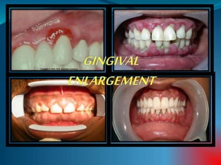
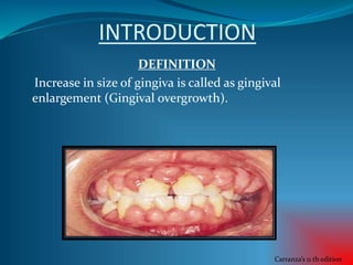
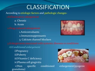
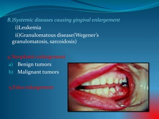
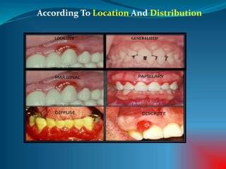
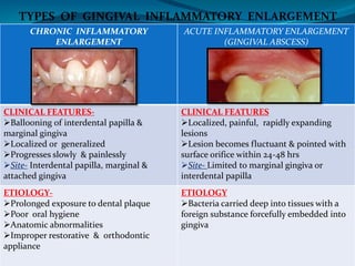
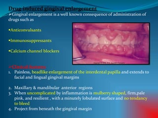
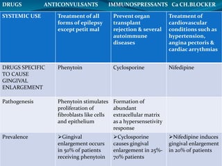
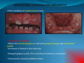
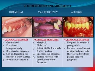
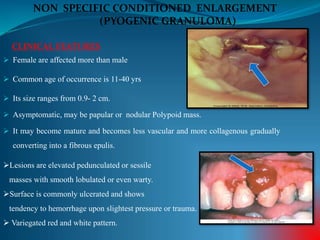
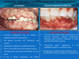
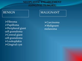
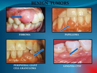
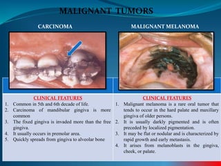
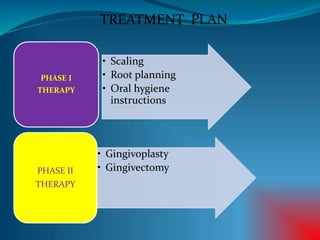
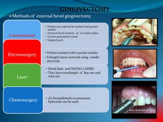
CONTENTS
 INTRODUCTION
-DEFINITION
-CLASSIFICATION
-GRADING
 INFLAMMATORY GINGIVAL
ENLARGEMENT
 DRUG-INDUCED
 IDIOPATHIC
 ENLARGEMENTS ASS. WITH
SYSTEMIC DISEASES
 NEOPLASTIC
 FALSE ENLARGEMENT
 TREATMENT
INTRODUCTION
DEFINITION
Increase in size of gingiva is called as gingival
enlargement (Gingival overgrowth).
Carranza’s 11 th edition
CLASSIFICATION
According to etiologic factors and pathologic changes-
1.Inflammatory enlargement
a. Chronic
b. Acute
2.Drug-induced enlargement
1.Anticonvulsants
2.Immunosuppressants
3. Calcium channel blockers
3.Enlargements associated with systemic diseases or conditions
A)Conditioned enlargement
i)Pregnancy
ii)Puberty
iii)Vitamin C deficiency
iv)Plasma cell gingivitis
v)Non specific conditioned enlargement(pyogenic
granuloma)
B.)Systemic diseases causing gingival enlargement
i)Leukemia
ii)Granulomatous disease(Wegener’s
granulomatosis, sarcoidosis)
4.Neoplastic enlargement
a) Benign tumors
b) Malignant tumors
5.False enlargement
According To Location And Distribution
LOCALIZED GENERALIZED
Degree Of Gingival Enlargement
Grade 0- No signs of gingival enlargement
Grade 1 - Enlargement confined to interdental papilla
Grade 2 - Enlargement involves papilla
and marginal gingiva
Grade 3 - Enlargement covers three quarters or
more of the crown
CHRONIC INFLAMMATORY
ENLARGEMENT
ACUTE INFLAMMATORY ENLARGEMENT
(GINGIVAL ABSCESS)
CLINICAL FEATURES-
Ballooning of interdental papilla &
marginal gingiva
Localized or generalized
Progresses slowly & painlessly
Site- Interdental papilla, marginal &
attached gingiva
CLINICAL FEATURES
Localized, painful, rapidly expanding
lesions
Lesion becomes fluctuant & pointed with
surface orifice within 24-48 hrs
Site- Limited to marginal gingiva or
interdental papilla
ETIOLOGY-
Prolonged exposure to dental plaque
Poor oral hygiene
Anatomic abnormalities
Improper restorative & orthodontic
appliance
ETIOLOGY
Bacteria carried deep into tissues with a
foreign substance forcefully embedded into
gingiva
TYPES OF GINGIVAL INFLAMMATORY ENLARGEMENT
Drug-induced gingival enlargement
Gingival enlargement is a well known consequence of administration of
drugs such as
Anticonvulsants
Immunosuppressants
Calcium channel blockers
Clinical features
1. Painless, beadlike enlargement of the interdental papilla and extends to
facial and lingual gingival margins
2. Maxillary & mandibular anterior regions
3. When uncomplicated by inflammation is mulberry shaped, firm,pale
pink, and resilient , with a minutely lobulated surface and no tendancy
to bleed
4. Project from beneath the gingival margin
SYSTEMIC USE Treatment of all
forms of epilepsy
except petit mal
Prevent organ
transplant
rejection & several
autoimmune
diseases
Treatment of
cardiovascular
conditions such as
hypertension,
angina pectoris &
cardiac arrythmias
DRUGS SPECIFIC
TO CAUSE
GINGIVAL
ENLARGEMENT
Phenytoin Cyclosporine Nifedipine
Pathogenesis Phenytoin stimulates
proliferation of
fibroblasts like cells
and epithelium
Formation of
abundant
extracellular matrix
as a hypersensetivity
response
Prevalence Gingival
enlargement occurs
in 50% of patients
receiving phenytoin
Cyclosporine
causes gingival
enlargement in 25%-
70% patients
Nifedipine induces
gingival enlargement
in 20% of patients
DRUGS ANTICONVULSANTS IMMUNOSPRESSANTS Ca CH.BLOCKER
IDIOPATHIC GINGIVAL ENLARGEMENT
Rare condition of undetermined cause.
CLINICAL FEATURES
-Affects the attached gingiva as well as the gingival margin and interdental
papilla
-Involment is limited to the either jaw
-Enlarged gingiva is pink, firm and almost leathery in consistency
-Characterstic minutely pebbled surface
ENLARGEMENTS ASSOCIATED WITH
SYSTEMIC DISEASES
Systemic diseases and conditions can affect the periodontium by two
different mechanisms-
1. Magnification of an existing inflammation initiated by dental
plaque(conditioned enlargement)
2. Manifestation of the systemic disease independently of the
inflammatory status of the gingiva.
CONDITIONED ENLARGEMENT
HORMONAL Vit.C DEFICIENCY ALLERGIC
CLINICAL FEATURES
1. Generalized
2. Prominent
interproximally
3. Bright red or magenta
4. Soft and friable & has
smooth & shiny surface
5. Bleeds spontaneously
CLINICAL FEATURES
1. Marginal
2. Bluish red
3. Soft & friable & smooth
& shiny surface
4. Spontaneous bleeding
5. Surface necrosis with
pseudomembrane
formation
CLINICAL FEATURES
1. Frequent in women &
young adults
2. Located on oral aspect
of attached gingiva &
therefore differs from
plaque induced
gingivitis.
 Female are affected more than male
 Common age of occurrence is 11-40 yrs
 Its size ranges from 0.9- 2 cm.
 Asymptomatic, may be papular or nodular Polypoid mass.
 It may become mature and becomes less vascular and more collagenous gradually
converting into a fibrous epulis.
CLINICAL FEATURES
NON SPECIFIC CONDITIONED ENLARGEMENT
(PYOGENIC GRANULOMA)
Lesions are elevated pedunculated or sessile
masses with smooth lobulated or even warty.
Surface is commonly ulcerated and shows
tendency to hemorrhage upon slightest pressure or trauma.
 Variegated red and white pattern.
SYSTEMIC DISEASES CAUSING GINGIVAL ENLARGEMENT
GRANULOMATOUS DISEASE
(WEGENER’S GRNULOMATOSIS)
CLINICAL FEATURES
1. Leukemic enlargement may be diffuse or
marginal localized or generalized.
2. The gingiva becomes soft, edematous and
swollen.
3. Appearance of gingiva is purplish and glossy.
4. There is also pallor in the surrounding mucosa.
5. Ulceration pain and severe hemorrhage can also
occur.
6. It has a spongy consistency and bleeds
CLINICAL FEATURES
1. It involve the orofacial region and include oral
mucosal ulceration, gingival enlargement,
abnormal tooth mobility, exfoliation of teeth,
and delayed healing response.
2. “Strawberry gums” appearance of the
mandibular gingiva is commonly seen.
3. Enlargement is reddish purple and bleeds easily
on stimulation but the condition is considered an
LEUKEMIA
NEOPLASTIC ENLARGEMENT
(GINGIVAL TUMORS)
BENIGN
Fibroma
Papilloma
Peripheral giant
cell granuloma
Central giant
cell granuloma
Leukoplakia
Gingival cyst
MALIGNANT
Carcinoma
Malignant
melanoma
BENIGN TUMORS
FIBROMA PAPILLOMA
PERIPHERAL GIANT
CELL GRANULOMA
GINGIVAL CYST
MALIGNANT TUMORS
CLINICAL FEATURES
1. Common in 5th and 6th decade of life.
2. Carcinoma of mandibular gingiva is more
common
3. The fixed gingiva is invaded more than the free
gingiva.
4. It usually occurs in premolar area.
5. Quickly spreads from gingiva to alveolar bone
CLINICAL FEATURES
1. Malignant melanoma is a rare oral tumor that
tends to occur in the hard palate and maxillary
gingiva of older persons.
2. It is usually darkly pigmented and is often
preceded by localized pigmentation.
3. It may be flat or nodular and is characterized by
rapid growth and early metastasis.
4. It arises from melanoblasts in the gingiva,
cheek, or palate.
CARCINOMA MALIGNANT MELANOMA
FALSE ENLARGEMENT
UNDERLYING OSSEOUS LESION
1. Enlargement of the bone subjacent to the
gingival area occur most often in tori and
exostoses.
2. It can also occur in paget’s disease,
fibrous dysplasia, cherubism, central
giant cell granuloma, ameloblastoma,
osteoma and osteosarcoma.
UNDERLYING DENTAL TISSUES
During various stages of eruption
particularly of the primary dentition, the
labial gingiva may show a bulbous
marginal distortion caused by
superimposition of the bulk of the
gingiva on the normal prominence of the
enamel in the gingival half of the crown.
• Scaling
• Root planning
• Oral hygiene
instructions
PHASE I
THERAPY
• Gingivoplasty
• GingivectomyPHASE II
THERAPY
TREATMENT PLAN
GINGIVECTOMY
• Pockets are explored & marked with pocket
marker
• External bevel incision- 45˚ to tooth surface
• Curette granulation tissue
• Surgical pack
Conventional
• Pockets marked with a pocket marker
• Enlarged tissue removed using needle
electrode.
Electrosurgery
• Diode laser and Nd:YAG LASERS
• They have wavelength of 890 nm and
1064 nm.Laser
Methods of external bevel gingivectomy
• 5% formaldehyde or potassium
hydroxide can be usedChemosurgery
Gingival enlargement

Gingival enlargement

  • 2.
    CONTENTS  INTRODUCTION -DEFINITION -CLASSIFICATION -GRADING  INFLAMMATORYGINGIVAL ENLARGEMENT  DRUG-INDUCED  IDIOPATHIC  ENLARGEMENTS ASS. WITH SYSTEMIC DISEASES  NEOPLASTIC  FALSE ENLARGEMENT  TREATMENT
  • 3.
    INTRODUCTION DEFINITION Increase in sizeof gingiva is called as gingival enlargement (Gingival overgrowth). Carranza’s 11 th edition
  • 4.
    CLASSIFICATION According to etiologicfactors and pathologic changes- 1.Inflammatory enlargement a. Chronic b. Acute 2.Drug-induced enlargement 1.Anticonvulsants 2.Immunosuppressants 3. Calcium channel blockers 3.Enlargements associated with systemic diseases or conditions A)Conditioned enlargement i)Pregnancy ii)Puberty iii)Vitamin C deficiency iv)Plasma cell gingivitis v)Non specific conditioned enlargement(pyogenic granuloma)
  • 5.
    B.)Systemic diseases causinggingival enlargement i)Leukemia ii)Granulomatous disease(Wegener’s granulomatosis, sarcoidosis) 4.Neoplastic enlargement a) Benign tumors b) Malignant tumors 5.False enlargement
  • 6.
    According To LocationAnd Distribution LOCALIZED GENERALIZED
  • 7.
    Degree Of GingivalEnlargement Grade 0- No signs of gingival enlargement Grade 1 - Enlargement confined to interdental papilla Grade 2 - Enlargement involves papilla and marginal gingiva Grade 3 - Enlargement covers three quarters or more of the crown
  • 8.
    CHRONIC INFLAMMATORY ENLARGEMENT ACUTE INFLAMMATORYENLARGEMENT (GINGIVAL ABSCESS) CLINICAL FEATURES- Ballooning of interdental papilla & marginal gingiva Localized or generalized Progresses slowly & painlessly Site- Interdental papilla, marginal & attached gingiva CLINICAL FEATURES Localized, painful, rapidly expanding lesions Lesion becomes fluctuant & pointed with surface orifice within 24-48 hrs Site- Limited to marginal gingiva or interdental papilla ETIOLOGY- Prolonged exposure to dental plaque Poor oral hygiene Anatomic abnormalities Improper restorative & orthodontic appliance ETIOLOGY Bacteria carried deep into tissues with a foreign substance forcefully embedded into gingiva TYPES OF GINGIVAL INFLAMMATORY ENLARGEMENT
  • 9.
    Drug-induced gingival enlargement Gingivalenlargement is a well known consequence of administration of drugs such as Anticonvulsants Immunosuppressants Calcium channel blockers Clinical features 1. Painless, beadlike enlargement of the interdental papilla and extends to facial and lingual gingival margins 2. Maxillary & mandibular anterior regions 3. When uncomplicated by inflammation is mulberry shaped, firm,pale pink, and resilient , with a minutely lobulated surface and no tendancy to bleed 4. Project from beneath the gingival margin
  • 10.
    SYSTEMIC USE Treatmentof all forms of epilepsy except petit mal Prevent organ transplant rejection & several autoimmune diseases Treatment of cardiovascular conditions such as hypertension, angina pectoris & cardiac arrythmias DRUGS SPECIFIC TO CAUSE GINGIVAL ENLARGEMENT Phenytoin Cyclosporine Nifedipine Pathogenesis Phenytoin stimulates proliferation of fibroblasts like cells and epithelium Formation of abundant extracellular matrix as a hypersensetivity response Prevalence Gingival enlargement occurs in 50% of patients receiving phenytoin Cyclosporine causes gingival enlargement in 25%- 70% patients Nifedipine induces gingival enlargement in 20% of patients DRUGS ANTICONVULSANTS IMMUNOSPRESSANTS Ca CH.BLOCKER
  • 11.
    IDIOPATHIC GINGIVAL ENLARGEMENT Rarecondition of undetermined cause. CLINICAL FEATURES -Affects the attached gingiva as well as the gingival margin and interdental papilla -Involment is limited to the either jaw -Enlarged gingiva is pink, firm and almost leathery in consistency -Characterstic minutely pebbled surface
  • 12.
    ENLARGEMENTS ASSOCIATED WITH SYSTEMICDISEASES Systemic diseases and conditions can affect the periodontium by two different mechanisms- 1. Magnification of an existing inflammation initiated by dental plaque(conditioned enlargement) 2. Manifestation of the systemic disease independently of the inflammatory status of the gingiva.
  • 13.
    CONDITIONED ENLARGEMENT HORMONAL Vit.CDEFICIENCY ALLERGIC CLINICAL FEATURES 1. Generalized 2. Prominent interproximally 3. Bright red or magenta 4. Soft and friable & has smooth & shiny surface 5. Bleeds spontaneously CLINICAL FEATURES 1. Marginal 2. Bluish red 3. Soft & friable & smooth & shiny surface 4. Spontaneous bleeding 5. Surface necrosis with pseudomembrane formation CLINICAL FEATURES 1. Frequent in women & young adults 2. Located on oral aspect of attached gingiva & therefore differs from plaque induced gingivitis.
  • 14.
     Female areaffected more than male  Common age of occurrence is 11-40 yrs  Its size ranges from 0.9- 2 cm.  Asymptomatic, may be papular or nodular Polypoid mass.  It may become mature and becomes less vascular and more collagenous gradually converting into a fibrous epulis. CLINICAL FEATURES NON SPECIFIC CONDITIONED ENLARGEMENT (PYOGENIC GRANULOMA) Lesions are elevated pedunculated or sessile masses with smooth lobulated or even warty. Surface is commonly ulcerated and shows tendency to hemorrhage upon slightest pressure or trauma.  Variegated red and white pattern.
  • 15.
    SYSTEMIC DISEASES CAUSINGGINGIVAL ENLARGEMENT GRANULOMATOUS DISEASE (WEGENER’S GRNULOMATOSIS) CLINICAL FEATURES 1. Leukemic enlargement may be diffuse or marginal localized or generalized. 2. The gingiva becomes soft, edematous and swollen. 3. Appearance of gingiva is purplish and glossy. 4. There is also pallor in the surrounding mucosa. 5. Ulceration pain and severe hemorrhage can also occur. 6. It has a spongy consistency and bleeds CLINICAL FEATURES 1. It involve the orofacial region and include oral mucosal ulceration, gingival enlargement, abnormal tooth mobility, exfoliation of teeth, and delayed healing response. 2. “Strawberry gums” appearance of the mandibular gingiva is commonly seen. 3. Enlargement is reddish purple and bleeds easily on stimulation but the condition is considered an LEUKEMIA
  • 16.
    NEOPLASTIC ENLARGEMENT (GINGIVAL TUMORS) BENIGN Fibroma Papilloma Peripheralgiant cell granuloma Central giant cell granuloma Leukoplakia Gingival cyst MALIGNANT Carcinoma Malignant melanoma
  • 17.
    BENIGN TUMORS FIBROMA PAPILLOMA PERIPHERALGIANT CELL GRANULOMA GINGIVAL CYST
  • 18.
    MALIGNANT TUMORS CLINICAL FEATURES 1.Common in 5th and 6th decade of life. 2. Carcinoma of mandibular gingiva is more common 3. The fixed gingiva is invaded more than the free gingiva. 4. It usually occurs in premolar area. 5. Quickly spreads from gingiva to alveolar bone CLINICAL FEATURES 1. Malignant melanoma is a rare oral tumor that tends to occur in the hard palate and maxillary gingiva of older persons. 2. It is usually darkly pigmented and is often preceded by localized pigmentation. 3. It may be flat or nodular and is characterized by rapid growth and early metastasis. 4. It arises from melanoblasts in the gingiva, cheek, or palate. CARCINOMA MALIGNANT MELANOMA
  • 19.
    FALSE ENLARGEMENT UNDERLYING OSSEOUSLESION 1. Enlargement of the bone subjacent to the gingival area occur most often in tori and exostoses. 2. It can also occur in paget’s disease, fibrous dysplasia, cherubism, central giant cell granuloma, ameloblastoma, osteoma and osteosarcoma. UNDERLYING DENTAL TISSUES During various stages of eruption particularly of the primary dentition, the labial gingiva may show a bulbous marginal distortion caused by superimposition of the bulk of the gingiva on the normal prominence of the enamel in the gingival half of the crown.
  • 21.
    • Scaling • Rootplanning • Oral hygiene instructions PHASE I THERAPY • Gingivoplasty • GingivectomyPHASE II THERAPY TREATMENT PLAN
  • 22.
    GINGIVECTOMY • Pockets areexplored & marked with pocket marker • External bevel incision- 45˚ to tooth surface • Curette granulation tissue • Surgical pack Conventional • Pockets marked with a pocket marker • Enlarged tissue removed using needle electrode. Electrosurgery • Diode laser and Nd:YAG LASERS • They have wavelength of 890 nm and 1064 nm.Laser Methods of external bevel gingivectomy • 5% formaldehyde or potassium hydroxide can be usedChemosurgery