2. Odontogenic infection
• Orofacial infections may be odontogenic or non
odontogenic in nature and the vast proportion of
odontogenic infections are caused by the endogenous
bacteria present in the oral cavity .
3. Odontogenic infection
• These infections may range from low-grade, well-
localized infections that require only minimal
treatment to severe life-threatening facial space
infections.
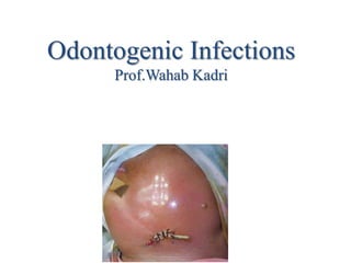
4. Odontogenic infection
• This imbalance, in turn, may lead to the
multiplication of micro-organisms followed by
invasion of different structures.
• The severity of infection is related to the number and
virulence of micro-organisms and resistance of the
host
5.
6. Microbiology
Odontogenic infections are multimicrobial:
• Gram (+) cocci, aerobic and anaerobic:
– Streptococci and their anaerobic counterpart,
peptostreptococci
– Staphylococci, and their anaerobic counterpart,
peptococci
• Gram (+) rods:
– Lactobacillus, diphtheroids, Actinomyces
• Gram (-) rods:
– Fusobacterium, Bacteroids, Eikenella, Psuedomonas
(occasional)
7. Odontogenic infection
• Odontogenic infections progress through 3
stages:
• Inoculation
• Cellulitis
• Abscess
• Sinus tract/fistula may be seen in neglected
cases
8. Inoculation
• Characterized by the entry of pathogenic
microbes into the body without disease
occurring.
• An infection involves the proliferation of
microbes resulting in triggering of the defense
mechanism, a process manifesting as
inflammation
9. Odontogenic infection
• Inflammation
• Inflammation is the series of changes which occurred
in the living tissue in response to an irritant. The
manifestation of inflammation is typical and is
characterized by: rubor (redness), calor (hotness),
tumor (swelling or edema), dolor (pain), and functio
laesa (loss of function). This reaction is protective
and aims at limiting or eliminating the irritant.
• Depending on the duration and severity,
inflammation is distinguished as acute, subacute or
chronic
10. Odontogenic infection
• Cellulitis
Is an acute diffuse painful indurated swelling of the
soft tissues resulting from a diffuse spreading of
purulent exudate along the fascial planes with or
without suppuration.
• Abscess
A collection of pus in a cavity formed by disintegration of
tissue as result of infection.
11. Abscess vs. Cellulitis
Abscess:
• Chronic
• Well-localized
• Fluid filled
(fluctuant)
• Amenable to
drainage and
removal of the
offending tooth
• Rapid improvement
Cellulitis
• Acute
• Diffuse, not well localized
• No pus or very little
pus
• Amenable to removal of
the offending tooth and
antibiotics
• Slower improvement
12.
13. Odontogenic infection
• Discharging Sinus
Some times abscess ruptures to produce a
draining sinus tract. Usually, infection recur
when the site of drainage closes. Sinus is thus a
one side tract of a single compartment
•
14. Odontogenic infection
• Fistulae
• A drainage pathway or abnormal communication
between two epithelium-lined surfaces due to
destruction of the intervening tissue. Fistula is thus an
epithelialized tract opening in both side of two
different compartments.
15. Acute dentoalvealar abscess
• The usual cause of odontogenic infections is necrosis of
dental pulp, which is followed by bacterial invasion
through the pulp chamber and into the deeper tissues.
• Necrosis of the pulp is the result of deep caries of a
tooth, to which the pulp responds with a typical
inflammatory reaction. Vasodilatation and edema cause
pressure in the tooth and severe pain as the rigid walls
of the tooth prevent swelling.
• If left untreated the pressure leads to strangulation of
the blood supply to the tooth through the apex and
consequent necrosis.
16. Acute dentoalvealar abscess
• The necrotic pulp then provides a perfect
setting for bacterial invasion into the bone
tissue. Pus is formed in the cancellous bone,
and spreads in various directions by way of the
tissues presenting the least resistance until a
cortical plate is encountered.
17. Acute dentoalvealar abscess
• Clinically, the condition has rapid onset.
Radiographically, changes in bone density may
not be noticeable (you have to wait for
approximately 10 days to detect bone
rarefaction). It is characterized by symptoms
that are classified as
• local and
• systemic
18. Local Symptoms
• Pain
The severity of the pain depends on the
degree of inflammation. Initially, the pain is dull
and continuous and worsens during percussion of
the responsible tooth or when it comes into
contact with antagonist teeth. There is a sense of
elongation of the responsible tooth and slight
mobility.
19. Acute dentoalveolar abscess
Local Symptoms
• Edema appears intraorally or extraorally and it
usually has a buccal and more rarely palatal or
lingual localization.
• This swelling presents before suppuration,
particularly in areas with loose tissue, such as the
sublingual region, lips, or eyelids. Usually the
edema is soft with redness of the skin.
• During the final stages, the swelling fluctuates,
especially at the mucosa of the oral cavity.
• This stage is considered the most suitable for
incision and drainage of the abscess.
20. Acute dentoalvealar abscess
• Systemic Symptoms
The systemic symptoms usually observed are: fever,
chills, malaise with pain in muscles and joints, insomnia,
nausea, and vomiting. Laboratory tests usually show
leukocytosis, an increased erythrocyte sedimentation rate,
and a raised C-reactive protein (CRP) level.
• Treatment
Extraction of the tooth (or removal of the necrotic pulp
by an endodontic procedure) results in resolution of the
infection.
21. Spread of odontogenic infection
Routes of Spread of Odontogenic Infection:
a. By direct continuity via the tissue
b. Via the lymphatics into the regional lymph nodes and
subsequently into the blood stream
c. Haematogenous spread leading to thrombophlebitis,
bacteremia or septicemia. Thrombus may propagate along
the veins, entering the cranial cavity via emissary veins to
produce cavernous sinus thrombosis.
22. Direct spread
• Whether the pus spreads buccally, palatally or
lingually depends mainly on the position of the
tooth in the dental arch, the thickness of the
bone, and the distance it must travel.
23. Direct spread
• The length of the root and the relationship
between the apex and the proximal and distal
attachments of various muscles also play a
significant role in the spread of pus.
24. Vestibular space
•Boundary
–Superior : buccinator muscle attachment at zygomatic
process
–Inferior : oral mucosa at upper vestibule
–Medial : lateral cortex of the maxilla
–Lateral : buccinator muscle
•Signs and symptom
–Swelling and shallow labial or buccal vestibule.
–Swelling of the cheek and lip commissure.
•Spreading
–Buccal and canine spaces; superiorly.
–Cavernous sinus; via facial, angular, ophthalmic veins.
25.
26. Fascial space infection
• Sometimes, infection may spreads towards the
fascial spaces, forming serious abscesses called
fascial space infection.
• The fascial spaces are potential areas and do not
exist in healthy individuals. Bone, muscle, fascia,
neurovascular bundles, and skin can all act as
barriers to the spread of infection.
• It should be remembered however, that no tissue
barrier or boundary is so restrictive to universally
prevent spread of infection into contiguous
anatomical spaces.
27. Classification of Fascial Spaces
• Based on mode of involvement-
Primary spaces.
Secondary spaces.
Primary maxillary- canine, buccal, infratemporal.
Primary mandibular- submental, sublingual, buccal,
submandibular.
Secondary spaces- masseteric, pterygomandibular,
superficial & deep temporal, lateral pharyngeal,
retropharyngeal, parotid, prevertebral.
28. • Based on clinical significance-
Face- Buccal, canine, parotid, masticatory.
Suprahyoid- Sublingual, submental, submandibular,
lateral pharyngeal, peritonsillar.
Infrahyoid- Pretracheal.
Spaces of total neck- Retropharyngeal, space of
carotid sheath.
35. Etiology-
Infected mandibular & maxillary premolars &
molars.
Clinical Features-
Obliteration of nasolabial fold.
Angle of mouth shifted to opposite side.
Swelling in cheek extending to corner of
mouth.
Buccal space associated with temporal space –
Dumb bell shaped appearance due to lack of
swelling over zygomatic arch.
37. Infratemporal Space
Boundaries-
Superiorly: infratemporal surface of
greater wing of sphenoid.
Inferiorly: lateral pterygoid muscle.
Laterally: temporalis tendon &
coronoid process.
Medially: lateral pterygoid plate &
lateral pharyngeal wall.
Posteriorly: condyle & lateral
pterygoid muscles.
Anteriorly: infratemporal surface of
maxilla & posterior surface of
zygomatic bone.
Infratemporal
space
38. Etiology-
Infected maxillary 3rd
molars.
Infected needles or
contaminated LA
solution.
Clinical Features-
Extra-oral swelling over
sigmoid notch area.
Intra-oral swelling in
tuberosity area.
Trismus.
39. Contents-
Pterygoid plexus of veins.
Internal maxillary artery.
Mandibular nerve & its branches.
Spread of Infection-
To temporal space.
Cavernous sinus thrombosis- infection spreads via pterygoid
plexus of veins.
40. Submental Space
Boundaries-
Roof: mylohyoid muscle.
Inferior: deep cervical fascia, platysma, superficial fascia & skin.
Laterally: anterior belly of digastric.
Posteriorly: submandibular space.
Contents-
Lymph nodes, anterior jugular vein.
Etiology-
Infected mandibular incisors.
Anterior extension of submandibular space.
Clinical Features-
• Chin appears glossy & swollen.
• Pain & discomfort on swallowing.
41.
42. Sublingual Space
Boundaries-
Superiorly: mucosa of floor of mouth.
Inferior: mylohyoid muscle.
Posteriorly: body of hyoid bone.
Anteriorly & laterally: inner aspect of mandibular body.
Medially: geniohyoid,styloglossus,genioglossus muscle.
Contents-
Deep part of Submandibular gland.
Wharton’s duct.
Sublingual gland.
Lingual & hypoglossal nerves.
Terminal branches of lingual artery.
43. Etiology-
Infected mandibular premolar & 1st molar.
Clinical Features-
Swelling of floor of mouth.
Elevated tongue.
Pain & discomfort on swallowing.
44. Submandibular Space
Boundaries-
Superiorly: mylohyoid muscle, inferior border of mandible.
Inferior: anterior & posterior belly of digastric.
Laterally: deep cervical fascia, platysma, superficial fascia & skin.
Medially: hyoglossus,styloglossus,mylohyoid muscle.
Posteriorly: to hyoid bone.
Anteriorly: submental space.
Contents-
Submandibular salivary gland.
Proximal portion of Wharton’s duct.
Lingual & hypoglossal nerves.
Branches of facial artery- palatine,tonsillar,glandular,submental.
45. Etiology-
Infected mandibular 2nd & 3rd molars.
From submental,sublingual spaces.
Clinical Features-
• Indurated swelling in submandibular region.
• Usually bulges over lower border of mandible.
Spread of Infection-
Across midline to contralateral space.
To contiguous pharyngeal spaces.
46.
47. Pterygomandibular Space
Boundaries-
Superiorly: lower head of lateral pterygoid muscle.
Laterally: medial surface of ramus.
Medially: medial pterygoid muscle.
Posteriorly: deep part of parotid.
Anteriorly: pterygomandibular raphe.
Contents-
Inferior alveolar neurovascular bundle.
Lingual & auriculotemporal nerves.
Mylohyoid nerve & vessels.
Pterygomandibular
space
48. Etiology-
Infected mandibular 3rd molars(mesioangular/horizontal)
Pericoronitis.
Infected needles or contaminated LA solution.
Clinical Features-
Absence of extra-oral swelling.
Severe trismus.
Difficulty in swallowing.
Anterior bulging of half of soft palate & tonsillar pillars with
deviation of uvula to unaffected side.
Spread of Infection-
Superiorly to infratemporal space.
Medially to lateral pharyngeal space.
To submandibular space.
49.
50. Masseteric Space
Boundaries-
Superiorly: zygomatic arch.
Inferiorly: inferior border of mandible.
Laterally: masseter muscle.
Medially: ramus of mandible.
Posteriorly: parotid gland & its fascia.
Anteriorly: buccal space & buccopharyngeal fascia.
Contents-
Masseteric artery & vein.
Etiology-
Mandibular 3rd molars(pericoronitis).
54. Lateral Pharyngeal Space
Boundaries-
Shape of an inverted cone or pyramid, the base is at sphenoid
bone and the apex at hyoid bone.
Anteriorly: pterygomandibular raphe.
Posteriorly: extends to prevertebral fascia.
Laterally: fascia covering medial pterygoid muscle, parotid &
mandible.
Medially: buccopharyngeal fascia on lateral surface of superior
constrictor muscle.
Styloid process divides the space into anterior muscular and
posterior vascular compartment.
55.
56. Etiology-
Infected mandibular 3rd molars.
Tonsillar infections.
Pharyngitis.
Parotitis.
Spread of Infection-
To retropharyngeal space.
To peritonsillar space.
57. Clinical Features-
Trismus.
Induration & swelling at angle of jaw.
Fever.
Pharyngeal bulging
Posterior tonsillar pillar deviation.
Neurological involvement.
Thrombosis of internal jugular vein.
Erosion of carotid vessels may occur.
58. Retropharyngeal Space
Posteromedial to lateral pharyngeal space and anterior to the
prevertebral space .
Boundaries-
Anterior: posterior pharyngeal wall.
Posterior: prevertebral fascia.
Superior: skull base.
Inferior: mediastinum.
Laterally: lateral pharyngeal space.
Etiology-
Nasal & pharygeal infections.
Spread from odontogenic infections.
59.
60. Clinical Features-
Stiffness of neck.
Dysponea.
Dysphagia.
Bulging of posterior pharyngeal wall.
Complications-
Airway obstruction.
Aspiration pneumonia.
Acute mediastinitis.
Can spread to Danger space.
61. Prevertebral Space
Potential space between two layers of prevertebral
fascia (alar and prevertebral layers).
Extends from skull base superiorly to the diaphragm
inferiorly.
Mediastinitis is concern with prevertebral space
infections similarly to retropharyngeal space
infections.
62.
63. Fascial space infection
• Facial spaces have been classified as either primary or
secondary spaces infection
• Primary maxillary spaces
Canine
Buccal
Infratemporal
• Primary mandibular spaces
Submental
Buccal
Submandibular
Sublingual
66. Palate
• The palate is usually involved in infections
originating from the maxillary lateral incisor or
the palatal roots of the posterior teeth. The
infection spreads from the apices of these
teeth, perforating the palatal alveolar bone, and
pus accumulates below the palatal
mucoperiosteum.
68. PRINCIPLES OF THERAPY OF
ODONTOGENIC INFECTIONS
I : Determine the severity of the infection.
–Complete history : chief complaint, onset,
duration, rapidity, previous treatment.
–Physical examination : vital signs, signs of
infection, characteristic of the swelling (soft,
doughy, indurated, fluctuant)
–Radiographic examination : intraoral or/and
extraoral film.
–Source of infection; specific tooth.
–Determine the cellulitis or abscess.
69. Signs and symptoms of infection :
•Pain and tenderness
•Swelling : cellulitis or abscess
•Redness of the covering mucosa or skin
•Increased temperature
•Trismus : masticatory muscle involvement
•Fever : phagocytic activity
70.
71. PRINCIPLES OF THERAPY OF ODONTOGENIC
INFECTIONSII :
Evaluate the state of the patient’s host defense
mechanisms.
1.Host defense mechanisms
–Local defenses
•Intact anatomic barrier
•Indigenous bacteria
–Humoral defenses
•Immunoglobulins
•Complement
–Cellular defenses
•Phagocytes : granulocytes, monocytes
•Lymphocytes
72. 2. Medical conditions that compromise host
defenses.
–Uncontrolled metabolic diseases
•Uremia, alcoholism, malnutrition, severe diabetes
–Suppressing diseases
•Leukemia, lymphoma, malignant tumors
–Suppressing drugs
•Cancer chemotherapeutics agents,
immunosuppressives
74. 2. Histopathologic examination
–Granulomatous infection : TB, Actinomycosis,
Syphilis, Fungus
3. Microbiological examination and testing
1.Gram stain : positive or negative, shape (cocci or
bacilli or spirochete), chain or cluster
2.Culture and sensitivity test : aerobe or anaerobe,
specific microorganism and antibiotic sensitivity
76. PRINCIPLES OF THERAPY OF
ODONTOGENIC INFECTIONS
III : Determine whether the patient should be treated by
a general dentist Criteria for referral to a specialist
1. Rapidly progressive infection*
2. Difficulty in breathing*
3. Difficulty in swallowing*
4. Fascial space involvement
5. Elevated temperature (>101 F)
6. Severe jaw trismus (<10 mm)
7. Toxic appearance
8. Compromised host defenses
ral practitioner or a specialist.
77. PRINCIPLES OF THERAPY OF
ODONTOGENIC INFECTIONSIV
Treat the infection surgically.
•Surgical drainage (primary method)
•Removal of the cause of the infection
•Obtaining a specimen of the pus for culture
and sensitivity test
78.
79.
80.
81.
82.
83.
84.
85.
86.
87.
88.
89.
90.
91.
92.
93.
94.
95.
96.
97.
98. Criteria for hospitalization
1. Rapidly progressive cellulitis
2. Dyspnea (shortness of breath or difficult breathing)
3. Dysphagia (difficulty in swallowing)
4. Spread to deep facial spaces
5. Fever of more than 38º C
6. Intense trismus ( inter-incisal distance less than 10 mm)
7. Failure of initial treatment
8. Severe involvement of general health status
9. Immunocompromised patients (diabetes, alcoholism or
drug addiction, malnutrition, treatment with
corticoids,….)
99. Ludwig's Angina
• Ludwig's Angina is a massive indurated
brawny cellulites, occurs bilaterally in the
submandibular, sublingual & submental
spaces. Infection is propagated by lymphatic
spread or directly through submandibular
space.
• Cellulitis may then rapidly spread to involve
bilaterally the parapharyngeal and pterygoid
spaces
100.
101.
102. Causes
• The cause is usually an infection with
Streptococcal bacteria, although other
bacteria can cause the condition. Since the
advent of antibiotics, Ludwig's angina has
become a rare disease.
• The route of infection in most cases is from
infected lower third molars or from
pericoronitis, Although the widespread
involvement seen in Ludwig's is usually
develops in immunocompromised persons, it
can also develop in otherwise healthy
individuals.
103. Ludwig's Angina
• Clinically, the condition is characterized by:
1. Painful bilateral swelling of floor of mouth and elevation of
tongue.
2. Bilateral firm, brawny painful, diffuse swelling of upper
part of neck
3. Difficulty in swallowing and breathing
4. Rapid pulse, high fever, fast respiration
5. Leucocytosis
Patient should be hospitalized. Conservative treatment
includes intravenous antibiotic therapy and close airway
observation . Pus is evacuated, when indicated, by through &
through drainage
(8)
104. Management
• Secure Airway
– Naso-tracheal intubation
– tracheostomy
• Incision & Drainage at multiple sites to
improve drainage
• Antibiotics
• Supportive therapy
• Check for immune status of the patient.
105.
106. Cavernous sinus thrombosis
Infections may spread via hematogenous route to
the cavernous sinus occurs from:
1- Anteriorly: a) Superior labial venous plexus to
b) Anterior facial vein, then via c) Superior or
inferior ophthalmic vein into the cavernous sinus
2- Posteriorly: from retromandibular vein to the
ptrygo- mandibular venous plexus, the emissary
vein passing through foramen ovale, spinosum, to
cavernous sinus
3- Superior petrosal sinus (inside the ear)
108. Dangerous triangle of the face
• Never squeeze infection boil in the dangerous
area
109. Osteomylitis
• Osteomylitis is defined as an inflammation of the bone
marrow with a tendency to progression to involve
adjacent cortical plates and often periosteal tissues.
• The incidence of osteomyelitis is much higher in the
mandible due to the dense cortical bone that prevents
the penetration of periosteal blood supply, and the
inferior alveolar artery is the only supply to the
mandible.
• It is much less common in the maxilla due to the
excellent blood supply from number of different
arteries. In addition the maxillary bone is much less
dense than the mandible
110. Classification of Osteomylitis
1- Acute suppurative
2- Subacute
3- Chronic suppurative
4- Rarely, a sclerotic nonpurulent form of osteomylitis
occurs; this is termed Garrès sclerosing osteomylitis.
Other related disorders are chronic recurrent multifocal
osteomylitis; tuberculous osteomylitis
Acute and chronic osteomylitis is distinguished by the
development of dead bone sequestra. Sequestra is an
island of dead bone that have not been resorbed
111. Radiographic Features
• The appearance of “moth-eaten” bone or
sequestrum of bone, is the classic feature
of chronic osteomylitis
112. Surgical Options
• Classic treatment is sequestrectomy and
saucerization. The aim is to débride the necrotic
bony sequestra in the infected area and improves
blood flow
• Decortication involves removal of the dense, often
chronically infected and poorly vascularized bony
cortex till reaching good bleeding bone, and
placement of the vascular periosteum adjacent to the
medullary bone to allow increased blood flow and
healing in the affected area