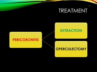
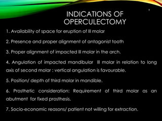
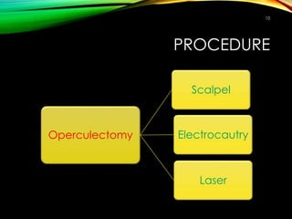
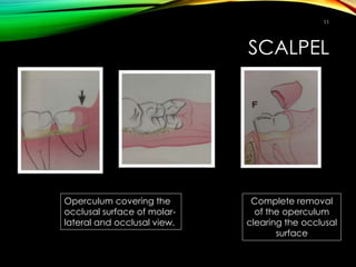
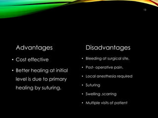
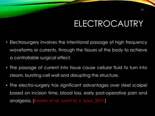
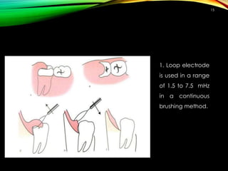
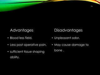
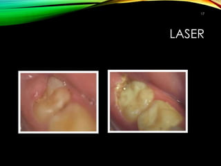
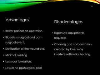
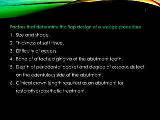
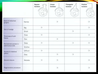
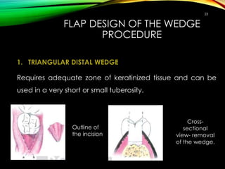
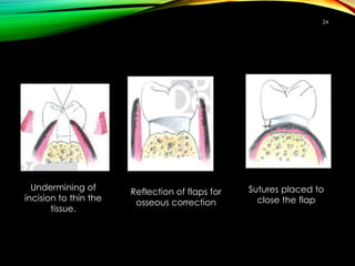
PERICORONITIS
Achi joshi
SAIMS, Indore
1
PERICORONITIS
• Pericoronitis is defined as
inflammation of the oral soft tissues
surrounding the crown of a
partially erupted tooth.
• The major cause is the microbial
flora that develops in the distally
located pseudopocket.
• The term pericoronitis was first
introduced to dental literature by
Bloch in 1921.
2
CLINICAL FEATURES
• Red, swollen, suppurating lesion that is exquisitely tender with
radiating pain to ear, throat and floor of mouth.
The diagnosis of pericoronitis is mainly clinical with three distinct
diagnostic categories recognised:
1) Acute pericoronitis,
2) Sub-acute pericoronitis, and
3) Chronic pericoronitis.
These classifications are empirically derived based on how
individual cases arbitrarily fall into the three distinct clinical
categories.
3
1. Acute Pericoronitis:
Trismus, pain, dysphagia, extraoral swelling, malaise, halitosis, pus
discharge, sore throat, and anorexia. Pain may disturb sleep,
lymphadenitis involving the deep cervical lymph nodes may be
present.
4
5
2. Subacute Pericoronitis:
Pain, dysphagia, intraoral swelling, halitosis, pus discharge, sore
throat. Associated pain is most often described as continuous,
dull, and is occasionally sharp or throbbing. Unlike acute attacks,
radiation of painful symptoms into adjacent muscles is rare. The
individual does not have limited mouth opening. This is a
distinguishing feature from acute pericoronitis (Nigerian Journal of Clinical Practice, 2014 )
3. Chronic pericoronitis:
It is diagnosed based on a history of temporary dull aching low
grade pain that typically lasts only 1-2 days. Signs include
palpable non-tender submandibular lymph nodes and
macerated buccal tissue consistent with cheek biting.
6
COMPLICATIONS
1. Pericoronal abscess
2. It may spreads posteriorly in orophrengeal area.
3. Dysphagia
4. Involvement of lymph nodes- posterior and deep
cervical.
5. Peritonsillar abscess.
6. Ludwigs angina
7
TREATMENT
8
PERICORONITIS
EXTRACTION
OPERCULECTOMY
INDICATIONS OF
OPERCULECTOMY
1. Availability of space for eruption of lll molar
2. Presence and proper alignment of antagonist tooth
3. Proper alignment of impacted lll molar in the arch.
4. Angulation of impacted mandibular lll molar in relation to long
axis of second molar : vertical angulation is favourable.
5. Position/ depth of third molar in mandible.
6. Prosthetic consideration: Requirement of third molar as an
abutment for fixed prosthesis.
7. Socio-economic reasons/ patient not willing for extraction.
9
PROCEDURE
Operculectomy
Scalpel
Laser
Electrocautry
10
SCALPEL
11
Operculum covering the
occlusal surface of molar-
lateral and occlusal view.
Complete removal
of the operculum
clearing the occlusal
surface
12
•Incisions distal to molar should
follow the area with greatest
amount of attached gingiva.
•It may be directed disto-
lingually or disto-facially
Advantages
• Cost effective
• Better healing at initial
level is due to primary
healing by suturing.
Disadvantages
• Bleeding at surgical site.
• Post- operative pain.
• Local anesthesia required
• Suturing
• Swelling ,scarring
• Multiple visits of patient
13
ELECTROCAUTRY
14
• Electrosurgery involves the intentional passage of high frequency
waveforms or currents, through the tissues of the body to achieve
a controllable surgical effect.
• The passage of current into tissue cause cellular fluid to turn into
steam, bursting cell wall and disrupting the structure.
• The electro-surgery has significant advantages over steel scalpel
based on incision time, blood loss, early post-operative pain and
analgesia. (Kearns et al, sumit M, k kaur, 2011)
15
1. Loop electrode
is used in a range
of 1.5 to 7.5 mHz
in a continuous
brushing method.
Advantages
• Blood less field.
• Less post operative pain.
• sufficient tissue shaping
ability.
Disadvantages
• Unpleasant odor.
• May cause damage to
bone .
16
LASER
17
Advantages
• Better patient co-operation.
• Bloodless surgical and post-
surgical event;
• Sterilization of the wound site.
• Minimal swelling
• Less scar formation.
• Less or no postsurgical pain
Disadvantages
• Expensive equipments
required.
• Charring and carbonization
created by laser may
interfere with initial healing.
18
DISTAL MOLAR SURGERY
• Treatment of periodontal pockets on the distal surface
of terminal molars is often complicated by the
presence of bulbous fibrous tissue over the maxillary
tuberosity or prominent retromolar pads in the
mandible.
•Operations for this purpose were described by Robinson
in 1966
19
• The procedure allows treatment of irregular osseous
defects and access to maxillary distal furcation area.
OBJECTIVES :
• Eliminate periodontal pocket.
• Maintain and preserve attached gingiva.
• Make area accessible for instrumentation.
20
Factors that determine the flap design of a wedge procedure
1. Size and shape.
2. Thickness of soft tissue.
3. Difficulty of access.
4. Band of attached gingiva of the abutment tooth.
5. Depth of periodontal pocket and degree of osseous defect
on the edentulous side of the abutment.
6. Clinical crown length required as an abutment for
restorative/prosthetic treatment.
21
22
FLAP DESIGN OF THE WEDGE
PROCEDURE
1. TRIANGULAR DISTAL WEDGE
Requires adequate zone of keratinized tissue and can be
used in a very short or small tuberosity.
23
Outline of
the incision
Cross-
sectional
view- removal
of the wedge.
24
Undermining of
incision to thin the
tissue.
Reflection of flaps for
osseous correction
Sutures placed to
close the flap
SQUARE , PARALLEL DISTAL WEDGE
• Indicated when tuberosity is longer.
• Allows conservation of keratinized tissue
• Provides greater access to tissues.
25
Cross-
sectional
view- proper
blade
angulations.
Outline of
the incision
26
Flap reflection and tissue
is removed
osseous
correction
Sutures placed to
close the flap
REFERENCES
1. Carranza. Clinical periodontology. 10th edition.
2. Edward cohen. Atlas of cosmetic and reconstructive periodontal surgery.
87-102.
3. N. Sato. Atlas of periodontal surgery.
4. Sumit Malhotra, Kamaljeet Kaur. Electro-surgery versus Conventional
Surgery for Excision of Pericoronal flaps. Indian J Stomatol 2012;3(4):236-40.
27
28
THANK YOU

Pericoronitis

  • 1.
  • 2.
    PERICORONITIS • Pericoronitis isdefined as inflammation of the oral soft tissues surrounding the crown of a partially erupted tooth. • The major cause is the microbial flora that develops in the distally located pseudopocket. • The term pericoronitis was first introduced to dental literature by Bloch in 1921. 2
  • 3.
    CLINICAL FEATURES • Red,swollen, suppurating lesion that is exquisitely tender with radiating pain to ear, throat and floor of mouth. The diagnosis of pericoronitis is mainly clinical with three distinct diagnostic categories recognised: 1) Acute pericoronitis, 2) Sub-acute pericoronitis, and 3) Chronic pericoronitis. These classifications are empirically derived based on how individual cases arbitrarily fall into the three distinct clinical categories. 3
  • 4.
    1. Acute Pericoronitis: Trismus,pain, dysphagia, extraoral swelling, malaise, halitosis, pus discharge, sore throat, and anorexia. Pain may disturb sleep, lymphadenitis involving the deep cervical lymph nodes may be present. 4
  • 5.
    5 2. Subacute Pericoronitis: Pain,dysphagia, intraoral swelling, halitosis, pus discharge, sore throat. Associated pain is most often described as continuous, dull, and is occasionally sharp or throbbing. Unlike acute attacks, radiation of painful symptoms into adjacent muscles is rare. The individual does not have limited mouth opening. This is a distinguishing feature from acute pericoronitis (Nigerian Journal of Clinical Practice, 2014 )
  • 6.
    3. Chronic pericoronitis: Itis diagnosed based on a history of temporary dull aching low grade pain that typically lasts only 1-2 days. Signs include palpable non-tender submandibular lymph nodes and macerated buccal tissue consistent with cheek biting. 6
  • 7.
    COMPLICATIONS 1. Pericoronal abscess 2.It may spreads posteriorly in orophrengeal area. 3. Dysphagia 4. Involvement of lymph nodes- posterior and deep cervical. 5. Peritonsillar abscess. 6. Ludwigs angina 7
  • 8.
  • 9.
    INDICATIONS OF OPERCULECTOMY 1. Availabilityof space for eruption of lll molar 2. Presence and proper alignment of antagonist tooth 3. Proper alignment of impacted lll molar in the arch. 4. Angulation of impacted mandibular lll molar in relation to long axis of second molar : vertical angulation is favourable. 5. Position/ depth of third molar in mandible. 6. Prosthetic consideration: Requirement of third molar as an abutment for fixed prosthesis. 7. Socio-economic reasons/ patient not willing for extraction. 9
  • 10.
  • 11.
    SCALPEL 11 Operculum covering the occlusalsurface of molar- lateral and occlusal view. Complete removal of the operculum clearing the occlusal surface
  • 12.
    12 •Incisions distal tomolar should follow the area with greatest amount of attached gingiva. •It may be directed disto- lingually or disto-facially
  • 13.
    Advantages • Cost effective •Better healing at initial level is due to primary healing by suturing. Disadvantages • Bleeding at surgical site. • Post- operative pain. • Local anesthesia required • Suturing • Swelling ,scarring • Multiple visits of patient 13
  • 14.
    ELECTROCAUTRY 14 • Electrosurgery involvesthe intentional passage of high frequency waveforms or currents, through the tissues of the body to achieve a controllable surgical effect. • The passage of current into tissue cause cellular fluid to turn into steam, bursting cell wall and disrupting the structure. • The electro-surgery has significant advantages over steel scalpel based on incision time, blood loss, early post-operative pain and analgesia. (Kearns et al, sumit M, k kaur, 2011)
  • 15.
    15 1. Loop electrode isused in a range of 1.5 to 7.5 mHz in a continuous brushing method.
  • 16.
    Advantages • Blood lessfield. • Less post operative pain. • sufficient tissue shaping ability. Disadvantages • Unpleasant odor. • May cause damage to bone . 16
  • 17.
  • 18.
    Advantages • Better patientco-operation. • Bloodless surgical and post- surgical event; • Sterilization of the wound site. • Minimal swelling • Less scar formation. • Less or no postsurgical pain Disadvantages • Expensive equipments required. • Charring and carbonization created by laser may interfere with initial healing. 18
  • 19.
    DISTAL MOLAR SURGERY •Treatment of periodontal pockets on the distal surface of terminal molars is often complicated by the presence of bulbous fibrous tissue over the maxillary tuberosity or prominent retromolar pads in the mandible. •Operations for this purpose were described by Robinson in 1966 19
  • 20.
    • The procedureallows treatment of irregular osseous defects and access to maxillary distal furcation area. OBJECTIVES : • Eliminate periodontal pocket. • Maintain and preserve attached gingiva. • Make area accessible for instrumentation. 20
  • 21.
    Factors that determinethe flap design of a wedge procedure 1. Size and shape. 2. Thickness of soft tissue. 3. Difficulty of access. 4. Band of attached gingiva of the abutment tooth. 5. Depth of periodontal pocket and degree of osseous defect on the edentulous side of the abutment. 6. Clinical crown length required as an abutment for restorative/prosthetic treatment. 21
  • 22.
  • 23.
    FLAP DESIGN OFTHE WEDGE PROCEDURE 1. TRIANGULAR DISTAL WEDGE Requires adequate zone of keratinized tissue and can be used in a very short or small tuberosity. 23 Outline of the incision Cross- sectional view- removal of the wedge.
  • 24.
    24 Undermining of incision tothin the tissue. Reflection of flaps for osseous correction Sutures placed to close the flap
  • 25.
    SQUARE , PARALLELDISTAL WEDGE • Indicated when tuberosity is longer. • Allows conservation of keratinized tissue • Provides greater access to tissues. 25 Cross- sectional view- proper blade angulations. Outline of the incision
  • 26.
    26 Flap reflection andtissue is removed osseous correction Sutures placed to close the flap
  • 27.
    REFERENCES 1. Carranza. Clinicalperiodontology. 10th edition. 2. Edward cohen. Atlas of cosmetic and reconstructive periodontal surgery. 87-102. 3. N. Sato. Atlas of periodontal surgery. 4. Sumit Malhotra, Kamaljeet Kaur. Electro-surgery versus Conventional Surgery for Excision of Pericoronal flaps. Indian J Stomatol 2012;3(4):236-40. 27
  • 28.