
More Related Content
Similar to Одонтогенные инфекции анг.pptx
Similar to Одонтогенные инфекции анг.pptx (20)
More from ssuser8923c6
Recently uploaded
Recently uploaded (20)
Одонтогенные инфекции анг.pptx
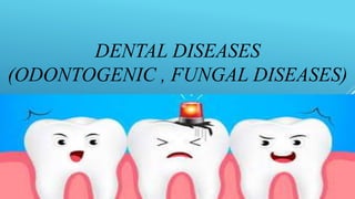
- 1. DENTAL DISEASES (ODONTOGENIC , FUNGAL DISEASES)
- 2. DENTAL DISEASES The nature disease damaged tissue disease Non-inflammatory odontogenic infections hard tissues of the tooth Caries Inflammatory odontogenic infections dental pulp Periodontium Periosteum bone tissue Soft tissues of the face and neck Maxillary sinus lymph nodes Generalized infection Pulpitis Periodontitis Periostitis Osteomyelitis Abscess, phlegmon Sinusitis Lymphadenitis Sepsis Inflammatory periodontal infections Periodontist gum tissue Periodontopathies Gingivitis, pericoronitis Inflammatory non-odontogenic infections mucous membrane Major salivary glands Skin and subcutaneous tissue Stamotite Mumps Furuncle, carbuncle, erysipelas, abscess, etc.
- 3. Odontogenic infections (OI) is a secondary infectious process , accompanied by acute or chronic purulent-inflammatory diseases, caused by pathological processes in the tissues of the tooth ( periodontium), due to incorrect or untimely treatment of caries, pulpitis, periodontal disease, inflammation of the bone tissue of the upper and lower jaws. OI caused in most cases by microorganisms that are part of the usual biocenosis of microbes of the skin of the face and oral mucosa, more often represented by cocci; - β-hemolytic streptococci - peptostreptococci - Staphylococcus aureus - Fusobacteria . Also bacteroids, actinomycetes, spirochetes, proteus, clostridia , candida can be sown from the foci of infection . The microbial landscape in odontogenic infections is usually mixed.
- 4. Odontogenic processes Localized Common (progressive) Pulpitis Periodontitis Alveolitis Abscess Phlegmon Periostitis Osteomyelitis sepsis
- 5. Pathogenesis The development of an odontogenic infection is influenced by the virulence and the number of microorganisms in the primary focus, as well as the state of the macroorganism . The spread of infectious pathogens from the primary stomatogenic focus in most cases occurs by contact. Under certain conditions (a high degree of pathogenicity, a decrease in local and general resistance), pathogens penetrate the lymphatic and bloodstream, migrate throughout the body. In the area of the infectious focus in the oral cavity, conditions are created for the unhindered reproduction of microorganisms (venous congestion, edema, tissue ischemia). Through the top of the tooth root, pathogens can go beyond the focus through the intermuscular, subperiosteal , and cellular space. So there are odontogenic periostitis, osteitis, sinusitis, osteomyelitis. In addition, microbial toxins cause an increase in vascular permeability, which, under conditions of good vascularization , perimaxillary tissues facilitates the penetration of bacterial agents into the vascular bed. In this way, perimaxillary abscesses and phlegmon are formed. Settling of microorganisms in the lymph nodes in violation of the barrier function of the latter is accompanied by the development of regional lymphadenitis.
- 6. CARIES Dental caries also known as tooth decay or a cavity, caries is a biofilm dependent disease, bacterial in origin, that causes demineralization and destruction of the hard tissues of the teeth (enamel, dentin and cementum). It is a result of the production of acid by bacterial fermentation of food debris accumulated on the tooth surface. Bacteria that live in plaque produce lactic acid, which demineralizes tooth hard tissues . The most important in the pathogenesis of caries are bacteria: - acid formers - streptococci (the leading role belongs to S. ( nutans ), - lactobacilli ; - proteolytic bacteria (Peptostreptococci , Bacteroids and other asporogenic anaerobes).
- 7. INFLAMMATORY ODONTOGENIC INFECTIONS Pulpitis is an inflammation of the soft tissue of the tooth (pulp), which is accompanied by severe pain and can lead to tooth loss. It is the most common consequence of caries. The microflora usually corresponds to the nature of pulpitis: with serous inflammation, streptococci, lactobacilli, bacteroids are more often found, with purulent - hemolytic streptococcus and Staphylococcus aureus ; with putrefactive - peptostreptococci , bacteroids, veillonella , protea, clostridia .
- 8. Periodontitis is a dental disease that affects the connective tissue between the bone of the hole in which the tooth is located and the cementum of its root. The main role in this process belongs to microorganisms that enter the periodontium through the canal of the tooth from the inflamed pulp. Less commonly, they penetrate between the wall of the alveolus and the root of the tooth (with periodontopathies ) or as a result of hematogenous infection. The microorganisms that cause this disease produce enzymes that destroy individual components of the connective tissue ( hyaluronidase , neuraminidase, collagenase ) and induce an inflammatory process. Among the most common microbes, one can distinguish Staphylococcus aureus, hemolytic and non- hemolytic streptococcus, spirochetes, fusobacteria , fungi. In acute periodontitis, streptococci and spirochetes are often isolated, as they become chronic anaerobes are the most important. In adults, periodontitis is dominated by gram- negative anaerobes ( Porphyromonas gingivalis , Bacteroides forsythus , Prevotella melaninogenica ), facultative anaerobes ( Actinobacillus actinomycetemcomitans ) and treponema (T. denticola ).
- 9. Periostitis and osteomyelitis of the jaw - inflammation, respectively, of the periosteum and bone tissue; can be odontogenic or non- odontogenic (traumatic, hematogenous). The etiological moment of this disease is S. aureus, often streptococci, however, anaerobic microflora prevails: peptococci (P. niger ), peptostreptococci , bacteroids. In traumatic osteomyelitis, enterobacteria , S. aureus are more often found. and Pseudomonas aeruginosa . Osteomyelitis of the jaw is an infectious purulent-necrotic process that develops in the bone marrow, in all structural parts of the bone and its surrounding soft tissues. It can be limited (within 2-3 teeth) and spilled (diffuse). It is often caused by pyogenic Staphylococcus aureus. An abscess is a limited purulent-inflammatory process in the soft tissues of the maxillofacial region. It can be caused by streptococci, staphylococci, pneumococci, diplococci, Escherichia coli, fusobacteria and other anaerobic microorganisms.
- 10. Phlegmon is a diffuse purulent-inflammatory process in the soft tissues of the maxillofacial region (in the subcutaneous, intermuscular, interfascial loose tissue). The direct cause of the purulent process in the vast majority of cases are pathogenic microorganisms that penetrate into the cellular spaces directly through a wound or abrasion, or through the lymphatic or blood vessels. Most often, phlegmon develops under the influence of Staphylococcus aureus, the second most common is streptococcus . The danger of the disease is determined by the proximity of the brain, the visual analyzer, the initial section of the upper respiratory tract and the digestive tract. The infection can spread along the neurovascular bundles of the neck, pharynx, and esophagus into the mediastinum.
- 11. Stomatitis is an inflammation of the oral mucosa . There are: catarrhal ( superficial ); ulcerative gangrenous (deep) in the development of catarrhal stomatitis as secondary etiological factors. - with superficial stomatitis, staphylococci, neisseria, hemophilic bacteria, opportunistic corynebacteria are detected - with deep - fusobacteria and treponema Vincent , bacteroids, peptostreptococci , veillonella , actinomycetes (anaerobic microflora predominates). In childhood, impetigious stomatitis is observed. The disease is characterized by the appearance of superficial erosions on the mucous membrane of the lips, cheeks, gums, hard palate and tongue, often merging together. Erosions are covered with a yellowish-gray coating, when it is scraped off, bleeding occurs. The gums, especially on the free edge, often ulcerate. Initially, streptococci (usually Streptococcus pyogenes ) are isolated from the lesions , and staphylococci ( Staphylococcus aureus ) in later ones . Impetigo tends to spread purulent process on the skin
- 12. Infectious diseases in humans caused by fungi are collectively referred to as mycoses. Most fungal infections of the oral mucosa are caused by saprophytic fungi, which are constantly present in the composition of the resident microflora of this biotope. With a decrease in the activity of immunobiological resistance factors, metabolic disorders, or with irrational antibiotic therapy, saprophytic fungi cause opportunistic mycosis of the mucous membrane. Most fungal diseases of the oral mucosa do not occur exogenously, but as a result of autoinfection, which develops only when conditions unfavorable for the body appear. Mycoses
- 13. ORAL CANDIDIASIS IS an infectious disease caused by opportunistic yeast-like fungi of the genus candida. It is often found in children (in the newborn and infancy, at a young age) up to 10 years, as well as in the elderly (over 60), which is associated with a decrease in immunity. The disease is caused by yeast-like microorganisms of the genus candida , most often candida albicans and candida tropicalis . Normally, they are present in the human body constantly and do not harm in any way. however, under the influence of certain factors, the fungi are activated, their concentration increases, which leads to inflammation of the mucosa and the formation of a white cheesy plaque.
- 14. Yeast -like fungi are a combined group of conditionally pathogenic microscopic fungi 6–10 µm in size that do not have a typical mycelium and are capable of existing in the form of single cells, pseudohyphae and hyphal forms . do not belong to true dimorphic fungi, since both yeast cells and hyphae can be detected in tissues. The transition to the mycelial phase can be observed when cultivating at a lower temperature (22-25 0 C) or when the nutrient medium is depleted. The transition of the yeast phase to the mycelial (mold) phase in vivo can be observed during germination in body tissues. Etiology of candidiasis of the oral mucosa
- 15. - Acute pseudomembranous candidal stomatitis - Acute erythematous candidal stomatitis - Chronic hyperplastic candidal stomatitis ( candidal leukoplakia, multiple type of chronic hyperplastic candidal stomatitis) - Chronic atrophic candidal stomatitis (stomatitis under a removable denture caused by candidal infection) - Mucocutaneous candidiasis - Candidal granuloma - Angular cheilitis - Other specified manifestations in the oral cavity - Manifestations in the oral cavity, unspecified ( candidiasis stomatitis, thrush) TYPES OF CANDIDIOSIS
- 16. It occurs more often in newborns (premature, with birth injuries) or in adults with immunodeficiency. Initially, the mucosal patches become darker and more lustrous (“lacquered mucosa”), then white or yellowish creamy or “curdled” plaques appear on them, which can coalesce to form large lesions (hence “thrush”). Plaques can be localized on the tongue, soft and hard palate, gums, cheeks, tonsils, pharynx. Plaques are easily removed, leaving bleeding erosion. With the localization of lesions on the tongue, patients complain of a change in taste sensations or an increase in sensitivity to spicy or hot food. The lesion is often associated with diffuse erythema and increased dryness of the mucous membrane. In severe immunodeficiencies, almost the entire mucous membrane of the oral cavity, tonsils, pharynx, esophagus, stomach, bronchi and lungs are affected. Pseudomembranous candidiasis (thrush)
- 17. It develops as a result of wearing prostheses or in pathology mediated by defects in T-lymphocytes. Manifested by damage to the skin and oral mucosa in the form of cheilitis, seizures, glossitis. Chronic candidiasis
- 18. On the mucous membranes, white confluent papules are formed. Considered as a precancerous condition. In chronic hyperplastic candidiasis, there is a slightly raised area of the mucous membrane with clear boundaries in the form of a plaque that does not separate from the underlying epithelium ( candidal leukoplakia). histologically, with this form of candidiasis, the penetration of fungal hyphae into the thickness of the epithelium is noted. If white areas of the mucous membrane alternate with erythematous ones , they are referred to as the nodular form of candidal leukoplakia. Hyperplastic candidiasis
- 19. microscopic method. They take a scraping from the mucous membrane, make a smear on a glass slide. Microscopically unstained preparations stained according to Gram, according to Romanovsky- Giemsa, methylene blue. The diagnosis is based on the detection of elements of the fungus: single budding cells, pseudomycelium, other morphological structures ( blastoconidia , pseudohyphae). Microbiological diagnostics
- 20. Ulcerative necrotic gingivostomatitis Vincent ( fusospirochetosis ) Fusospirochetosis is an acute inflammation of the gums with severe alterations. Ulcerative-necrotic gingivitis develops against the background of catarrhal inflammation. This disease occurs mainly in young people, when as a result of SARS, tonsillitis, influenza, hypothermia, stress, malnutrition, hypovitaminosis, immunity decreases.
- 21. Among microorganisms, anaerobic forms predominate - FUSOBACTERIA ( fusobacterium plautii ), spirochetes ( treponema vincentii ). often the development of the disease is preceded by inflammation caused by staphylococci and streptococci. The pathogenetic significance of fusiform bacteria is associated with the presence of histolytic enzymes such as collagenase, proteinase, hyaluronidase, which cause destruction of connective tissue. At the same time, nitrogen-containing low molecular weight products formed as a result of the breakdown of collagen can be absorbed by spirochetes. Ulcerative necrotic gingivostomatitis Vincent ( fusospirochetosis )
- 22. The development of ulcerative necrotic gingivitis of Vincent is facilitated by poor individual oral hygiene, dental deposits, the presence of carious, decayed teeth, and difficulty in the eruption of a wisdom tooth (the presence of a "hood"). The patient complains of pain in the gums, which makes it difficult to eat and speak. During the examination, hyperemia, the presence of ulcers and necrosis of the gums (grayish plaque), a significant amount of soft plaque and hard dental deposits, there is an unpleasant smell from the mouth. Often, lesions of the tonsils and larynx are observed with the development of a condition known as Simanovsky - Vincent - Plaut 's angina . Ulcerative necrotic gingivostomatitis Vincent (fusospirochetosis)
- 23. confirm the diagnosis, a bacterioscopic examination is of great importance: When microscopy of the discharge of ulcers, fusobacteria and spirochetes are found in large numbers, therefore this disease is called fusospirochetosis: fusobacterium necrophorum, fusobacterium nucleatum + treponema vincentii, t. denticola , t. orale , t. macrodentium. . Perhaps the presence of peptostreptococci and bacteroids. Laboratory diagnostics The figure shows a pure culture of Fusobacterium
- 24. Erysipelas, erysipelas, is an acute, often recurrent infectious disease caused by group A beta-hemolytic streptococcus. Lesions can be a continuation of inflammation on the skin of the face or begin with small cracks and abrasions on the mucous membranes of the mouth and pockets. Sometimes erysipelas develops after surgical and orthopedic interventions in the oral cavity. Serous-hemorrhagic inflammation with severe edema develops on the oral mucosa. Leukocyte infiltration develops in the deep layers of the mucous membrane. The mucous membrane acquires a dark crimson color. In severe cases, blisters and areas of necrosis appear on it. Local manifestations are accompanied by symptoms of general intoxication. In weakened individuals, the process can be generalized with the development of sepsis.
- 25. ANGULUS INFECTIOSUS Angulus Infectiosus is a disease of the mucous membrane and skin of the corners of the mouth caused by streptococci (streptococcal zaeda) or yeast- like fungi of the genus Candida (yeast, or candidamicotic zaeda).The disease begins with the appearance in the corner of the mouth of a small streptococcal pustule, which quickly transforms into erosion with fragments of the epidermis along the edges. In the absence of treatment and non- compliance with the basic rules of hygiene, as well as due to stretching of the skin when opening the mouth and minor injuries, a crack forms in the center of erosion, passing to the mucous membrane of the cheek. The crack bleeds easily and becomes covered with a bloody or purulent crust. Increased salivation and untidy oral maintenance contribute to the constant irritation of streptococcal erosion, which can lead to streptococcal impetigo on the skin of the face.