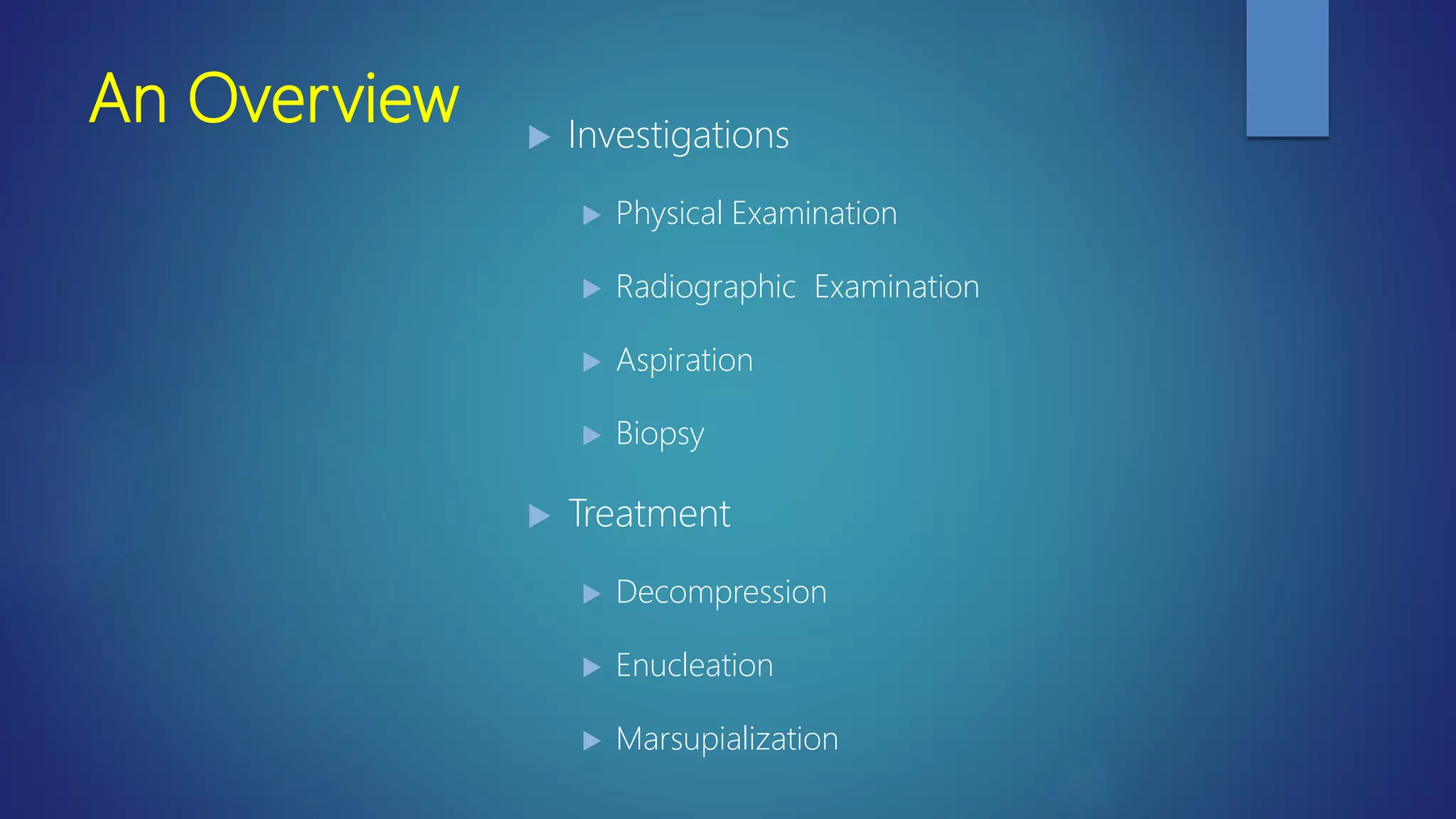
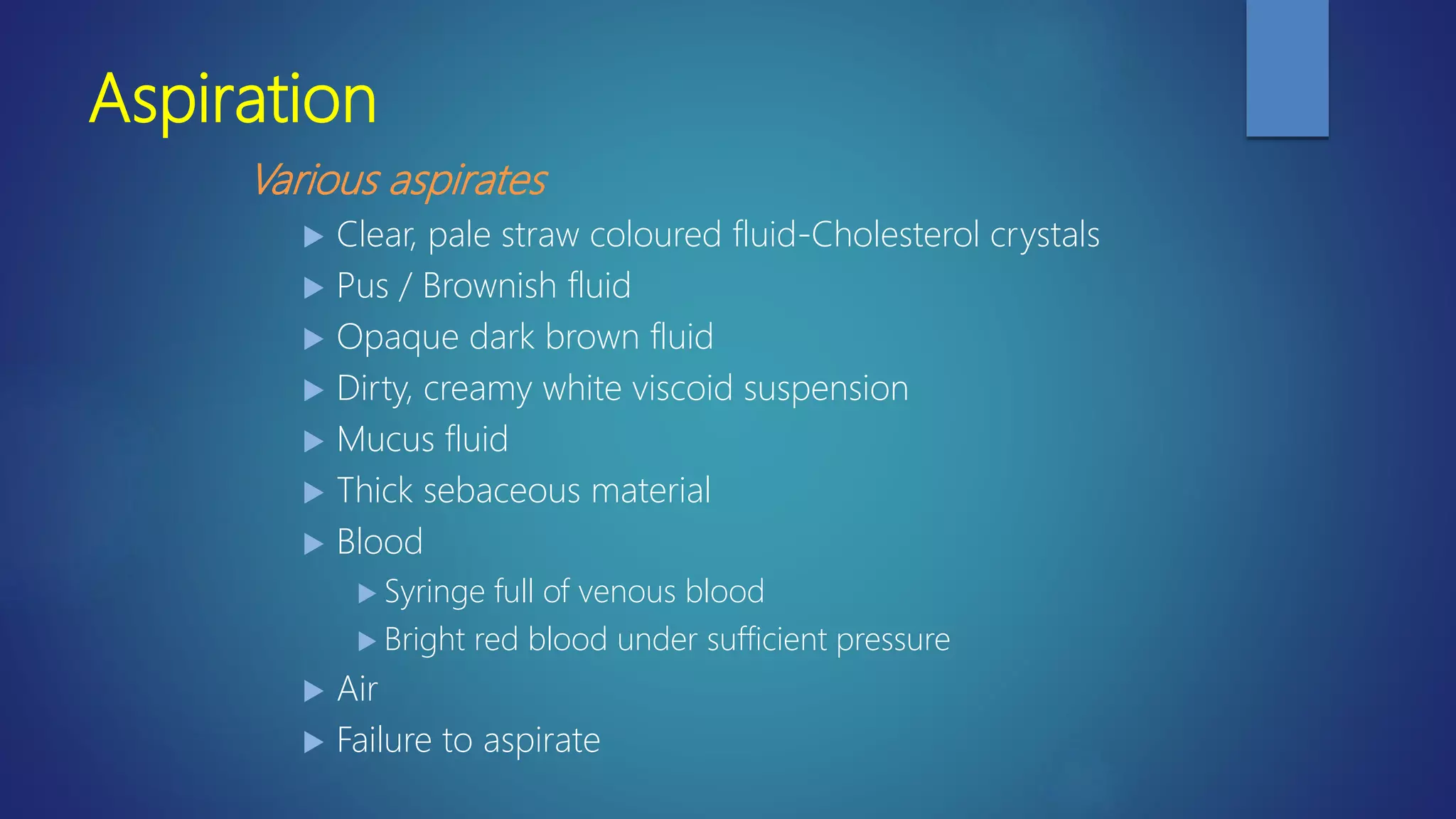
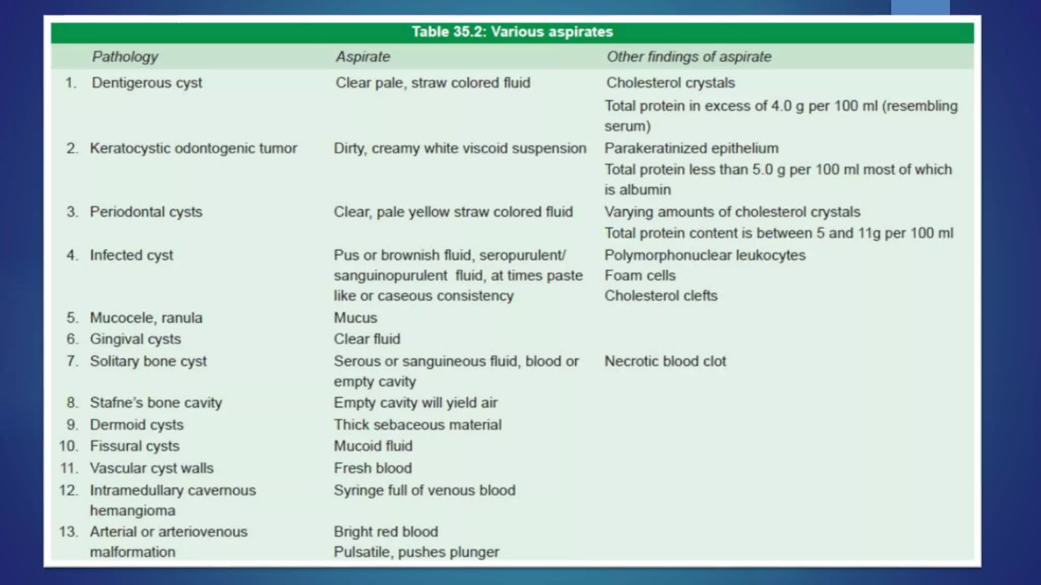
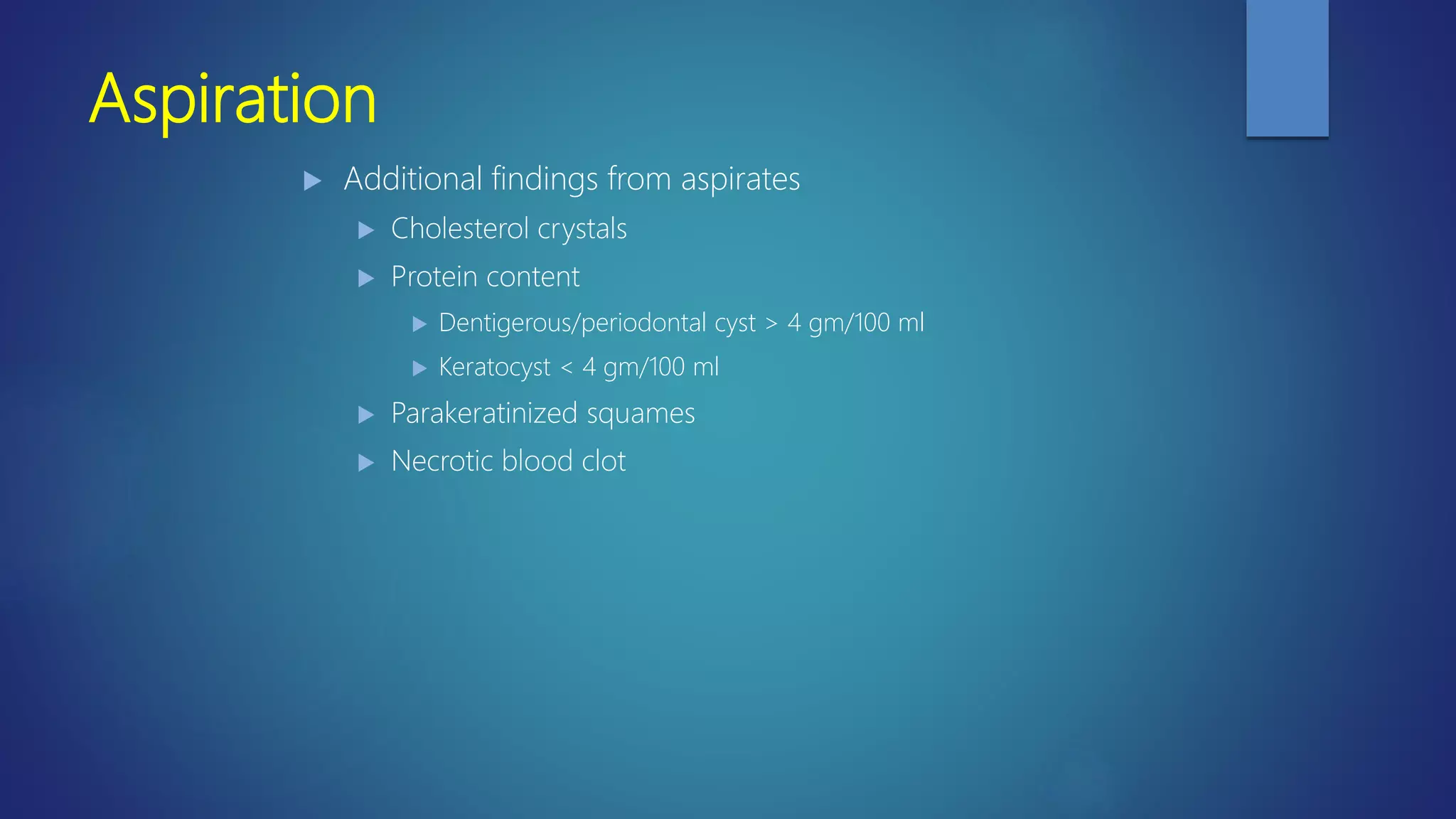
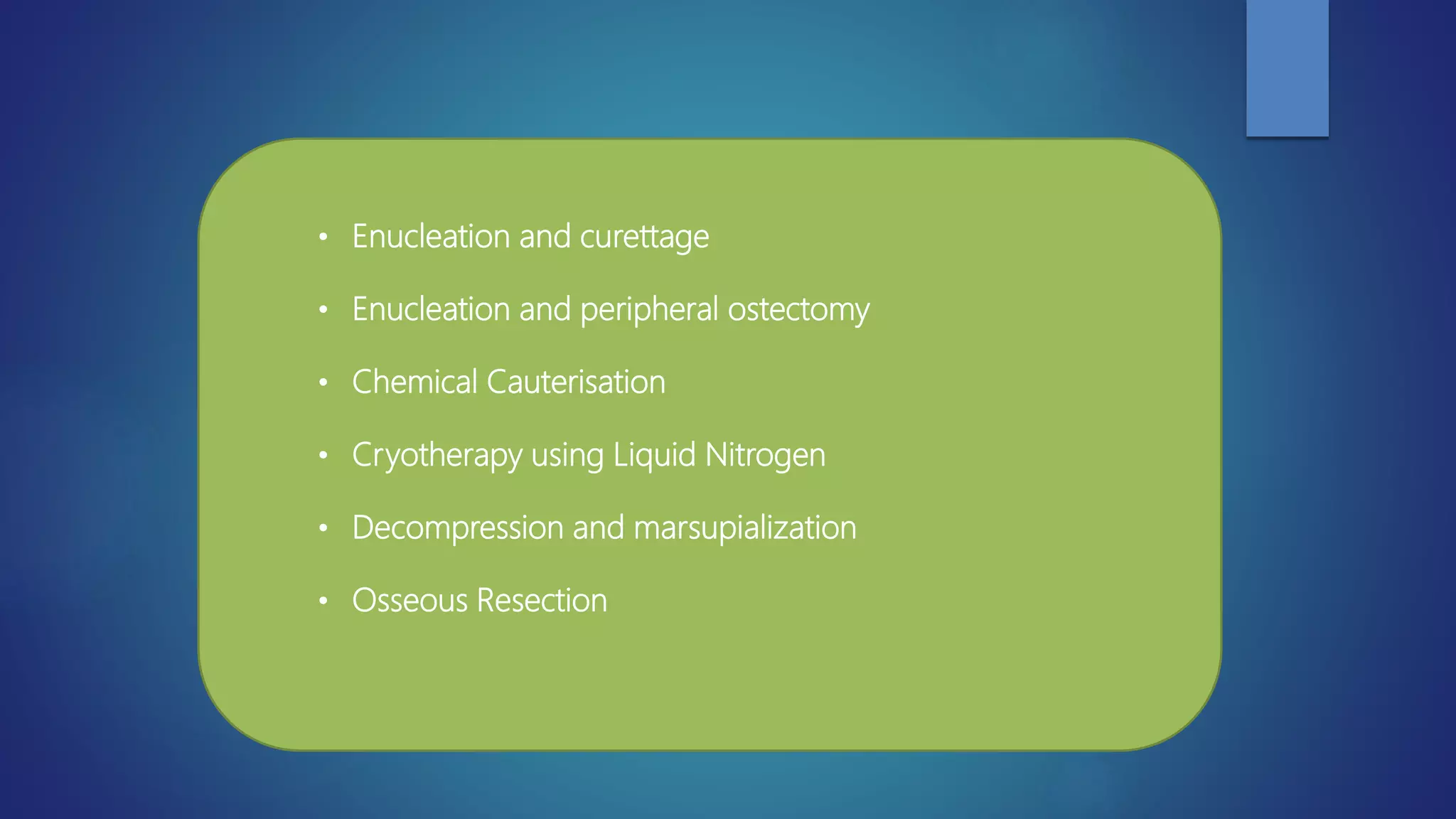
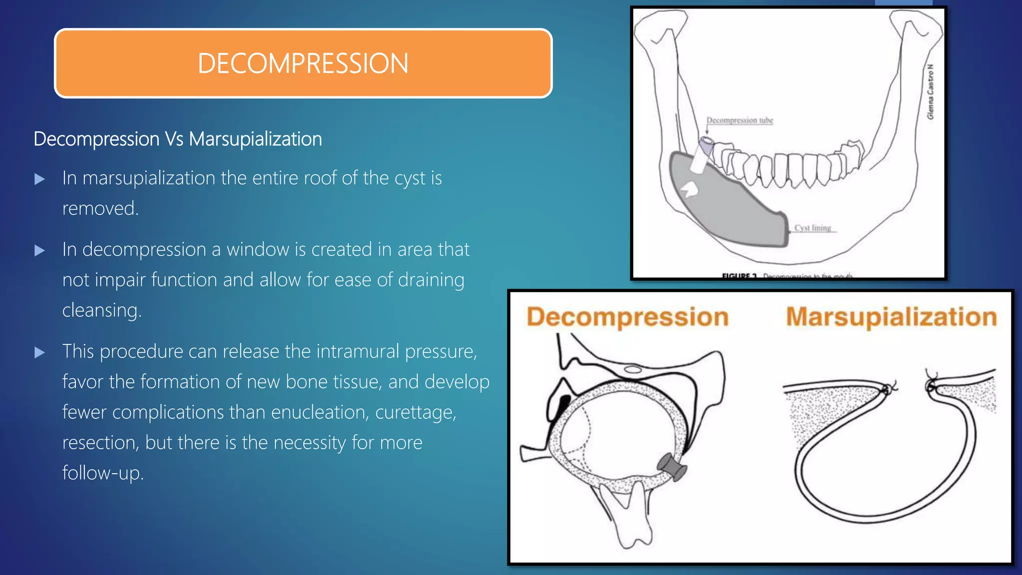
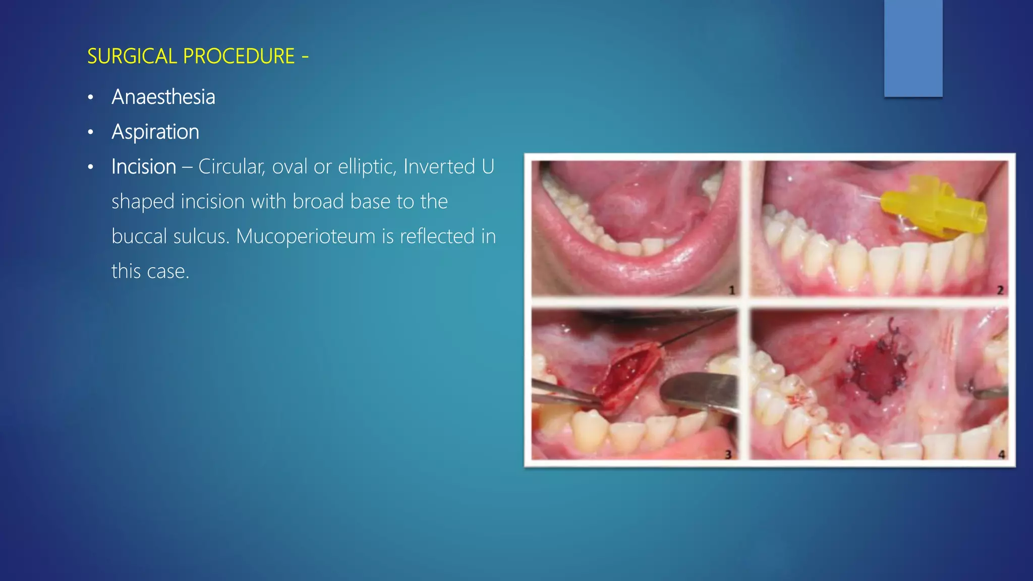
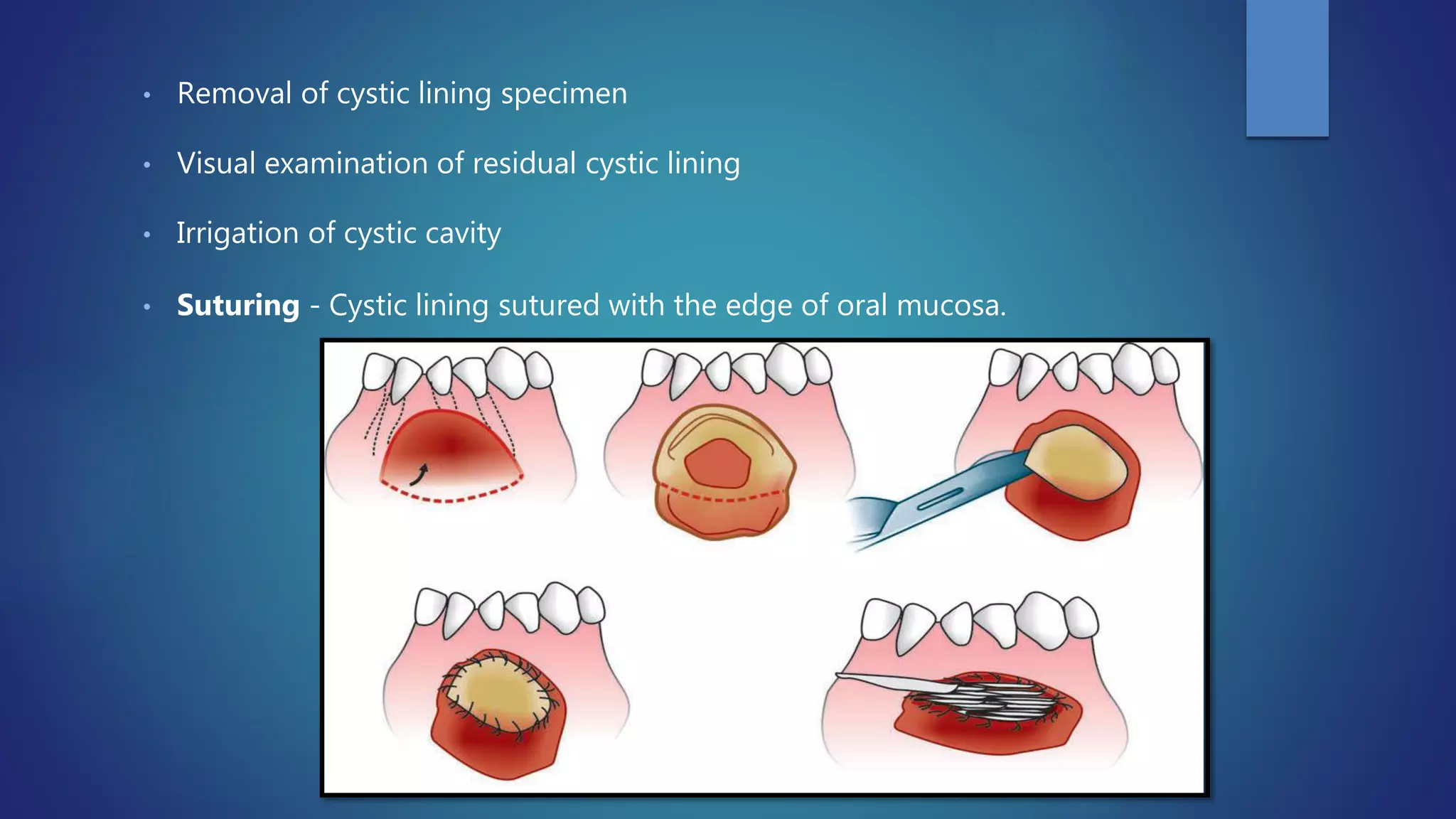
PRINCIPLES OF
MANAGEMENT OF
ODONTOGENIC
CYSTS
BY- DR. SWATI SAHU, OMFS
An Overview  Investigations
 Physical Examination
 Radiographic Examination
 Aspiration
 Biopsy
 Treatment
 Decompression
 Enucleation
 Marsupialization
Physical Examination
Radiographic Examination
Aspiration
Biopsy
Vitality
Excision
INVESTIGATIONS
Physical Examination
 Inspection and Palpation
 Bone expansion and asymmetric features
 Smooth, hard painless prominence
 Elastic effect
 Egg shell crackling
 Fluctuation
 Tooth Pathology / Absence of tooth from its place
 Tilting of Crowns
Physical Examination
 Site of occurrence
 Periodontal cyst
 Dentigerous cyst
 Keratocyst
 Solitary bone cyst and Stafnes bone cyst
 Percussion of teeth
 Neurological Examination
 A change in fitting of denture
Radiographic Examination
 IOPA
 Maxillary / Mandibular Occlusal radiographs
 Orthopantomograph
 Lateral Oblique view
 PA Mandible
 Water’s projection
 CT and MRI
IOPA and Occlusal radiographs
 Tooth pathology
 Location of pathology
 Character of pathology
 Lamina dura
 Adjacent vital structures
 Amount of palatal bone
involved
 Cortical expansion
Orthopantomograph
 Display the whole of lesion
 Multiple cysts in different location
can be viewed
 Anterioposterior and
superoinferior extent.
Lateral Oblique view & PA Mandible
 Cyst cavity encroaching on
or perforating the lower
border.
 Degree of involvement of
ramus and displacement of
inferior alveolar canal.
 Incorporates symphysis,
body and rami.
 Lateral and medial
expansion of ramus.
Water’s projection & Ultrasonography
 Anterior cyst of antrum
 Bilateral sinus comparison.
 Define the limits of cyst
invading the antrum.
 Location and extent in
depth of cyst.
 Diagnosis on the basis of
echo of an ultrasonic
impulse and the marginal
bone.
 For the diagnosis of
conditions in maxillary
sinus.
CT and MRI
 Helpful in large cystic and
multiple cystic lesion.
 3D reconstruction.
Radiopaque Dyes
 Size & relation of cyst in doubt
 To follow progess of regression of
marsupilized lesion.
 2 needles inserted – to prevent
suddent change in pressure.
 Radiopaque dyes
 Lipiodol
 Triosil
 Iopamidol.
 Care during injection, to avoid
overflow in soft tissues.
 After radiographs, remove the
contrast medium.
Aspiration
 Wide bore needle 18 gauge and 5 cc or 10 cc syringe.
 Local anesthesia infiltrated
 Failure to aspirate – solid tumor
Aspiration
Various aspirates
 Clear, pale straw coloured fluid-Cholesterol crystals
 Pus / Brownish fluid
 Opaque dark brown fluid
 Dirty, creamy white viscoid suspension
 Mucus fluid
 Thick sebaceous material
 Blood
 Syringe full of venous blood
 Bright red blood under sufficient pressure
 Air
 Failure to aspirate
Aspiration
 Additional findings from aspirates
 Cholesterol crystals
 Protein content
 Dentigerous/periodontal cyst > 4 gm/100 ml
 Keratocyst < 4 gm/100 ml
 Parakeratinized squames
 Necrotic blood clot
Biopsy
 To provide definitive, optimal therapy.
 Obtain a representative tissue for examination.
 Must not compromise a subsequent surgical
approach.
 The bone window created should not be directly
beneath the flap margin.
 Incisional biopsy specimen may not reveal all the
characteristics of the lesion.
Vitality Of Teeth
 Pre and Postoperative vitality of teeth.
 Non-vital teeth – Apical periodontal cyst
 Vital teeth – All other cyst
 Temporary absence of vitality response.
 RCT – within 24 hrs prior to surgery and over filled.
SURGICAL MANAGEMENT OF
CYSTS OF THE JAWS
• Enucleation and curettage
• Enucleation and peripheral ostectomy
• Chemical Cauterisation
• Cryotherapy using Liquid Nitrogen
• Decompression and marsupialization
• Osseous Resection
Regression of cyst without surgical
treatment (Oehlers)
 Removal of necrotic pulp remnants and/or removal of bacteria
from root canal of causative tooth and canal effectively filled.
 Reduced intracystic pressure.
 Reversal of chemical mechanisms of enlargement.
Decompression Vs Marsupialization
 In marsupialization the entire roof of the cyst is
removed.
 In decompression a window is created in area that
not impair function and allow for ease of draining
cleansing.
 This procedure can release the intramural pressure,
favor the formation of new bone tissue, and develop
fewer complications than enucleation, curettage,
resection, but there is the necessity for more
follow-up.
DECOMPRESSION
ENUCLEATION AND CURETTAGE
• Enucleation is the process by which the total removal of a cystic lesion is achieved.
• By definition, it means a shelling- out of the entire cystic lesion without rupture.
• Enucleation of cysts should be performed with care, in an attempt to remove the cyst
in one piece without fragmentation, which reduces the chances of recurrence by
increasing the likelihood of total removal.
INDICATIONS -
PRINCIPLE -
Enucleation allows for the cystic cavity to be covered by a mucoperiosteal flap and the
space fills with blood clot, which will eventually organize and form normal bone.
• Small cyst < 2cm
• Recurrent cyst of any type
• Keratocyst
• Malignant transformation
DISADVANTAGES-
ADVANTAGES-
• Primary closure of the wound
• Healing is rapid
• Postoperative care is reduced
• Thorough histological examination of the entire cystic lining can be done
• After primary closure, it is not possible to directly observe the healing as with marsupilization
• In young persons, the unerupted teeth in a dentigerous cyst will be removed with the lesion
• Removal of large cysts will weaken the mandible, making it prone to jaw fracture
• Damage to adjacent vital structures
• Pulpal necrosis
Modifications
 Enucleation and Packing.
 Enucleation and primary closure.
 Enucleation and Primary closure with bone grafts / reconstruction
ENUCLEATION AND PACKING
 Primary closure unsuccessful.
 Infection
 Expected wound dehiscence
 Secondary measure when there is dehiscence after
primary closure.
 Wound heals with granulation tissue until
epithelization is complete.
SURGICAL TECHNIQUE -
• Aspiration Biopsy of Radiolucent Lesions
• Mucoperiosteal Flaps
• Osseous Window
• Removal of Specimen
ENUCLEATION AND PRIMARY CLOSURE
ASPIRATION BIOPSY OF RADIOLUCENT LESIONS
• Any radiolucent lesion should be aspirated before surgical exploration.
• This provides the surgeon with valuable diagnostic information regarding the nature
of the lesion
MUCOPERIOSTEAL FLAPS
• Incision is carried around the necks of the involved teeth and the
adjoining teeth on either side, so that the flap will lie on sound bone.
• In case of the edentulous patient, incision is placed on the alveolar
crest, down to bone.
• Releasing incisions are given at either ends, which extend into the
buccal sulcus, so that the base of the flap is broader.
• Depending upon the location of the cyst, incision is placed buccally or
palatally.
OSSEOUS WINDOW -
• Once the flap has been elevated, a rotating bur should be used to remove an osseous
window.
• The size of the window depends on the size of the lesion and the proximity of the window to
normal anatomic structures such as roots and neurovascular bundles.
Technique :
• A dental curette is used to peel the connective tissues wall of the specimen
from surrounding bone.
• The concave surface of the instrument should always be kept in contact with
the osseous surfaces of the bone cavity.
• The bony cavity is inspected after irrigation with sterile saline.
• Any residual fragments of soft tissue within the cavity should be removed with curettes.
• Once the cavity is devoid of residual pathologic tissue, it is irrigated and the flap
is replaced and sutured in its proper location.
Other Surgical Procedures
 Nasteff - Rosenthal technique
 Use of suction drainage – Sucks in buccal soft
tissue including masseter muscle and fill bony
cavity
 Disadvantage
 Secondary deformity.
 Rhinocystostomy
 Anterior maxillary cyst joined with nasal cavity
 Cyst becomes invagination of nasal cavity
ENUCLEATION AND PRIMAY CLOSURE WITH
RECONSTRUCTION/BONE GRAFTING
 In large cystic lesions that have perforated and destroyed the cortical plates and inferior
border of the mandible that is beyond salvage or is nonexistent.
 It is advisable to reconstruct primarily with a stainless steel or titanium reconstructive
plate.
 Occasionally, autogenous bone grafts, e.g.iliac crest or costochondral grafts can be
used for reconstruction procedures, replacing the lost bone.
 A water tight closure has to be achieved both intra and extraorally.
 Intermaxillary ligation is necessary to help provide immobilization during the healing
phase for 4 to 6 weeks.
Postoperative Treatment Of Cyst Cavity
 Cavity greater than 4 cm should be grafted.
 Aim of replacement
 Restoration of morphological contour
 Mechanical strength and function
 Eliminate dead space and infection
 Prevention of ingrowth of soft tissue
 Enhance retention of prosthetic devices
ENUCLEATION AND PERIPHERAL OSTECTOMY
• Removal of 1 to 2 mm of bone beyond the visible margin of the lesion is adequate to
improve the cure rate.
• However, it is difficult to estimate how much bone to remove with a drill.
• This process is made easier by the use of a vital staining technique.
• Methylene blue or crystal violet (or any other vital stain) can be painted on the bony walls
of the enucleated cyst and allowed to penetrate into the bone.
• The cavity is then washed out and any bone retaining the stain is removed with a
drill .
• This process usually removes around 2mm of bone in the marrow and about 1
mm of cortical bone.
(A)The cavity remaining after a cyst has been enucleated, and stained with
methylene blue.
(B) The same cavity after removing the methylene blue with a peripheral ostectomy
using a pineapple-type bur.
Oral Maxillofacial Surg Clin N Am 25 (2013) 21–30
MARSUPILIZATION
Partsch I procedure (Decompression and marsupialization)
PRINCIPLE –
Marsupialization, (Partsch) or decompression, refers to creating a surgical window in the wall of the cyst,
and evacuation of the cystic contents. This process decreases Intracystic pressure and promotes shrinkage
of the cyst and bone fill. The only portion that is removed is the piece removed to produce the window.
The marsupialization technique:-
• It was described by Pogrel (2005)
• A window at least 1 cm in diameter is made into a cyst, and an attempt is
made to suture the cyst lining to the oral mucosa.
• In the maxilla, the cyst is then often packed open with the packing
protruding through the opening.
•The packing consists of iodoform gauze impregnated with bacitracin
ointment.
INDICATIONS -
1. Amount of tissue injury :
Proximity of a cyst to vital structures can mean unnecessary sacrifice of tissue if enucleation
is used.
2. Surgical access :
If access to all portions of the cyst is difficult, portions of the cystic wall may be left behind,
which could result in recurrence.
3. Assistance in eruption of teeth :
If an unerupted tooth that is needed in the dental arch is involved with the cyst (i.e., a
dentigerous cyst), marsupialization may allow its continued eruption into the oral cavity.
4. Extent of surgery :
Marsupialization is a reasonable alternative to enucleation, because it is simple and may be
less stressful for the patient.
5. Size of cyst :
In very large cysts, a risk of jaw fracture during enucleation is possible. It may be better to
marsupialize the cyst and defer enucleation until after considerable bone filling has
CONTRAINDICATIONS -
 Poor patient compliance
 Keratocyst (Controversy)
 Malignant changes in lining
ADVANTAGES -
• Large cyst – technically simple
• Dealt under LA
• Adjacent vital structures preserved
• Conserve tooth involved
• Prevent fracture complication – Endosteal bone formation
• Pathologic tissue is left in situ, without thorough histologic examination.
• Patient is inconvenienced in several respects
• The cystic cavity must be kept clean to prevent infection, because the cavity
frequently traps food debris.
• In most instances this means that the patient must irrigate the cavity several times
everyday with a syringe
DISADVANTAGES -
SURGICAL PROCEDURE -
• Anaesthesia
• Aspiration
• Incision – Circular, oval or elliptic, Inverted U
shaped incision with broad base to the
buccal sulcus. Mucoperioteum is reflected in
this case.
• Removal of bone –
 Thin bone — When the bone is expanded and thinned out, the initial incision
can be extended through the mucoperiosteum, bone and cystic lining into the
cystic cavity. The cystic lining and contents are then submitted for histological
examination.
 Thick bone: When the overlying bone is thick, bur holes are drilled in a circular
shape, which are then connected and the overlying bone is removed carefully
with a pair of rongeurs or mosquito forceps.
• Removal of cystic lining specimen
• Visual examination of residual cystic lining
• Irrigation of cystic cavity
• Suturing - Cystic lining sutured with the edge of oral mucosa.
• In U shaped incision the mucoperiosteal flap can be turned into cystic cavity covering the
margin. The remaining is sutured to oral mucosa.
• Packing - Prevents food contamination & covers wound margins.
• Done with ribbon gauze soaked with WHITEHEAD VARNISH.
 COMPOSTION OF WHITEHEAD VARNISH :
 Benzoin -10g
 Iodoform - 10g
 Storax -7.5g
 Balsam of Tolu - 5g
 Solvent ether to 100ml
 Pack changed for every 2 days.
 Maintenance of cystic cavity - Instruct the patient to clean and irrigate the
cavity regularly with oral antiseptic rinse with a disposable syringe.
• Use of plug
 Prevents contamination. Preserves patency of cyst orifice.
 Plug should be stable, retentive and safe design.
 Should be made of resilient material ( avoid irritation) like acrylic.
• Healing - Cavity may or may not obliterate totally. Depression remains in the
alveolar process.
MODIFICATION OF MARSUPILIZATION
Waldron’s method (1941) / PARTSCH II -
 Two stage technique
 Combination of two standard technique
 First marsupialization, Second enucleation, when the cavity becomes smaller
INDICATIONS -
• When bone has covered the adjacent vital structures
• Adequate bone fill has strengthened the jaw to prevent fracture during enucleation
• Patient finds difficult to clean cavity
• For detection of any occult pathologic condition
ADVANTAGES -
• Development of a thickened cystic lining which makes enucleation easier
• Spares adjacent vital structures
• Combined approach reduces morbidity
• Accelerated healing process
• Allows histopathological examination of residual tissue
DISADVANTAGES -
• Patient has to undergo secondary surgery and possible complications that are involved with the
surgical procedure.
Marsupialization into nose or antrum
 Extensive cysts of maxilla,
entirely occupying a large
portion of antrum or nose.
 Surgical technique
 Buccal flap
 Bone window
 Second unroofing removing
antral lining
 Intranasal antrostomy
Marsupialization in to nose or antrum
 Cavity packed with
 Ribbon gauze with tincture of benzoin
 Antibiotic ointment
 Antral balloon
 Balloon on Foleys catheter
 Draining with 0.75 cm dia sterile polyethylene tube
 Pack removed after 7 days
 Cyst cavity is lined by normal ciliated and mucous secreting epithelium from
respiratory mucosa.
CHEMICAL CAUTERIZATION
• Carnoy’s solution was first used as a medicament in surgery by Cutler and Zollinger in 1933.
• It is a powerful fixative, haemostatic and cauterizing agent which penetrates cancellous spaces in the
bone and devitalizes and fixes the left out tumour cells.
• Success of the application of this medicament after enucleation of KOT is thought to be due to both
penetration and fixation action.
• Carnoy’s solution may be used in the bony region but preferably not in close proximity to the neural
structures and maxillary sinus region to avoid damage to the neural tissue and necrosis of the sinus
wall.
Int J Pharm Bio Sci 2014 Jan; 5(1): (B/P) 492 - 495
• Its average depth of penetration is 1.54 mm after 5 mins of application (Stolinga
2005).
• But the duration of application is not clarified in the existing literature.
• However Blanas et al state that application of carnoy’s solution to cyst cavity
for 3 mins after enucleation should not damage the inferior alveolar nerve.
• Recurrence rate –
 7.8 % - stolinga 2003
 2.5% - Chapelle 2004
Int J Pharm Bio Sci 2014 Jan; 5(1): (B/P) 492 - 495
COMPOSITION OF CARNOYS SOLUTION
• Carnoy’s solution II (Recommended by Cutler and Zollinger - 1933) :
 Ferric chloride - 1 gram
 Chloroform - 3ml
 Glacial acetic acid - 1ml
 Absolute alcohol - 6ml
• Carnoy’s solution I ( Farmers solution ) :
 Absolute alcohol - 3ml
 Glacial acetic acid - 1ml
• Modified Carnoy’s solution :
 Ferric chloride - 1gram
 Glacial acetic acid - 1ml
 Absolute alcohol - 6ml Int J Pharm Bio Sci 2014 Jan; 5(1): (B/P) 492 - 495
ENUCLEATION AND LIQUID NITROGEN CRYOTHERAPY
• The ideal treatment for the KCOT would be enucleation or curettage followed by treatment of
the cavity with an agent that would kill the epithelial remnants or satellite cysts.
• The osseous framework should be left intact to allow for osteoconduction.
• KCOT <1.5 cm lesions are treated with this technique
• Liquid nitrogen has the ability to devitalize bone in situ while leaving the inorganic framework
untouched.
• As a result of this, cryotherapy has been used for a number of locally aggressive jaw lesions
including :-
KCOT
Ameloblastoma
Ossifying fibroma
• Cell death with cryosurgery occurs by direct damage from intracellular and extracellular ice
crystal formation plus osmotic and electrolyte disturbances
According to Schmidt and Pogrel (2001),the standardized technique is as follows -
Enucleation of the cyst.
The surrounding tissues are then protected with sterile wooden tongue blades and gauze
The cavity is sprayed with liquid nitrogen twice for 1 min
(5-min interval)
Bone graft can be inserted
Mucosa is closed with tight sutures
(A) Technique of placing a liquid nitrogen probe in it and freezing the whole
cavity.
(B) The cryoprobe has been removed, showing the frozen and
surrounding bony walls of the cyst cavity.
Cryotherapy preparation. Malleable
retractors are positioned so as to
self-retain gauze sponges to protect
and insulate the soft tissues.
Cryo-applicator in water-soluble jelly.
Open funnel cryo-application system.
ADVANTAGES -
• The bone matrix is left in place to act as a clean scaffold for new bone formation
• A bone graft can be placed immediately to accelerate healing and minimize the risk of a
pathologic fracture
• Decrease of bleeding and scarring.
DISADVANTAGES -
• Difficulty in controlling the amount of liquid nitrogen applied to the cavity.
• The resultant necrosis and swelling can be unpredictable (Pogrel, 1993; Salmassy and
Pogrel, 1995)
• When the liquid nitrogen cryotherapy is given around the inferior alveolar nerve, it is
affected and patients will suffer paraesthesia or anaesthesia.
• Using this cryotherapy technique seems to be associated with a recurrence rate of around
10%
CONCLUSION
o In summary, there are a multitude of odontogenic cysts that may present in head
and neck patients. The key to diagnosis is a careful history and physical
examination accompanied by radiographic evidence and pathologic
confirmation.
o Many of these entities represent benign lesions, however significant pathologic
disease may be lacking which necessitates prompt treatment and immediate
consultation as necessary.
References
1. Cysts of the Oral and Maxillofacial Regions ,Mervyn
Shear ,Fourth edition.
2. Shafer’s text book of Oral pathology, 6th edition
3. Contemporary OMFP,2nd edi,Sapp Eversole Wyso.
4. Clinical Pathological Correlation. Regezi, 4th edi.
5. Oral Pathology. Somes &Southam, 4th edi.
6. R.M.Browne;The Pathogenesis Of Odontogenic Cysts:
A Review.
Journal of Oral Pathology 1975;4:31-46
7. Sunitha Jacob;Rushton bodies or hyaline bodies in
radicular cyst:A morphologic curiosity.Indian Journal
of Pathology and Microbiology,2010.
8. P. R. MORGAN;Histological, histochemical and ultrastructural
studies on the nature of hyalin bodies in odontogenic cysts. Journal
of Oral Pathology 1974: 3: 127-147
9. Akira Yamaguchi;Hyaline bodies of odontogenic cysts:
Histologieal, histochemieal and electron microscopic studies.
Journal of Orat Pathotogy 1980:9: 221-234.
10. Monica Mehendiratta ;Ghost cells: A journey in the
dark.Dental Research Journal,Dec 2012:Vol .9,Issue 7.
11. Blanas N, Freund B, Schwartz M, Furst IM (2000), Systematic
review of the treatment and prognosis of the odontogenic
keratocyst, Oral Surg Oral Med Oral Pathol Oral Radiol Endod
90:553-8.
12.I Kaplan1.Glandular odontogenic cyst: a challenge in diagnosis
and treatment.Journal of Oral diseases (2008), 14.
MARGINAL RESECTION / RESECTION WITHOUT CONTINUITY
DEFECT / PERIPHERAL OSTEOTOMY / EN BLOC RESECTION
• Indicated in lesions which are known for recurrence
• Lesions that tend to grow beyond their surgically
apparent capsule
• When the lesion does not extent closer than 1 cm to
the inferior border of the mandible.
• Margin of 1 to 2 cm - minimum acceptable margin.
INDICATIONS -
• Ameloblastoma
• Calcifying epithelial odontogenic tumor
• Myxoma
• Ameloblastic odontoma
• Squamous odontogenic tumor
Procedure allows complete excision of the tumor but at the same time a continuity of the jaw bone
is retained thus deformity, disfigurement & need for secondary cosmetic surgery & prosthetic
rehabilitation are avoided .
OPERATIVE PROCEDURE -
• Intra-oral / extra-oral approach
• Intra-oral - good access and when the lesion is anterior to third molar region
• Extra-oral approach - lesion involves the ramus of the mandible or when immediate
reconstruction is planned
INTRAORAL APPROACH -
• Large mandibular lesions - a midline lip-splitting incision
• Connecting vertical incisions are made on the buccal and lingual
• Incisions - extend deep into buccal and lingual folds.
• The teeth bordering the surgical margin should be extracted
• Horizontal incisions connecting the lower ends of vertical incisions are made.
• The buccal and lingual mucoperiosteal flaps are then developed, but not reflected
superiorly over the region of bone to be removed.
• On exposure of the mandible, the bony segment is sectioned with an air-driven saw or bur,
at least 1 to 1.5 cm from the radiographic margin of the lesion Haemorrhage - controlled
by crushing the bone over small blood vessels with a blunt instrument or by using bone
wax .
• The mucoperiosteum is then undermined both lingually and facially to relieve tension.
• They are approximated with interrupted silk sutures.
SEGMENTAL (PARTIAL ) MANDIBULAR RESECTION /
HEMIMANDIBULECTOMY
• Least number of recurrences.
• Indications:
 Infiltrative lesions
 Lesions – posterior/ inferior border of mandible
 Lesions with high recurrence rate
OPERATIVE PROCEDURE -
• Depending on the size - a lip-splitting incision may or may not be necessary
• A submandibular incision - join the vertical lip incision
• Intra-orally - horizontal incision is made through the mucoperiosteum
• The facial and lingual flaps are advanced below the horizontal incision using a
periosteal elevator.
• The lingual flap is raised as deep as to expose the mylohyoid attachment.
• A vertical mucoperiosteal incision is made 0.5 cm proximal to the anticipated
anterior bony cut.
• Expose the mental neurovascular bundle, which is ligated and sectioned. Preservation of the
marginal mandibular branch of the facial nerve
• Using an air-driven saw, bur or a Gigli saw, a vertical cut is made through the mandible
anterior to the lesion.
• Using bone forceps, the proximal part of the mandible is rotated laterally, exposing the
inferior alveolar nerve and vessels, at the lingula of the mandible. They are ligated and cut
adjacent to the mandibular foramen.
• The capsule is cut with a scalpel and the segment of mandible is disarticulated and removed
using bone holding forceps.
• Bleeding - controlled by digital pressure, coagulation or ligation, depending on the size of
the bleeding vessel.
Keratocyst
 Recurrence
 Pindborg & Hansen 5 – 62 % and in 1st 5 years.
 Possible reasons
 Satellite cyst
 Thin lining
 Intrinsic growth potential
 Basal cells of oral mucosa
Keratocyst
 Bramley
 Small single cyst & regular outline
 Enucleation intra orally
 Large less accessible & regular outline
 Enucleation extre orally
 Unilocular with scalloped or loculated perphery & small
multiocular lesions
 Marginal excision & excision of overlying mucosa
 Marginal multilocular lesion
 Resection & reconstruction
Keratocyst - Chemical cautery
 Stoelinga & Van Hoelst
 Carnoy’s solution -chemical tissue fixative
 Absolute alcohol – 6 ml
 Chloroform – 3 ml
 Glacial acetic acid – 1 ml
 Ferric chloride – 1 g
 5 minutes application penetrates
 Bone – 1.54 mm
 Nerve – 0.15 mm
 Mucosa – 0.51 mm
 Voorsmiths’- Tanning effect
 Coating neurovascular bundle with petroleum jelly.
Keratocyst – Chemical cautery
 Technique of application
 Contraindication
 Maxillary sinus
 Developing teeth
 Near orbit
Keratocyst - Cryotherapy
 Cellular necrosis in bone maintaining inorganic
osseous framework
 Predictable cell lysis -20°C
 Effect of cryotherapy
 Direct damage
 Osmotic disturbances
 Electrolyte imbalance
 Single minute freeze – 1-3mm bone necrosis
 Immediate bone grafting for defect greater than 4
cm to decrease
 Wound dehiscence
 Pathologic #
 Provide greater residual bone height and density
Keratocyst - Cryotherapy
 Water soluble conducting medium (KY jelly)
 Cryoprobe and freezing – Equall conduction to all bone margins
 3 freezes for 1 min each with a 5 minutes thaw between freezes.
 Liquid Nitrogen Spray
 More profound freezing effect
 2 freezes
Large Keratocyst – Brosch Technique
(Farmand & Makek)
 Alternative to resection & grafting or marsupialization
 Large cyst that occupy molar, angle and ramal region with
cortical perforation.
 Removal of lateral cortical plate along with entire cyst and
coronoid process.
Large Keratocyst – Brosch Technique
(Farmand & Makek)
 Technique.
 Osteotomy cut
 Removal in toto
 Suturing.
 Advantage
 Entire pathology removed
 Mandibular anatomy maintained
 Disadvantage
 Access
 Time consuming
Postoperative Follow up
 Early detection and deal with any recurrence
 Follow up at 3 weeks, 1, 3, 6, 12 months and then after annually for next 5
yrs at least.
 Vitality of associated and adjacent teeth and endodontic treatment.
 Exposure and orthodontic treatment for unerupted teeth.
Reference
 Oral & maxillofacial surgery – LASKIN, Vol. II
 Contemporary oral & maxillofacial surgery – PETERSON
 Cysts – SHEAR
 Oral pathology – SHAFERS
 Outline of oral surgery – KILLEY & KAY
 Text book of oral medicine - BURKITT
 Oral and maxillofacial pathology - FONSECA
 Maxillofacial surgery – Peter Ward Booth , Vol. II
 Clinics of North America on Management Of Odontogenic Keratocyst
 Electronic media
THANKYOU

Management of cyst

  • 1.
  • 2.
    An Overview Investigations  Physical Examination  Radiographic Examination  Aspiration  Biopsy  Treatment  Decompression  Enucleation  Marsupialization
  • 3.
  • 4.
    Physical Examination  Inspectionand Palpation  Bone expansion and asymmetric features  Smooth, hard painless prominence  Elastic effect  Egg shell crackling  Fluctuation  Tooth Pathology / Absence of tooth from its place  Tilting of Crowns
  • 5.
    Physical Examination  Siteof occurrence  Periodontal cyst  Dentigerous cyst  Keratocyst  Solitary bone cyst and Stafnes bone cyst  Percussion of teeth  Neurological Examination  A change in fitting of denture
  • 6.
    Radiographic Examination  IOPA Maxillary / Mandibular Occlusal radiographs  Orthopantomograph  Lateral Oblique view  PA Mandible  Water’s projection  CT and MRI
  • 7.
    IOPA and Occlusalradiographs  Tooth pathology  Location of pathology  Character of pathology  Lamina dura  Adjacent vital structures  Amount of palatal bone involved  Cortical expansion
  • 8.
    Orthopantomograph  Display thewhole of lesion  Multiple cysts in different location can be viewed  Anterioposterior and superoinferior extent.
  • 9.
    Lateral Oblique view& PA Mandible  Cyst cavity encroaching on or perforating the lower border.  Degree of involvement of ramus and displacement of inferior alveolar canal.  Incorporates symphysis, body and rami.  Lateral and medial expansion of ramus.
  • 10.
    Water’s projection &Ultrasonography  Anterior cyst of antrum  Bilateral sinus comparison.  Define the limits of cyst invading the antrum.  Location and extent in depth of cyst.  Diagnosis on the basis of echo of an ultrasonic impulse and the marginal bone.  For the diagnosis of conditions in maxillary sinus.
  • 11.
    CT and MRI Helpful in large cystic and multiple cystic lesion.  3D reconstruction.
  • 12.
    Radiopaque Dyes  Size& relation of cyst in doubt  To follow progess of regression of marsupilized lesion.  2 needles inserted – to prevent suddent change in pressure.  Radiopaque dyes  Lipiodol  Triosil  Iopamidol.  Care during injection, to avoid overflow in soft tissues.  After radiographs, remove the contrast medium.
  • 13.
    Aspiration  Wide boreneedle 18 gauge and 5 cc or 10 cc syringe.  Local anesthesia infiltrated  Failure to aspirate – solid tumor
  • 14.
    Aspiration Various aspirates  Clear,pale straw coloured fluid-Cholesterol crystals  Pus / Brownish fluid  Opaque dark brown fluid  Dirty, creamy white viscoid suspension  Mucus fluid  Thick sebaceous material  Blood  Syringe full of venous blood  Bright red blood under sufficient pressure  Air  Failure to aspirate
  • 16.
    Aspiration  Additional findingsfrom aspirates  Cholesterol crystals  Protein content  Dentigerous/periodontal cyst > 4 gm/100 ml  Keratocyst < 4 gm/100 ml  Parakeratinized squames  Necrotic blood clot
  • 17.
    Biopsy  To providedefinitive, optimal therapy.  Obtain a representative tissue for examination.  Must not compromise a subsequent surgical approach.  The bone window created should not be directly beneath the flap margin.  Incisional biopsy specimen may not reveal all the characteristics of the lesion.
  • 18.
    Vitality Of Teeth Pre and Postoperative vitality of teeth.  Non-vital teeth – Apical periodontal cyst  Vital teeth – All other cyst  Temporary absence of vitality response.  RCT – within 24 hrs prior to surgery and over filled.
  • 19.
  • 20.
    • Enucleation andcurettage • Enucleation and peripheral ostectomy • Chemical Cauterisation • Cryotherapy using Liquid Nitrogen • Decompression and marsupialization • Osseous Resection
  • 21.
    Regression of cystwithout surgical treatment (Oehlers)  Removal of necrotic pulp remnants and/or removal of bacteria from root canal of causative tooth and canal effectively filled.  Reduced intracystic pressure.  Reversal of chemical mechanisms of enlargement.
  • 22.
    Decompression Vs Marsupialization In marsupialization the entire roof of the cyst is removed.  In decompression a window is created in area that not impair function and allow for ease of draining cleansing.  This procedure can release the intramural pressure, favor the formation of new bone tissue, and develop fewer complications than enucleation, curettage, resection, but there is the necessity for more follow-up. DECOMPRESSION
  • 24.
    ENUCLEATION AND CURETTAGE •Enucleation is the process by which the total removal of a cystic lesion is achieved. • By definition, it means a shelling- out of the entire cystic lesion without rupture. • Enucleation of cysts should be performed with care, in an attempt to remove the cyst in one piece without fragmentation, which reduces the chances of recurrence by increasing the likelihood of total removal.
  • 25.
    INDICATIONS - PRINCIPLE - Enucleationallows for the cystic cavity to be covered by a mucoperiosteal flap and the space fills with blood clot, which will eventually organize and form normal bone. • Small cyst < 2cm • Recurrent cyst of any type • Keratocyst • Malignant transformation
  • 26.
    DISADVANTAGES- ADVANTAGES- • Primary closureof the wound • Healing is rapid • Postoperative care is reduced • Thorough histological examination of the entire cystic lining can be done • After primary closure, it is not possible to directly observe the healing as with marsupilization • In young persons, the unerupted teeth in a dentigerous cyst will be removed with the lesion • Removal of large cysts will weaken the mandible, making it prone to jaw fracture • Damage to adjacent vital structures • Pulpal necrosis
  • 27.
    Modifications  Enucleation andPacking.  Enucleation and primary closure.  Enucleation and Primary closure with bone grafts / reconstruction
  • 28.
    ENUCLEATION AND PACKING Primary closure unsuccessful.  Infection  Expected wound dehiscence  Secondary measure when there is dehiscence after primary closure.  Wound heals with granulation tissue until epithelization is complete.
  • 29.
    SURGICAL TECHNIQUE - •Aspiration Biopsy of Radiolucent Lesions • Mucoperiosteal Flaps • Osseous Window • Removal of Specimen ENUCLEATION AND PRIMARY CLOSURE
  • 30.
    ASPIRATION BIOPSY OFRADIOLUCENT LESIONS • Any radiolucent lesion should be aspirated before surgical exploration. • This provides the surgeon with valuable diagnostic information regarding the nature of the lesion
  • 31.
    MUCOPERIOSTEAL FLAPS • Incisionis carried around the necks of the involved teeth and the adjoining teeth on either side, so that the flap will lie on sound bone. • In case of the edentulous patient, incision is placed on the alveolar crest, down to bone. • Releasing incisions are given at either ends, which extend into the buccal sulcus, so that the base of the flap is broader. • Depending upon the location of the cyst, incision is placed buccally or palatally.
  • 32.
    OSSEOUS WINDOW - •Once the flap has been elevated, a rotating bur should be used to remove an osseous window. • The size of the window depends on the size of the lesion and the proximity of the window to normal anatomic structures such as roots and neurovascular bundles.
  • 33.
    Technique : • Adental curette is used to peel the connective tissues wall of the specimen from surrounding bone. • The concave surface of the instrument should always be kept in contact with the osseous surfaces of the bone cavity. • The bony cavity is inspected after irrigation with sterile saline. • Any residual fragments of soft tissue within the cavity should be removed with curettes. • Once the cavity is devoid of residual pathologic tissue, it is irrigated and the flap is replaced and sutured in its proper location.
  • 36.
    Other Surgical Procedures Nasteff - Rosenthal technique  Use of suction drainage – Sucks in buccal soft tissue including masseter muscle and fill bony cavity  Disadvantage  Secondary deformity.  Rhinocystostomy  Anterior maxillary cyst joined with nasal cavity  Cyst becomes invagination of nasal cavity
  • 37.
    ENUCLEATION AND PRIMAYCLOSURE WITH RECONSTRUCTION/BONE GRAFTING  In large cystic lesions that have perforated and destroyed the cortical plates and inferior border of the mandible that is beyond salvage or is nonexistent.  It is advisable to reconstruct primarily with a stainless steel or titanium reconstructive plate.
  • 38.
     Occasionally, autogenousbone grafts, e.g.iliac crest or costochondral grafts can be used for reconstruction procedures, replacing the lost bone.  A water tight closure has to be achieved both intra and extraorally.  Intermaxillary ligation is necessary to help provide immobilization during the healing phase for 4 to 6 weeks.
  • 39.
    Postoperative Treatment OfCyst Cavity  Cavity greater than 4 cm should be grafted.  Aim of replacement  Restoration of morphological contour  Mechanical strength and function  Eliminate dead space and infection  Prevention of ingrowth of soft tissue  Enhance retention of prosthetic devices
  • 40.
    ENUCLEATION AND PERIPHERALOSTECTOMY • Removal of 1 to 2 mm of bone beyond the visible margin of the lesion is adequate to improve the cure rate. • However, it is difficult to estimate how much bone to remove with a drill. • This process is made easier by the use of a vital staining technique. • Methylene blue or crystal violet (or any other vital stain) can be painted on the bony walls of the enucleated cyst and allowed to penetrate into the bone.
  • 41.
    • The cavityis then washed out and any bone retaining the stain is removed with a drill . • This process usually removes around 2mm of bone in the marrow and about 1 mm of cortical bone.
  • 42.
    (A)The cavity remainingafter a cyst has been enucleated, and stained with methylene blue. (B) The same cavity after removing the methylene blue with a peripheral ostectomy using a pineapple-type bur. Oral Maxillofacial Surg Clin N Am 25 (2013) 21–30
  • 43.
    MARSUPILIZATION Partsch I procedure(Decompression and marsupialization) PRINCIPLE – Marsupialization, (Partsch) or decompression, refers to creating a surgical window in the wall of the cyst, and evacuation of the cystic contents. This process decreases Intracystic pressure and promotes shrinkage of the cyst and bone fill. The only portion that is removed is the piece removed to produce the window.
  • 44.
    The marsupialization technique:- •It was described by Pogrel (2005) • A window at least 1 cm in diameter is made into a cyst, and an attempt is made to suture the cyst lining to the oral mucosa. • In the maxilla, the cyst is then often packed open with the packing protruding through the opening. •The packing consists of iodoform gauze impregnated with bacitracin ointment.
  • 45.
    INDICATIONS - 1. Amountof tissue injury : Proximity of a cyst to vital structures can mean unnecessary sacrifice of tissue if enucleation is used. 2. Surgical access : If access to all portions of the cyst is difficult, portions of the cystic wall may be left behind, which could result in recurrence. 3. Assistance in eruption of teeth : If an unerupted tooth that is needed in the dental arch is involved with the cyst (i.e., a dentigerous cyst), marsupialization may allow its continued eruption into the oral cavity.
  • 46.
    4. Extent ofsurgery : Marsupialization is a reasonable alternative to enucleation, because it is simple and may be less stressful for the patient. 5. Size of cyst : In very large cysts, a risk of jaw fracture during enucleation is possible. It may be better to marsupialize the cyst and defer enucleation until after considerable bone filling has
  • 47.
    CONTRAINDICATIONS -  Poorpatient compliance  Keratocyst (Controversy)  Malignant changes in lining
  • 48.
    ADVANTAGES - • Largecyst – technically simple • Dealt under LA • Adjacent vital structures preserved • Conserve tooth involved • Prevent fracture complication – Endosteal bone formation
  • 49.
    • Pathologic tissueis left in situ, without thorough histologic examination. • Patient is inconvenienced in several respects • The cystic cavity must be kept clean to prevent infection, because the cavity frequently traps food debris. • In most instances this means that the patient must irrigate the cavity several times everyday with a syringe DISADVANTAGES -
  • 50.
    SURGICAL PROCEDURE - •Anaesthesia • Aspiration • Incision – Circular, oval or elliptic, Inverted U shaped incision with broad base to the buccal sulcus. Mucoperioteum is reflected in this case.
  • 51.
    • Removal ofbone –  Thin bone — When the bone is expanded and thinned out, the initial incision can be extended through the mucoperiosteum, bone and cystic lining into the cystic cavity. The cystic lining and contents are then submitted for histological examination.  Thick bone: When the overlying bone is thick, bur holes are drilled in a circular shape, which are then connected and the overlying bone is removed carefully with a pair of rongeurs or mosquito forceps.
  • 52.
    • Removal ofcystic lining specimen • Visual examination of residual cystic lining • Irrigation of cystic cavity • Suturing - Cystic lining sutured with the edge of oral mucosa.
  • 53.
    • In Ushaped incision the mucoperiosteal flap can be turned into cystic cavity covering the margin. The remaining is sutured to oral mucosa. • Packing - Prevents food contamination & covers wound margins. • Done with ribbon gauze soaked with WHITEHEAD VARNISH.
  • 54.
     COMPOSTION OFWHITEHEAD VARNISH :  Benzoin -10g  Iodoform - 10g  Storax -7.5g  Balsam of Tolu - 5g  Solvent ether to 100ml  Pack changed for every 2 days.  Maintenance of cystic cavity - Instruct the patient to clean and irrigate the cavity regularly with oral antiseptic rinse with a disposable syringe.
  • 55.
    • Use ofplug  Prevents contamination. Preserves patency of cyst orifice.  Plug should be stable, retentive and safe design.  Should be made of resilient material ( avoid irritation) like acrylic. • Healing - Cavity may or may not obliterate totally. Depression remains in the alveolar process.
  • 56.
    MODIFICATION OF MARSUPILIZATION Waldron’smethod (1941) / PARTSCH II -  Two stage technique  Combination of two standard technique  First marsupialization, Second enucleation, when the cavity becomes smaller
  • 57.
    INDICATIONS - • Whenbone has covered the adjacent vital structures • Adequate bone fill has strengthened the jaw to prevent fracture during enucleation • Patient finds difficult to clean cavity • For detection of any occult pathologic condition
  • 58.
    ADVANTAGES - • Developmentof a thickened cystic lining which makes enucleation easier • Spares adjacent vital structures • Combined approach reduces morbidity • Accelerated healing process • Allows histopathological examination of residual tissue DISADVANTAGES - • Patient has to undergo secondary surgery and possible complications that are involved with the surgical procedure.
  • 60.
    Marsupialization into noseor antrum  Extensive cysts of maxilla, entirely occupying a large portion of antrum or nose.  Surgical technique  Buccal flap  Bone window  Second unroofing removing antral lining  Intranasal antrostomy
  • 61.
    Marsupialization in tonose or antrum  Cavity packed with  Ribbon gauze with tincture of benzoin  Antibiotic ointment  Antral balloon  Balloon on Foleys catheter  Draining with 0.75 cm dia sterile polyethylene tube  Pack removed after 7 days  Cyst cavity is lined by normal ciliated and mucous secreting epithelium from respiratory mucosa.
  • 62.
    CHEMICAL CAUTERIZATION • Carnoy’ssolution was first used as a medicament in surgery by Cutler and Zollinger in 1933. • It is a powerful fixative, haemostatic and cauterizing agent which penetrates cancellous spaces in the bone and devitalizes and fixes the left out tumour cells. • Success of the application of this medicament after enucleation of KOT is thought to be due to both penetration and fixation action. • Carnoy’s solution may be used in the bony region but preferably not in close proximity to the neural structures and maxillary sinus region to avoid damage to the neural tissue and necrosis of the sinus wall. Int J Pharm Bio Sci 2014 Jan; 5(1): (B/P) 492 - 495
  • 63.
    • Its averagedepth of penetration is 1.54 mm after 5 mins of application (Stolinga 2005). • But the duration of application is not clarified in the existing literature. • However Blanas et al state that application of carnoy’s solution to cyst cavity for 3 mins after enucleation should not damage the inferior alveolar nerve. • Recurrence rate –  7.8 % - stolinga 2003  2.5% - Chapelle 2004 Int J Pharm Bio Sci 2014 Jan; 5(1): (B/P) 492 - 495
  • 64.
    COMPOSITION OF CARNOYSSOLUTION • Carnoy’s solution II (Recommended by Cutler and Zollinger - 1933) :  Ferric chloride - 1 gram  Chloroform - 3ml  Glacial acetic acid - 1ml  Absolute alcohol - 6ml • Carnoy’s solution I ( Farmers solution ) :  Absolute alcohol - 3ml  Glacial acetic acid - 1ml • Modified Carnoy’s solution :  Ferric chloride - 1gram  Glacial acetic acid - 1ml  Absolute alcohol - 6ml Int J Pharm Bio Sci 2014 Jan; 5(1): (B/P) 492 - 495
  • 66.
    ENUCLEATION AND LIQUIDNITROGEN CRYOTHERAPY • The ideal treatment for the KCOT would be enucleation or curettage followed by treatment of the cavity with an agent that would kill the epithelial remnants or satellite cysts. • The osseous framework should be left intact to allow for osteoconduction. • KCOT <1.5 cm lesions are treated with this technique • Liquid nitrogen has the ability to devitalize bone in situ while leaving the inorganic framework untouched.
  • 67.
    • As aresult of this, cryotherapy has been used for a number of locally aggressive jaw lesions including :- KCOT Ameloblastoma Ossifying fibroma • Cell death with cryosurgery occurs by direct damage from intracellular and extracellular ice crystal formation plus osmotic and electrolyte disturbances
  • 68.
    According to Schmidtand Pogrel (2001),the standardized technique is as follows - Enucleation of the cyst. The surrounding tissues are then protected with sterile wooden tongue blades and gauze The cavity is sprayed with liquid nitrogen twice for 1 min (5-min interval) Bone graft can be inserted Mucosa is closed with tight sutures
  • 69.
    (A) Technique ofplacing a liquid nitrogen probe in it and freezing the whole cavity. (B) The cryoprobe has been removed, showing the frozen and surrounding bony walls of the cyst cavity.
  • 70.
    Cryotherapy preparation. Malleable retractorsare positioned so as to self-retain gauze sponges to protect and insulate the soft tissues. Cryo-applicator in water-soluble jelly. Open funnel cryo-application system.
  • 71.
    ADVANTAGES - • Thebone matrix is left in place to act as a clean scaffold for new bone formation • A bone graft can be placed immediately to accelerate healing and minimize the risk of a pathologic fracture • Decrease of bleeding and scarring.
  • 72.
    DISADVANTAGES - • Difficultyin controlling the amount of liquid nitrogen applied to the cavity. • The resultant necrosis and swelling can be unpredictable (Pogrel, 1993; Salmassy and Pogrel, 1995) • When the liquid nitrogen cryotherapy is given around the inferior alveolar nerve, it is affected and patients will suffer paraesthesia or anaesthesia. • Using this cryotherapy technique seems to be associated with a recurrence rate of around 10%
  • 73.
    CONCLUSION o In summary,there are a multitude of odontogenic cysts that may present in head and neck patients. The key to diagnosis is a careful history and physical examination accompanied by radiographic evidence and pathologic confirmation. o Many of these entities represent benign lesions, however significant pathologic disease may be lacking which necessitates prompt treatment and immediate consultation as necessary.
  • 74.
    References 1. Cysts ofthe Oral and Maxillofacial Regions ,Mervyn Shear ,Fourth edition. 2. Shafer’s text book of Oral pathology, 6th edition 3. Contemporary OMFP,2nd edi,Sapp Eversole Wyso. 4. Clinical Pathological Correlation. Regezi, 4th edi. 5. Oral Pathology. Somes &Southam, 4th edi. 6. R.M.Browne;The Pathogenesis Of Odontogenic Cysts: A Review. Journal of Oral Pathology 1975;4:31-46 7. Sunitha Jacob;Rushton bodies or hyaline bodies in radicular cyst:A morphologic curiosity.Indian Journal of Pathology and Microbiology,2010.
  • 75.
    8. P. R.MORGAN;Histological, histochemical and ultrastructural studies on the nature of hyalin bodies in odontogenic cysts. Journal of Oral Pathology 1974: 3: 127-147 9. Akira Yamaguchi;Hyaline bodies of odontogenic cysts: Histologieal, histochemieal and electron microscopic studies. Journal of Orat Pathotogy 1980:9: 221-234. 10. Monica Mehendiratta ;Ghost cells: A journey in the dark.Dental Research Journal,Dec 2012:Vol .9,Issue 7. 11. Blanas N, Freund B, Schwartz M, Furst IM (2000), Systematic review of the treatment and prognosis of the odontogenic keratocyst, Oral Surg Oral Med Oral Pathol Oral Radiol Endod 90:553-8. 12.I Kaplan1.Glandular odontogenic cyst: a challenge in diagnosis and treatment.Journal of Oral diseases (2008), 14.
  • 76.
    MARGINAL RESECTION /RESECTION WITHOUT CONTINUITY DEFECT / PERIPHERAL OSTEOTOMY / EN BLOC RESECTION • Indicated in lesions which are known for recurrence • Lesions that tend to grow beyond their surgically apparent capsule • When the lesion does not extent closer than 1 cm to the inferior border of the mandible. • Margin of 1 to 2 cm - minimum acceptable margin.
  • 77.
    INDICATIONS - • Ameloblastoma •Calcifying epithelial odontogenic tumor • Myxoma • Ameloblastic odontoma • Squamous odontogenic tumor Procedure allows complete excision of the tumor but at the same time a continuity of the jaw bone is retained thus deformity, disfigurement & need for secondary cosmetic surgery & prosthetic rehabilitation are avoided .
  • 78.
    OPERATIVE PROCEDURE - •Intra-oral / extra-oral approach • Intra-oral - good access and when the lesion is anterior to third molar region • Extra-oral approach - lesion involves the ramus of the mandible or when immediate reconstruction is planned
  • 79.
    INTRAORAL APPROACH - •Large mandibular lesions - a midline lip-splitting incision • Connecting vertical incisions are made on the buccal and lingual • Incisions - extend deep into buccal and lingual folds. • The teeth bordering the surgical margin should be extracted • Horizontal incisions connecting the lower ends of vertical incisions are made. • The buccal and lingual mucoperiosteal flaps are then developed, but not reflected superiorly over the region of bone to be removed.
  • 80.
    • On exposureof the mandible, the bony segment is sectioned with an air-driven saw or bur, at least 1 to 1.5 cm from the radiographic margin of the lesion Haemorrhage - controlled by crushing the bone over small blood vessels with a blunt instrument or by using bone wax . • The mucoperiosteum is then undermined both lingually and facially to relieve tension. • They are approximated with interrupted silk sutures.
  • 81.
    SEGMENTAL (PARTIAL )MANDIBULAR RESECTION / HEMIMANDIBULECTOMY • Least number of recurrences. • Indications:  Infiltrative lesions  Lesions – posterior/ inferior border of mandible  Lesions with high recurrence rate
  • 82.
    OPERATIVE PROCEDURE - •Depending on the size - a lip-splitting incision may or may not be necessary • A submandibular incision - join the vertical lip incision • Intra-orally - horizontal incision is made through the mucoperiosteum • The facial and lingual flaps are advanced below the horizontal incision using a periosteal elevator. • The lingual flap is raised as deep as to expose the mylohyoid attachment. • A vertical mucoperiosteal incision is made 0.5 cm proximal to the anticipated anterior bony cut.
  • 83.
    • Expose themental neurovascular bundle, which is ligated and sectioned. Preservation of the marginal mandibular branch of the facial nerve • Using an air-driven saw, bur or a Gigli saw, a vertical cut is made through the mandible anterior to the lesion. • Using bone forceps, the proximal part of the mandible is rotated laterally, exposing the inferior alveolar nerve and vessels, at the lingula of the mandible. They are ligated and cut adjacent to the mandibular foramen. • The capsule is cut with a scalpel and the segment of mandible is disarticulated and removed using bone holding forceps. • Bleeding - controlled by digital pressure, coagulation or ligation, depending on the size of the bleeding vessel.
  • 85.
    Keratocyst  Recurrence  Pindborg& Hansen 5 – 62 % and in 1st 5 years.  Possible reasons  Satellite cyst  Thin lining  Intrinsic growth potential  Basal cells of oral mucosa
  • 86.
    Keratocyst  Bramley  Smallsingle cyst & regular outline  Enucleation intra orally  Large less accessible & regular outline  Enucleation extre orally  Unilocular with scalloped or loculated perphery & small multiocular lesions  Marginal excision & excision of overlying mucosa  Marginal multilocular lesion  Resection & reconstruction
  • 87.
    Keratocyst - Chemicalcautery  Stoelinga & Van Hoelst  Carnoy’s solution -chemical tissue fixative  Absolute alcohol – 6 ml  Chloroform – 3 ml  Glacial acetic acid – 1 ml  Ferric chloride – 1 g  5 minutes application penetrates  Bone – 1.54 mm  Nerve – 0.15 mm  Mucosa – 0.51 mm  Voorsmiths’- Tanning effect  Coating neurovascular bundle with petroleum jelly.
  • 88.
    Keratocyst – Chemicalcautery  Technique of application  Contraindication  Maxillary sinus  Developing teeth  Near orbit
  • 89.
    Keratocyst - Cryotherapy Cellular necrosis in bone maintaining inorganic osseous framework  Predictable cell lysis -20°C  Effect of cryotherapy  Direct damage  Osmotic disturbances  Electrolyte imbalance  Single minute freeze – 1-3mm bone necrosis  Immediate bone grafting for defect greater than 4 cm to decrease  Wound dehiscence  Pathologic #  Provide greater residual bone height and density
  • 90.
    Keratocyst - Cryotherapy Water soluble conducting medium (KY jelly)  Cryoprobe and freezing – Equall conduction to all bone margins  3 freezes for 1 min each with a 5 minutes thaw between freezes.  Liquid Nitrogen Spray  More profound freezing effect  2 freezes
  • 91.
    Large Keratocyst –Brosch Technique (Farmand & Makek)  Alternative to resection & grafting or marsupialization  Large cyst that occupy molar, angle and ramal region with cortical perforation.  Removal of lateral cortical plate along with entire cyst and coronoid process.
  • 92.
    Large Keratocyst –Brosch Technique (Farmand & Makek)  Technique.  Osteotomy cut  Removal in toto  Suturing.  Advantage  Entire pathology removed  Mandibular anatomy maintained  Disadvantage  Access  Time consuming
  • 93.
    Postoperative Follow up Early detection and deal with any recurrence  Follow up at 3 weeks, 1, 3, 6, 12 months and then after annually for next 5 yrs at least.  Vitality of associated and adjacent teeth and endodontic treatment.  Exposure and orthodontic treatment for unerupted teeth.
  • 94.
    Reference  Oral &maxillofacial surgery – LASKIN, Vol. II  Contemporary oral & maxillofacial surgery – PETERSON  Cysts – SHEAR  Oral pathology – SHAFERS  Outline of oral surgery – KILLEY & KAY  Text book of oral medicine - BURKITT  Oral and maxillofacial pathology - FONSECA  Maxillofacial surgery – Peter Ward Booth , Vol. II  Clinics of North America on Management Of Odontogenic Keratocyst  Electronic media
  • 95.