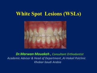
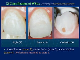
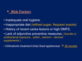
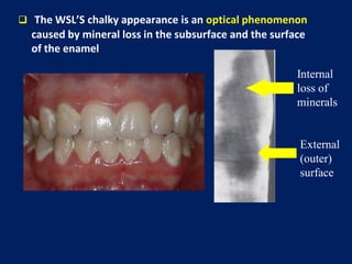
White Spot Lesions (WSLs)
Dr.Marwan Mouakeh , Consultant Orthodontist
Academic Adviser & Head of Department ,Al-Hokail Polclinic
Khobar-Saudi Arabia
White spot lesions (WSLs)
Definition :
• Decalcification of the enamel surface adjacent to
fixed orthodontic appliances .
 White spot lesions ( WSLs )
• A Prevalent iatrogenic effect of orthodontic therapy
• Incipient carious lesions develop around brackets
and bands usually near the gingival margin .
 Classifications of White Lesions on Enamel
• Dental Fluorosis
• Opacities
• White Spot Lesions
• Dental Fluorosis
 White / yellowish lesion
 Not well defined
 Symmetrical distribution
 Associated with cumulative
fluoride intake during enamel
development .
 Characteristics
 Dental Fluorosis
 Mild Fluorosis
 Severe Fluorosis
• Affected teeth are less
susceptible to dental caries .
• Enamel opacities (VS , Fluorosis)
 More defined shape.
 Well differentiated from
surrounding enamel.
 Often located in the middle of
the crown.
 Randomly distributed .
 Enamel Hypomineralization
 Well demarcated opacities
on the labial surface, due to
injury or infection of the
deciduous teeth, which has
affected mineralization of the
permanent teeth .
White spot lesions ( WSLs )
• Subsurface enamel
porosity from carious
demineralization .
• Manifesting as a milky
white opacity on the
enamel.
 Initial lesions :
• white decalcification with
beginning enamel breakdown
• affecting the primary teeth , not
associated with orthodontic
treatment .
 Early Childhood Caries
 White spot lesions (WSLs)
to be differentiated from
• A small lesion (score 2), severe lesion (score 3), and cavitation
(score 4). No lesion is recorded as score 1.
 Classification of WSLs according to Gorelick and coworkers
• Overall prevalence of WSLs related to fixed appliance
treatment ranges widely from 2 % - 96 %
• Significant increase in prevalence with orthodontic
treatment (72 % - 84 %)
 Prevalence of WSLs
• 49.6 % in orthodontic patients
( Ogaard et al AJO-O 1989)
• 24 % in an untreated controls
( Gorelick et al AJO-O 1982)
•Increased with Age and Treatment duration .
 Etiopathogenesis of WSLs
• Areas of Demineralized Enamel developed due to
prolonged plaque accumulation .
 Factors Necessary for Caries Development
 Formation of WSLs
Fixed orthodontic appliance
Rapid increase in dental plaque
Low pH
Increased cariogenic risk of S.mutans
Acid by-products
Additional lowering of pH
Decalcification of enamel
Development of WSLs
Plaque accumulation
Low pH adjacent to
orthodontic brackets
Inhibition of remineralization
and consequently decalcification
Streptococcus mutans
•Etiopathogenesis of WSLs
 Risk Factors
• Inadequate oral hygiene .
• Inappropriate diet (refined sugar, frequent snacks)
• History of recent caries lesions or high DMFS.
• Lack of adjunctive preventive measures ( fluoride or
antibacterial exposure , xylitol , calcium – derived
supplements ) .
• Orthodontic treatment time( fixed appliances) > 36 months
Diagnostic Methods
External
(outer)
surface
Internal
loss of
minerals
 The WSL’S chalky appearance is an optical phenomenon
caused by mineral loss in the subsurface and the surface
of the enamel
• Alteration of the enamel refractive index is the
consequence of both :
- surface roughness & loss of surface shine , plus
- alteration of the internal reflection
•Porous enamel scatters more light than
sound enamel .
• Visual Enamel Opacity ??
Wet
Dry
•Porous enamel scatters more
light than sound enamel .
 Fluorescence methods
– QLF
– Infra-red Fluorescence
 Transillumination
 Electrical Conductance
– ECM
 Digital Radiography
– DDR
• Diagnostic Methods
•Fluorescence Example
White Spot
Reflections obscuring image
 Location of WSL
 High prevalence on the cervical and middle thirds of the
crowns :
 1st Molars
 maxillary lateral incisors
 mandibular incisors and canines.
 Mainly on the vestibular surfaces
• The highest incidence of WSLs at the labio – gingival area of the
maxillary lateral incisors .
• A strong relationship between resistance to WSL formation and
the rate of salivary flow .
 Location of WSLs
 Differential Diagnosis of WSLs
- Carious , vs , Non carious Lesions
 Procedure :
 Clean & dry the teeth
 Carefully evaluate the lesion ( magnification &
lightning )
 Differential Diagnosis of WSLs
Carious , vs , Non carious Lesions
 Carious lesions : appear Rough ,Opaque ,and Porous
 Noncarious lesions : Smooth & Shiny appearance
 Evolution of WSLs
• WSL left untreated after removal
of a fixed appliance will naturally
reduce in size with no intervention .
• About 75 % of the small lesions
will regress during 6 months after
debonding provided the application
of caries – preventive program .
• Remineralization that could occur a few weeks following the
completion of orthodontic treatment ,it is the result of improved
oral hygiene and from the available minerals in saliva, fluoridated
toothpaste ….
• Discoloration of white
spot lesions .
• Evolution of WSLs
• Cavitation due to white
spot lesions .
Prevention & Treatment of WSLs
 Decalcification of enamel
 Prevention
• Oral hygiene
• Dentifrices
• Mouth rinses
• Varnishes
• Decalcification of enamel
 Prevention
• Good Oral hygiene
• Mouth rinses
 White Spot Lesions…
Can be Remineralized with
• Changes in diet
• Fluoride varnish
• Daily brushing with fluoride toothpaste.
 Fluoride
• Inhibits demineralization
• Enhances remineralization
• Inhibits plaque bacteria
• Prevention of WSLs
Demineralization <------------ > Remineralization
•Frequent carbohydrate intake
•Frequent exposure to acids
•Plaque presence
•Decreased salivary flow
•Exposure to fluoride
•Removal of plaque
•Balanced diet
•Limited exposure to carbohydrates
• Ways to Obtain Fluoride: Topical
 Fluoride Toothpaste
 Fluoride Varnish
 Fluoride Mouthrinses
Decalcification of enamel
 Prevention :
 F . Dentifrices
 Sodium Fluoride
Amine Fluoride
Monofluorophosphate
Stannous Fluoride ( +
Plaque inhibiting effect )
•Decalcification of enamel
 Prevention :
 F. Dentifrices
• Fluoride concentration ,
0.11% - 0.15% twice daily :
 Reduce demineralization and
 Enhance remineralization
• Fluoridated mouth rinses ( Na.F 0.05 %) :
An improved cariostatic effect if combined with Antibacterial
agents :
- Chlorhexidine : inhibits plaque formation
( - ) : Tooth discoloration , metallic taste
- Triclosan ( with Zinc ) : antibacterial + anti- inflammatory
effects .
• Prevention of WSLs
 Highly concentrated topical fluoride
treatment (22,600 ppm)
 Can reverse early white spot lesions.
• Temporary discoloration of teeth
and gingival tissue
• Decrease enamel decalcification
44.3 %
• Varnishes
• Prevention of WSLs
Fluoridated Sealants , Primers and Adhesives
• G.I Cements: Sustained F. Release in plaque around
brackets .
• Prevention of WSLs
Fluoridated Sealants , Primers and Adhesives
• Resin reinforced GIC = Better bond strength
• Prevention of WSLs
• Complicated designed appliances make oral hygiene difficult to carry out.
• Prevention of WSLs
• Use of S.S ligatures > Elastic modules
• Coil spring traction > Elastic chains …
• Prevention of WSLs
• Self-ligating Brackets …..
• Prevention of WSLs
• Use of Bondable Buccal Tubes …..
• Prevention of WSLs
•Excess bonding material retains plaque.
• Prevention of WSLs
• Indirect Bonding and / or Light-cure Technique…..
• Prevention of WSLs
Treatment protocols for
WSLs
•First , Allow Natural
Remineralization.
 Treatment of WSLs
• Avoid high concentration of F agents since they arrest
enamel remineralization .
• Allow Remineralization of enamel by Saliva (less
visible lesions) .
 Treatment of WSLs
• Shows a lower right canine, which had an orthodontic white
spot lesion that was treated at debond with strong fluoride
varnish. The lesion has not regressed and has stained brown.
 Treatment of WSLs
• Low doses of F applications (50 – ppm F. mouth rinses).
• Tooth Whitening “ Bleaching “
• Use of CPP –ACP “ Casein derivates “
• Enamel Microabrasion
• Cosmetic Restorations ( Veneers ) .
 Treatment methods
• F. mouth rinses : 0.05% Na F + Chlorhexidine .
• Topical F. gel or varnishes application
Topical Application of Titanium Tetrafluoride ( solution ) :
the mechanism of interaction with the enamel
 Strong Ti-O-Ti chains on the
enamel surface .
 A titanium-rich , glaze –like coating
formed on the enamel surface following
the application of Ti. Tetra F.
 Treatment of WSLs
• Chlorhexidine mouthwash
• Antimicrobial therapy .
• To achieve a shift from :
Unfavorably biofilm favorably biofilm
• Maybe used as a complement to F. therapy.
 Drawback : the tendency to stain the teeth
• Casein Phosphopeptide Amorphous
Calcium Phosphate (CPP-ACP )
 Treatment of WSLs
• Casein Phosphopeptide Amorphous Calcium
Phosphate (CPP-ACP )
• Trade name : Recaldent
• Derived from milk caseine
• Enhances formation of calcium phosphate crystals
 Recaldent CPP-ACP :
• Natural milk derived product containing casein phosphopeptides
CPP and amorphous calcium phosphates ACP
 Treatment of post-orthodontic white spot lesions with
casein phosphopeptide-stabilised amorphous calcium
phosphate .
Brochner et al, Clin Oral Invest, April 2010
 The mean area of the WS lesions
decreased by 58% in the CPP- ACP
group and 26% in the fluoride group .
• Casein Phosphopeptide Amorphous Calcium Phosphate (CPP-ACP )
7 days
4 weeks 12 weeks
 Recaldent CPP-ACP
Chewing Gum :
This gum contains the active CPP-ACP .
 Recaldent also contains Xylitol, which is
a natural sweetener shown to help control
mouth bacteria and improve saliva quality.
•The in vivo studies of the remineralization
properties of Recaldent (CPP-ACP) Gum
have shown subsurface mineral gain can
occur by chewing the gum for periods of 15
minutes for 2 weeks.
• Vital Tooth Bleaching
 Treatment of WSLs
•Vital Tooth Bleaching
•View of the same maxillary
anterior teeth following 4 weeks
of overnight vital bleaching with
10% carbamide peroxide
delivered in a custom tray.
 Treatment of WSLs
• View of the same maxillary
anterior teeth following 4
weeks of bleaching , 30 minutes
twice per day, with a hydrogen
peroxide gel impregnated on
polyethylene strips.
•Vital Tooth Bleaching
Microabrasion of the enamel
 Treatment of WSLs
 Microabrasion
• A technique to remove the superficial non- carious
superficial enamel defects as well as WSL .
• Topical application of an 18 % hydrochloric acid and
pumice , 1 – 2 minutes .
 Microabrasion
• 5 to 10 applications of the microabrasion compound
should be effective .
• The abraded enamel surface is less susceptible to
demineralization than natural enamel .
• Following the microabrasion technique , a 4-minute 2 % Na F
treatment is recommended .
• Cosmetic Restorations
in Severe Cases
 Treatment of WSLs
 Decalcification restored with composite resin
.
• Cosmetic Restorations
 ( ICon ): Caries Infiltration with light-curable
resin for non-cavitated lesions
• Patient who needed composite
buildups on the maxillary anterior
teeth to esthetically improve areas
of severe decalcification following
orthodontic treatment .
• Cosmetic Restorations
Clinical Test …..
 How Do You Proceed ?
 Problem : Inadequate oral hygiene, generalized
gingivitis, plaque accumulation, and white spot
lesions at the bucco-cervical surfaces.
 Discuss with the patient the risk factors,
 Make recommendations according to the problems (eg, diet
and oral hygiene behavioral assessment),
 Prescribe high-fluoride (5000 ppm) toothpaste and 0.12%
chlorhexidine rinses, and
 Reevaluate in few months to assess whether patient has
been compliant and able to proceed with orthodontic
treatment .
 How Do You Proceed ?
 What do you recommend?
Reevaluate the risk factors
Reinforce oral hygiene instructions,
Prescribe high-fluoride toothpaste and chlorhexidine rinses,
Apply fluoride varnish at least 2 or 3 times a year, and also
Recommend frequent use of xylitol or sugar-free gum.
 Problem : at the debonding appointment , you
note white spot lesions and areas of cavitation on
the patient.
What do you do?
 Summary : Treatment protocols for WSLs
• First , allow natural remineralization.
• Low doses of F applications (50 – ppm F. mouth rinses).
•Use of CPP –ACP “ Casein derivates “
• If the lesions persist , professional bleaching is
indicated.
• If the effect of bleaching is inadequate , Microabrasion
is an option.
• Lastly , direct or indirect Veneers could be considered.
 Conclusions :
• To prevent development of white spot lesions, orthodontists
should assess each patient’s risk factors before and during
treatment.
• Oral hygiene instructions are important, but patients
might need to be assisted with additional measures, including
fluoride varnish, chlorhexidine , dietary modification,
or calcium-containing remineralization products that can
help prevent enamel demineralization, enhance remineralization,
and modify patient and biofilm factors.
 Conclusions :
• Restorative treatment for established white spot
lesions can range from the most conservative
(remineralization with fluoride, calcium, and
phosphate) to the most aggressive (tooth reduction
and porcelain veneers).
 Conclusions :
It is crucial to establish a caries risk
assessment and recommendation
protocol for patients before, during, and
after treatment to be able to provide
overall successful orthodontic
treatments for them.
Thank You … Dr.Marwan Mouakeh
Syria –Aleppo, the public park

White spot lesions

  • 1.
    White Spot Lesions(WSLs) Dr.Marwan Mouakeh , Consultant Orthodontist Academic Adviser & Head of Department ,Al-Hokail Polclinic Khobar-Saudi Arabia
  • 2.
    White spot lesions(WSLs) Definition : • Decalcification of the enamel surface adjacent to fixed orthodontic appliances .
  • 3.
     White spotlesions ( WSLs ) • A Prevalent iatrogenic effect of orthodontic therapy • Incipient carious lesions develop around brackets and bands usually near the gingival margin .
  • 4.
     Classifications ofWhite Lesions on Enamel • Dental Fluorosis • Opacities • White Spot Lesions
  • 5.
    • Dental Fluorosis White / yellowish lesion  Not well defined  Symmetrical distribution  Associated with cumulative fluoride intake during enamel development .  Characteristics
  • 6.
     Dental Fluorosis Mild Fluorosis  Severe Fluorosis • Affected teeth are less susceptible to dental caries .
  • 7.
    • Enamel opacities(VS , Fluorosis)  More defined shape.  Well differentiated from surrounding enamel.  Often located in the middle of the crown.  Randomly distributed .
  • 8.
     Enamel Hypomineralization Well demarcated opacities on the labial surface, due to injury or infection of the deciduous teeth, which has affected mineralization of the permanent teeth .
  • 9.
    White spot lesions( WSLs ) • Subsurface enamel porosity from carious demineralization . • Manifesting as a milky white opacity on the enamel.
  • 10.
     Initial lesions: • white decalcification with beginning enamel breakdown • affecting the primary teeth , not associated with orthodontic treatment .  Early Childhood Caries  White spot lesions (WSLs) to be differentiated from
  • 11.
    • A smalllesion (score 2), severe lesion (score 3), and cavitation (score 4). No lesion is recorded as score 1.  Classification of WSLs according to Gorelick and coworkers
  • 12.
    • Overall prevalenceof WSLs related to fixed appliance treatment ranges widely from 2 % - 96 % • Significant increase in prevalence with orthodontic treatment (72 % - 84 %)  Prevalence of WSLs • 49.6 % in orthodontic patients ( Ogaard et al AJO-O 1989) • 24 % in an untreated controls ( Gorelick et al AJO-O 1982) •Increased with Age and Treatment duration .
  • 13.
     Etiopathogenesis ofWSLs • Areas of Demineralized Enamel developed due to prolonged plaque accumulation .
  • 14.
     Factors Necessaryfor Caries Development
  • 15.
     Formation ofWSLs Fixed orthodontic appliance Rapid increase in dental plaque Low pH Increased cariogenic risk of S.mutans Acid by-products Additional lowering of pH Decalcification of enamel
  • 16.
    Development of WSLs Plaqueaccumulation Low pH adjacent to orthodontic brackets Inhibition of remineralization and consequently decalcification Streptococcus mutans
  • 17.
  • 18.
     Risk Factors •Inadequate oral hygiene . • Inappropriate diet (refined sugar, frequent snacks) • History of recent caries lesions or high DMFS. • Lack of adjunctive preventive measures ( fluoride or antibacterial exposure , xylitol , calcium – derived supplements ) . • Orthodontic treatment time( fixed appliances) > 36 months
  • 19.
  • 20.
    External (outer) surface Internal loss of minerals  TheWSL’S chalky appearance is an optical phenomenon caused by mineral loss in the subsurface and the surface of the enamel
  • 21.
    • Alteration ofthe enamel refractive index is the consequence of both : - surface roughness & loss of surface shine , plus - alteration of the internal reflection •Porous enamel scatters more light than sound enamel . • Visual Enamel Opacity ??
  • 22.
    Wet Dry •Porous enamel scattersmore light than sound enamel .
  • 23.
     Fluorescence methods –QLF – Infra-red Fluorescence  Transillumination  Electrical Conductance – ECM  Digital Radiography – DDR • Diagnostic Methods
  • 24.
  • 25.
     Location ofWSL  High prevalence on the cervical and middle thirds of the crowns :  1st Molars  maxillary lateral incisors  mandibular incisors and canines.  Mainly on the vestibular surfaces
  • 26.
    • The highestincidence of WSLs at the labio – gingival area of the maxillary lateral incisors . • A strong relationship between resistance to WSL formation and the rate of salivary flow .  Location of WSLs
  • 27.
     Differential Diagnosisof WSLs - Carious , vs , Non carious Lesions  Procedure :  Clean & dry the teeth  Carefully evaluate the lesion ( magnification & lightning )
  • 28.
     Differential Diagnosisof WSLs Carious , vs , Non carious Lesions  Carious lesions : appear Rough ,Opaque ,and Porous  Noncarious lesions : Smooth & Shiny appearance
  • 29.
     Evolution ofWSLs • WSL left untreated after removal of a fixed appliance will naturally reduce in size with no intervention . • About 75 % of the small lesions will regress during 6 months after debonding provided the application of caries – preventive program .
  • 30.
    • Remineralization thatcould occur a few weeks following the completion of orthodontic treatment ,it is the result of improved oral hygiene and from the available minerals in saliva, fluoridated toothpaste ….
  • 31.
    • Discoloration ofwhite spot lesions . • Evolution of WSLs • Cavitation due to white spot lesions .
  • 32.
  • 33.
     Decalcification ofenamel  Prevention • Oral hygiene • Dentifrices • Mouth rinses • Varnishes
  • 34.
    • Decalcification ofenamel  Prevention • Good Oral hygiene • Mouth rinses
  • 35.
     White SpotLesions… Can be Remineralized with • Changes in diet • Fluoride varnish • Daily brushing with fluoride toothpaste.
  • 36.
     Fluoride • Inhibitsdemineralization • Enhances remineralization • Inhibits plaque bacteria • Prevention of WSLs
  • 37.
    Demineralization <------------ >Remineralization •Frequent carbohydrate intake •Frequent exposure to acids •Plaque presence •Decreased salivary flow •Exposure to fluoride •Removal of plaque •Balanced diet •Limited exposure to carbohydrates
  • 38.
    • Ways toObtain Fluoride: Topical  Fluoride Toothpaste  Fluoride Varnish  Fluoride Mouthrinses
  • 39.
    Decalcification of enamel Prevention :  F . Dentifrices  Sodium Fluoride Amine Fluoride Monofluorophosphate Stannous Fluoride ( + Plaque inhibiting effect )
  • 40.
    •Decalcification of enamel Prevention :  F. Dentifrices • Fluoride concentration , 0.11% - 0.15% twice daily :  Reduce demineralization and  Enhance remineralization
  • 41.
    • Fluoridated mouthrinses ( Na.F 0.05 %) : An improved cariostatic effect if combined with Antibacterial agents : - Chlorhexidine : inhibits plaque formation ( - ) : Tooth discoloration , metallic taste - Triclosan ( with Zinc ) : antibacterial + anti- inflammatory effects . • Prevention of WSLs
  • 42.
     Highly concentratedtopical fluoride treatment (22,600 ppm)  Can reverse early white spot lesions. • Temporary discoloration of teeth and gingival tissue • Decrease enamel decalcification 44.3 % • Varnishes • Prevention of WSLs
  • 43.
    Fluoridated Sealants ,Primers and Adhesives • G.I Cements: Sustained F. Release in plaque around brackets . • Prevention of WSLs
  • 44.
    Fluoridated Sealants ,Primers and Adhesives • Resin reinforced GIC = Better bond strength • Prevention of WSLs
  • 45.
    • Complicated designedappliances make oral hygiene difficult to carry out. • Prevention of WSLs
  • 46.
    • Use ofS.S ligatures > Elastic modules • Coil spring traction > Elastic chains … • Prevention of WSLs
  • 47.
    • Self-ligating Brackets….. • Prevention of WSLs
  • 48.
    • Use ofBondable Buccal Tubes ….. • Prevention of WSLs
  • 49.
    •Excess bonding materialretains plaque. • Prevention of WSLs
  • 50.
    • Indirect Bondingand / or Light-cure Technique….. • Prevention of WSLs
  • 51.
    Treatment protocols for WSLs •First, Allow Natural Remineralization.
  • 52.
     Treatment ofWSLs • Avoid high concentration of F agents since they arrest enamel remineralization . • Allow Remineralization of enamel by Saliva (less visible lesions) .
  • 53.
     Treatment ofWSLs • Shows a lower right canine, which had an orthodontic white spot lesion that was treated at debond with strong fluoride varnish. The lesion has not regressed and has stained brown.
  • 54.
     Treatment ofWSLs • Low doses of F applications (50 – ppm F. mouth rinses). • Tooth Whitening “ Bleaching “ • Use of CPP –ACP “ Casein derivates “ • Enamel Microabrasion • Cosmetic Restorations ( Veneers ) .
  • 55.
     Treatment methods •F. mouth rinses : 0.05% Na F + Chlorhexidine . • Topical F. gel or varnishes application
  • 56.
    Topical Application ofTitanium Tetrafluoride ( solution ) : the mechanism of interaction with the enamel  Strong Ti-O-Ti chains on the enamel surface .  A titanium-rich , glaze –like coating formed on the enamel surface following the application of Ti. Tetra F.  Treatment of WSLs
  • 57.
    • Chlorhexidine mouthwash •Antimicrobial therapy . • To achieve a shift from : Unfavorably biofilm favorably biofilm • Maybe used as a complement to F. therapy.  Drawback : the tendency to stain the teeth
  • 58.
    • Casein PhosphopeptideAmorphous Calcium Phosphate (CPP-ACP )  Treatment of WSLs
  • 59.
    • Casein PhosphopeptideAmorphous Calcium Phosphate (CPP-ACP ) • Trade name : Recaldent • Derived from milk caseine • Enhances formation of calcium phosphate crystals
  • 60.
     Recaldent CPP-ACP: • Natural milk derived product containing casein phosphopeptides CPP and amorphous calcium phosphates ACP
  • 61.
     Treatment ofpost-orthodontic white spot lesions with casein phosphopeptide-stabilised amorphous calcium phosphate . Brochner et al, Clin Oral Invest, April 2010  The mean area of the WS lesions decreased by 58% in the CPP- ACP group and 26% in the fluoride group .
  • 62.
    • Casein PhosphopeptideAmorphous Calcium Phosphate (CPP-ACP ) 7 days 4 weeks 12 weeks
  • 63.
     Recaldent CPP-ACP ChewingGum : This gum contains the active CPP-ACP .  Recaldent also contains Xylitol, which is a natural sweetener shown to help control mouth bacteria and improve saliva quality. •The in vivo studies of the remineralization properties of Recaldent (CPP-ACP) Gum have shown subsurface mineral gain can occur by chewing the gum for periods of 15 minutes for 2 weeks.
  • 64.
    • Vital ToothBleaching  Treatment of WSLs
  • 65.
    •Vital Tooth Bleaching •Viewof the same maxillary anterior teeth following 4 weeks of overnight vital bleaching with 10% carbamide peroxide delivered in a custom tray.  Treatment of WSLs
  • 66.
    • View ofthe same maxillary anterior teeth following 4 weeks of bleaching , 30 minutes twice per day, with a hydrogen peroxide gel impregnated on polyethylene strips. •Vital Tooth Bleaching
  • 67.
    Microabrasion of theenamel  Treatment of WSLs
  • 68.
     Microabrasion • Atechnique to remove the superficial non- carious superficial enamel defects as well as WSL . • Topical application of an 18 % hydrochloric acid and pumice , 1 – 2 minutes .
  • 69.
     Microabrasion • 5to 10 applications of the microabrasion compound should be effective . • The abraded enamel surface is less susceptible to demineralization than natural enamel . • Following the microabrasion technique , a 4-minute 2 % Na F treatment is recommended .
  • 70.
    • Cosmetic Restorations inSevere Cases  Treatment of WSLs
  • 71.
     Decalcification restoredwith composite resin . • Cosmetic Restorations  ( ICon ): Caries Infiltration with light-curable resin for non-cavitated lesions
  • 72.
    • Patient whoneeded composite buildups on the maxillary anterior teeth to esthetically improve areas of severe decalcification following orthodontic treatment . • Cosmetic Restorations
  • 73.
  • 74.
     How DoYou Proceed ?  Problem : Inadequate oral hygiene, generalized gingivitis, plaque accumulation, and white spot lesions at the bucco-cervical surfaces.
  • 75.
     Discuss withthe patient the risk factors,  Make recommendations according to the problems (eg, diet and oral hygiene behavioral assessment),  Prescribe high-fluoride (5000 ppm) toothpaste and 0.12% chlorhexidine rinses, and  Reevaluate in few months to assess whether patient has been compliant and able to proceed with orthodontic treatment .  How Do You Proceed ?
  • 76.
     What doyou recommend? Reevaluate the risk factors Reinforce oral hygiene instructions, Prescribe high-fluoride toothpaste and chlorhexidine rinses, Apply fluoride varnish at least 2 or 3 times a year, and also Recommend frequent use of xylitol or sugar-free gum.
  • 77.
     Problem :at the debonding appointment , you note white spot lesions and areas of cavitation on the patient. What do you do?
  • 78.
     Summary :Treatment protocols for WSLs • First , allow natural remineralization. • Low doses of F applications (50 – ppm F. mouth rinses). •Use of CPP –ACP “ Casein derivates “ • If the lesions persist , professional bleaching is indicated. • If the effect of bleaching is inadequate , Microabrasion is an option. • Lastly , direct or indirect Veneers could be considered.
  • 79.
     Conclusions : •To prevent development of white spot lesions, orthodontists should assess each patient’s risk factors before and during treatment. • Oral hygiene instructions are important, but patients might need to be assisted with additional measures, including fluoride varnish, chlorhexidine , dietary modification, or calcium-containing remineralization products that can help prevent enamel demineralization, enhance remineralization, and modify patient and biofilm factors.
  • 80.
     Conclusions : •Restorative treatment for established white spot lesions can range from the most conservative (remineralization with fluoride, calcium, and phosphate) to the most aggressive (tooth reduction and porcelain veneers).
  • 81.
     Conclusions : Itis crucial to establish a caries risk assessment and recommendation protocol for patients before, during, and after treatment to be able to provide overall successful orthodontic treatments for them.
  • 82.
    Thank You …Dr.Marwan Mouakeh Syria –Aleppo, the public park