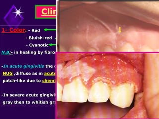
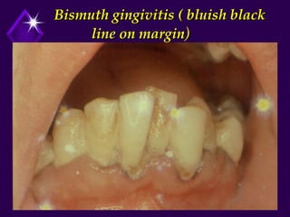
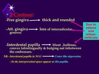
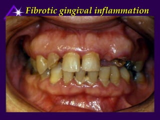
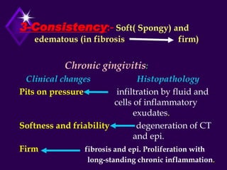
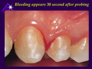
Clinical Features of Gingivitis
(Plaque-induced gingivitis)
Khalid S. Hassan
BDS,MSc,PhD ,Assist. Prof.
Department of PDS, Periodontics
Clinical Criteria of Healthy Gingiva
coral pink (alveolar mucosa is red )Color:-1
-Variation in color [degree of keratinization, vascularity, pigmentation and thickness of epi).
-Contour:-2
-Free gingiva thin and end in a knife like edge
-Attach gingiva scalloped elevations related to roots and depression in the
interradicular areas interradicular groove.
-Interdental papillae pointed and fill the inter proximal space under contact area.
-papillae (OrangeinterdentalStippling in attach. gingiva and base of-:textureSuface-3
peel appearance)
(Firmly attached to the teeth and underling alveolarresillentFirm and-:Consistency-4
bone except free gingiva).
mm-3mm not exceed2-5.0-Depth of gingival sulcus:-5
.transudate:Type of gingival fluid-6
•Course and Duration:
Acute gingivitis: painful condition,
sudden onset, and short duration.
Sub-acute gingivitis: less severe than
acute gingivitis.
Recurrent gingivitis: reappear after
treatment.
Chronic gingivitis: comes in slowly, is
of long duration, and painless.
Chronic Gingivitis
Def. It is a simple and long-standing inflammation of
the gingiva (gingivitis may exist for years
without change to periodontitis)
Types. 1- Localized 2- Generalized
Distribution of inflammation:-
1- Papillary (localized or generalized)
2- Marginal (localized or generalized)
3- Diffuse (localized or generalized)
Chronic localized marginal
gingivitis
Chronic generalized marginal
gingivitis
Generalized diffuse gingivitis
Etiology:-
1-Local initiating factor (Dental Plaque).
2- Local predisposing factors.
3- Systemic factors
* Puberty
* Pregnancy
* Diabetes
* Vit. C deficiency
Etiology of Periodontal Diseases
Bacterial plaque Host response Reparative tissue capacity
Factors upset the
balance By
Increasing aggression of bacterial plaque
Decreasing defense mechanisms
Local Predisposing Factors Systemic Factors
1- Calculus
2- Material alba
3- Food debris
4- Food impaction
5- Food retention
6- Faulty dentistry
7- Malocclusion
8- Mouth breathing
9- Trauma from occlusion
* Overhanging filling
* Over and under contoured crowns
* Occlusal disharmony
* Orthodontic wires
* Rough fillings
* Open contact
1- Hormonal imbalance
2- Nutritional deficiency
3- Blood diseases
4- Genetics
5- Immunologic
6- Metallic intoxication
7- Debilitating diseases
8- Psychologic
Bacteria associated with
Gingivitis
Gr+ve species
S. Sangus
S. Mitis
S. Intermedius
S. Oralis
A . Viscosus
A . Naeslundii
Peptostrepococcus
Gr-ve species
F. Nucleatum
P.intermedia
Haemophilus
Capnocytophaga
Clinical Features
1- Color: - Red V.D. of capillary bed, keratinization
- Bluish-red b1. stasis
- Cyanotic 02 tension
N.B:- in healing by fibrosis pale pink.
-In acute gingivitis the color change may be marginal as in
NUG ,diffuse as in acute herpetic gingivostomatitis or
patch-like due to chemical irritation.
-In severe acute gingivitis, the red color change to a shiny
gray then to whitish gray due to necrosis.
Factors affecting the color
• Endogenous Factors ( systemic factors):
- melanin , bilirubin or iron.
- examples : Addison disease bluish black to brown.
- Peutz-jeghers syndrome melanin pigmentation.
- Albright’s syndrome melanin pigmentation.
• Exogenous Factors: - Metal dust.
- Coloring agents in food.
- Lozenges.
- Tobacco.
- Amalgam tattoo.
Metallic Pigmentation
Bismuth Lead Mercury Others
Bluish-black
pigmentation
of gingival
margin(liner)
-Liner pig. of
gingival margin.
(Burtonian line)
-Steel gray
-Liner
to diffuse
grayish
gingival
pigmentation
-Ulceration
of gingiva
-e.g.: phosphorous,
arsenic & chromium
-Bluish gray line.
-Necrosis of alveolar
bone loosening of
teeth
-Inflammation and
ulceration of gingiva.
Bismuth gingivitis ( bluish black
line on margin)
Due to
edema
and
inflam.
infiltrate
•2-Contour:
-Free gingiva thick and rounded
-Att. gingiva loss of interradicular
grooves
-Interdental papilla blunt ,bulbous,
convex labiolingually & bulging out inbetween
the embrasure.
NB.- Interdental papilla in NUG Crater like depression.
- In the interproximal space appear as flat papilla.
Plaque-induced gingivitis
Fibrotic gingival inflammation
Crater-licke depression papilla (NUG)
3-Consistency:- Soft( Spongy) and
edematous (in fibrosis firm)
Chronic gingivitis:
Clinical changes Histopathology
Pits on pressure infiltration by fluid and
cells of inflammatory
exudates.
Softness and friability degeneration of CT
and epi.
Firm fibrosis and epi. Proliferation with
long-standing chronic inflammation.
4- Surface texture:- Loss of stippling due to
-Stagnation of circulation
-Accumulation of inflammatory exudates
-Degeneration of gingival fibers
5- Position of gingiva:- gingival margin change to
coronal position deeping of gingival sulcus
without apical migration of epi. attachment
gingival pocket (pseudo-pocket- relative pocket).
-Actual position: is the level of the epi. attachment on the tooth.
-Apparent position: is the level of the crest of the gingival margin.
-The position of gingiva is determined by the actual position, not
the apparent position.
•6- Gingival Bleeding on Probing:-
- indicates an inflammatory lesion in the
epith. and CT.
- due to - Ulceration of sulcular epithelium.
- Thinning and degeneration of epi.
- Increase vascularity.
- Dilatation of the capillaries.
Acute gingival bleeding
• Caused by injury or occur spontaneously in
acute gingival disease.
• Laceration of the gingiva by aggressive
tooth brushing or sharp pieces of hard food
causes bleeding even in the absence of
gingival diseases.
• Spontaneous bleeding or on minor trauma
occurs in NUG.
Gingival Bleeding Associated with
Systemic Factors
• Occurs spontaneously or after irritation.
• Excessive and difficult to control.
• Occurs in other organs e.g. skin , internal organs.
• Examples : Vit. C deficiency.
- Thrombocytopenic purura.
- Vit K deficiency.
- Hemophilia .
- Leukemia.
- Some medications.
How do to determine
bleeding on probing
Insertion of a probe in gingival
sulcus
Bleeding appears 30 second after probing
Generalized gingival bleeding on
probing
•7-Pain:
- Chronic gingivitis is usually painless
unless complicated by acute
exacerbation.
- Chronically inflamed gingiva may be
painful on instrumentation (scaling
and root planning).
•Stillman’s Cleft:
-Described by Stillman.
-Is apostrophe-shaped indentation on
gingival margin.
- Is a specific type of gingival recession.
-Generally occur on the facial surface.
-Considered to be the result of occlusal
trauma.
-The margins of the clefts are rolled.
- Clinical significant: difficult to
maintain plaque control.
•McCall’s Festoons:
Enlargement of the gingival margin in the
canine and premolar area on the facial
surface.
In the early stages, the color and
consistency are normal.
Clinical significance: accumulation of food
debris leads to secondary inflammatory changes.
Histopathology:-
1- Inflammatory cell infiltration
2- Ulceration of sulcular epi.
3-Inter and intra-cellular edema.
4- Fibrosis in longstanding inflammation
Treatment:-
1- Thorough scaling.
2- Removal of local factors.
3- Plaque control and oral hygiene instruction.
Prognosis:- Excellent
Chronic gingivitis

Chronic gingivitis

  • 2.
    Clinical Features ofGingivitis (Plaque-induced gingivitis) Khalid S. Hassan BDS,MSc,PhD ,Assist. Prof. Department of PDS, Periodontics
  • 3.
    Clinical Criteria ofHealthy Gingiva coral pink (alveolar mucosa is red )Color:-1 -Variation in color [degree of keratinization, vascularity, pigmentation and thickness of epi). -Contour:-2 -Free gingiva thin and end in a knife like edge -Attach gingiva scalloped elevations related to roots and depression in the interradicular areas interradicular groove. -Interdental papillae pointed and fill the inter proximal space under contact area. -papillae (OrangeinterdentalStippling in attach. gingiva and base of-:textureSuface-3 peel appearance) (Firmly attached to the teeth and underling alveolarresillentFirm and-:Consistency-4 bone except free gingiva). mm-3mm not exceed2-5.0-Depth of gingival sulcus:-5 .transudate:Type of gingival fluid-6
  • 4.
    •Course and Duration: Acutegingivitis: painful condition, sudden onset, and short duration. Sub-acute gingivitis: less severe than acute gingivitis. Recurrent gingivitis: reappear after treatment. Chronic gingivitis: comes in slowly, is of long duration, and painless.
  • 5.
    Chronic Gingivitis Def. Itis a simple and long-standing inflammation of the gingiva (gingivitis may exist for years without change to periodontitis) Types. 1- Localized 2- Generalized Distribution of inflammation:- 1- Papillary (localized or generalized) 2- Marginal (localized or generalized) 3- Diffuse (localized or generalized)
  • 6.
  • 7.
  • 8.
  • 9.
    Etiology:- 1-Local initiating factor(Dental Plaque). 2- Local predisposing factors. 3- Systemic factors * Puberty * Pregnancy * Diabetes * Vit. C deficiency
  • 10.
    Etiology of PeriodontalDiseases Bacterial plaque Host response Reparative tissue capacity Factors upset the balance By Increasing aggression of bacterial plaque Decreasing defense mechanisms Local Predisposing Factors Systemic Factors 1- Calculus 2- Material alba 3- Food debris 4- Food impaction 5- Food retention 6- Faulty dentistry 7- Malocclusion 8- Mouth breathing 9- Trauma from occlusion * Overhanging filling * Over and under contoured crowns * Occlusal disharmony * Orthodontic wires * Rough fillings * Open contact 1- Hormonal imbalance 2- Nutritional deficiency 3- Blood diseases 4- Genetics 5- Immunologic 6- Metallic intoxication 7- Debilitating diseases 8- Psychologic
  • 11.
    Bacteria associated with Gingivitis Gr+vespecies S. Sangus S. Mitis S. Intermedius S. Oralis A . Viscosus A . Naeslundii Peptostrepococcus Gr-ve species F. Nucleatum P.intermedia Haemophilus Capnocytophaga
  • 12.
    Clinical Features 1- Color:- Red V.D. of capillary bed, keratinization - Bluish-red b1. stasis - Cyanotic 02 tension N.B:- in healing by fibrosis pale pink. -In acute gingivitis the color change may be marginal as in NUG ,diffuse as in acute herpetic gingivostomatitis or patch-like due to chemical irritation. -In severe acute gingivitis, the red color change to a shiny gray then to whitish gray due to necrosis.
  • 13.
    Factors affecting thecolor • Endogenous Factors ( systemic factors): - melanin , bilirubin or iron. - examples : Addison disease bluish black to brown. - Peutz-jeghers syndrome melanin pigmentation. - Albright’s syndrome melanin pigmentation. • Exogenous Factors: - Metal dust. - Coloring agents in food. - Lozenges. - Tobacco. - Amalgam tattoo.
  • 14.
    Metallic Pigmentation Bismuth LeadMercury Others Bluish-black pigmentation of gingival margin(liner) -Liner pig. of gingival margin. (Burtonian line) -Steel gray -Liner to diffuse grayish gingival pigmentation -Ulceration of gingiva -e.g.: phosphorous, arsenic & chromium -Bluish gray line. -Necrosis of alveolar bone loosening of teeth -Inflammation and ulceration of gingiva.
  • 15.
    Bismuth gingivitis (bluish black line on margin)
  • 17.
    Due to edema and inflam. infiltrate •2-Contour: -Free gingivathick and rounded -Att. gingiva loss of interradicular grooves -Interdental papilla blunt ,bulbous, convex labiolingually & bulging out inbetween the embrasure. NB.- Interdental papilla in NUG Crater like depression. - In the interproximal space appear as flat papilla.
  • 18.
  • 19.
  • 20.
  • 21.
    3-Consistency:- Soft( Spongy)and edematous (in fibrosis firm) Chronic gingivitis: Clinical changes Histopathology Pits on pressure infiltration by fluid and cells of inflammatory exudates. Softness and friability degeneration of CT and epi. Firm fibrosis and epi. Proliferation with long-standing chronic inflammation.
  • 22.
    4- Surface texture:-Loss of stippling due to -Stagnation of circulation -Accumulation of inflammatory exudates -Degeneration of gingival fibers 5- Position of gingiva:- gingival margin change to coronal position deeping of gingival sulcus without apical migration of epi. attachment gingival pocket (pseudo-pocket- relative pocket). -Actual position: is the level of the epi. attachment on the tooth. -Apparent position: is the level of the crest of the gingival margin. -The position of gingiva is determined by the actual position, not the apparent position.
  • 23.
    •6- Gingival Bleedingon Probing:- - indicates an inflammatory lesion in the epith. and CT. - due to - Ulceration of sulcular epithelium. - Thinning and degeneration of epi. - Increase vascularity. - Dilatation of the capillaries.
  • 24.
    Acute gingival bleeding •Caused by injury or occur spontaneously in acute gingival disease. • Laceration of the gingiva by aggressive tooth brushing or sharp pieces of hard food causes bleeding even in the absence of gingival diseases. • Spontaneous bleeding or on minor trauma occurs in NUG.
  • 25.
    Gingival Bleeding Associatedwith Systemic Factors • Occurs spontaneously or after irritation. • Excessive and difficult to control. • Occurs in other organs e.g. skin , internal organs. • Examples : Vit. C deficiency. - Thrombocytopenic purura. - Vit K deficiency. - Hemophilia . - Leukemia. - Some medications.
  • 26.
    How do todetermine bleeding on probing
  • 27.
    Insertion of aprobe in gingival sulcus
  • 28.
    Bleeding appears 30second after probing
  • 29.
  • 30.
    •7-Pain: - Chronic gingivitisis usually painless unless complicated by acute exacerbation. - Chronically inflamed gingiva may be painful on instrumentation (scaling and root planning).
  • 31.
    •Stillman’s Cleft: -Described byStillman. -Is apostrophe-shaped indentation on gingival margin. - Is a specific type of gingival recession. -Generally occur on the facial surface. -Considered to be the result of occlusal trauma. -The margins of the clefts are rolled. - Clinical significant: difficult to maintain plaque control.
  • 32.
    •McCall’s Festoons: Enlargement ofthe gingival margin in the canine and premolar area on the facial surface. In the early stages, the color and consistency are normal. Clinical significance: accumulation of food debris leads to secondary inflammatory changes.
  • 33.
    Histopathology:- 1- Inflammatory cellinfiltration 2- Ulceration of sulcular epi. 3-Inter and intra-cellular edema. 4- Fibrosis in longstanding inflammation Treatment:- 1- Thorough scaling. 2- Removal of local factors. 3- Plaque control and oral hygiene instruction. Prognosis:- Excellent