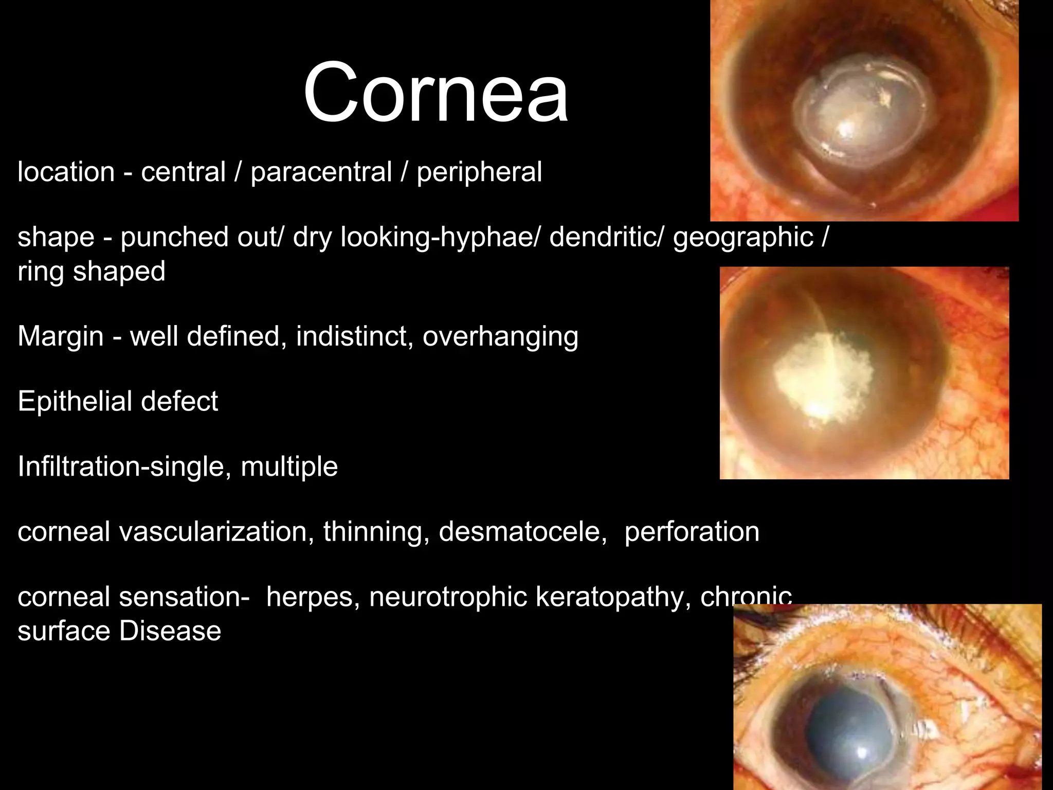
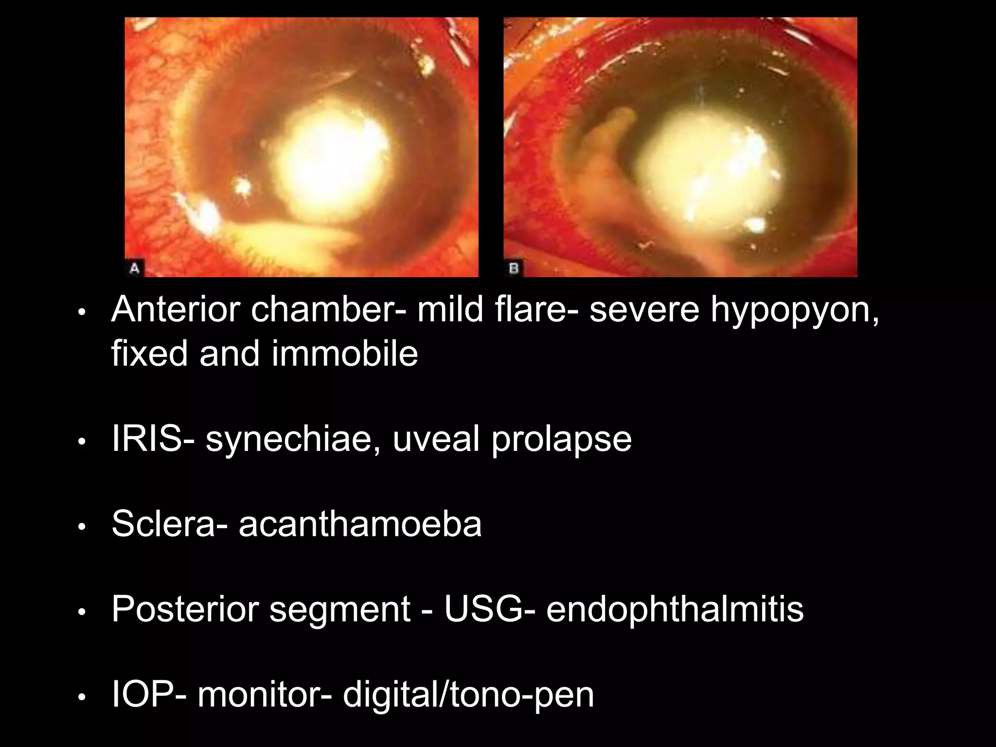
CORNEAL ULCER
Dr SHREEJI SHRESTHA.
Definition
• Tissue excavation associated with
epithelial defect usually with infiltration
and necrosis characterized by edema,
cellular infiltration and ciliary
congestion
(kanski)
Injury and foreign material
binds to iron used in microbial metabolism
Lysis of bacterial cell wall
Degrades CW of GPB
Prevent bacterial adherence
Activation of T cell and secretion of IgA
Microbiology
Eukaryotes Prokaryotes
protozoa
fungi
toxoplasma,
acanthamoeba
yeast like
Filamentous
candida, cryptococcous, rhinosporidium
Septate
Aseptate
Aspergillous, fusarium
Mucor, rhizopus
Gram positive cocci
gram negative cocci
Gram positive rods
gram negative rods
True bacteria
Filamentous bacteria
Actinomyces, mycobacterium
nocardia
Virus
HSV, HZV, EBV, CMV, Kaposi, HHV8
Enterococcus- endophthalmitis
P. Acne-post op endophthalmitis
B cerus- explosive endophthalmitis(soil)
H influenza- glaucoma Sx
M chelonei-immunocompromised patient
P aeruginosa- Contact lens
Acanthamoeba- swimming pool, hot tubs, tap water
Streptococcus Pneumococcus- dacryocystitis
Actinomyces - cannaliculits
Atypical mycobacteria- post LASIK sx
Moraxella - chronic alcoholism
Pathogensis
• corneal insult- PMN and phagocytes-
collagenase, elastase, cathepsin, oxygen free
radicals- destruction of cornea
• Reactive fibroblast- collagen - repair of cornea
Stages of corneal ulcer
• stage 1- Progressive stage
Saucer shaped, gray zone of infiltration.
Microbes adhere- PMN (tears & limbal vessel)-
invasion of cornea- necrosis & sloughing of
epithelium, BM, Stroma- ulcer progress by lateral
extension- deep penetration
stage 2 - Regressive stage
• Natural defense mechanism and antimicrobial
treatment
line of demarcation forms & margin and floor -
smooth & transparent
Enlargement of ulcer(digestion of necrotic material)
Superficial vascularization
Stage 3- healing stage
• Epithelization
• Kerotocyte - fibroblast- scar (ghost vessel)
• Cicatrization - collagen repair & fibrous tissue
(fibers arenot laid in regular manner- scar)
Complications
Pneucoccous
Staphyloma
Cicatrix
Hemorrhage
risk factor
Systemic
• Diabetes mellitus
• Sjögren’s syndrome
• Steven-Johnson syndrome
• AIDS
• Advanced malignancies
• Connective tissue disorders
• Alcoholics
• Extremes of age
• Measles malnutrition
Occupational
• Farmers
• Animal handlers
• Gardeners
Ocular
• Trauma
• Contact lenses
• Lid and adnexal
infections
• Ocular surface disease
• Allergic eye disorders
• Bullous keratopathy
• Topical medications
• Prior ocular surgery
Symptoms
• pain- superficial ulcer > deep ulcer
Onset
Acanthamoeba (radial keratoneuritis)> herpes simplex/fungal
relief pain-perforation
• Redness and photophobia
• Blurring of vision- central (staph,pseudomonas,fusarium)-
hypopyon, cataract, glaucoma
• discharge(mucopururlent, membranous)
clinical examination
• lacrimal sac- R/O dacryocystitis
• Eyelid - blisters, abnormal blink rate, lid swelling,
trichiasis, ectropion, lagophthalmous, blepharitis,
foreign body
• Conjunctiva -Circumciliary congestion,
hemophilus, pneumococcal, gonococcal-
chemosis,
• Precorneal tear film- filaments, dry eye
Cornea
location - central / paracentral / peripheral
shape - punched out/ dry looking-hyphae/ dendritic/ geographic /
ring shaped
Margin - well defined, indistinct, overhanging
Epithelial defect
Infiltration-single, multiple
corneal vascularization, thinning, desmatocele, perforation
corneal sensation- herpes, neurotrophic keratopathy, chronic
surface Disease
Grading
Feature Mild Moderate Severe
Size of ulcer (mm) < 2 2-5 > 5
Depth of ulcer (%) < 20 20-50 > 50
Infiltrate
Density Dense Dense Dense
Extent Superficial Extension upto mid
stroma
Deeper than mid
stroma
Scleral involvement Not involved Not involved May be involved
• Anterior chamber- mild flare- severe hypopyon,
fixed and immobile
• IRIS- synechiae, uveal prolapse
• Sclera- acanthamoeba
• Posterior segment - USG- endophthalmitis
• IOP- monitor- digital/tono-pen
work out- microbiological
• corneal scrapping(anesthesia-proparacaine)-
Kimura spatula/23-gauge needle/, edges(strep)
and base(moraxella)avoid contact with lash- gm
stain , giemsa, PAS, GMS,KOH, blood,
chocolate, SDA( row of c)
Blood agar Expect neisseia, hemophilus
Chocolate Neisseria, hemophilus,, moraxella
SDA Fungi
Non-nutrient with ecoli Acanthamoeba
Cooked meat Anaerobic
LJ Mycobacterium
Thioglycolate Diphtheria
Thayer Martin Neisseria
Types of
stain
Organism
Color
Gm Bacteria Purple, pink
Calcoflour
white
Fungi Bright green
acid fast Mycobacteria
Acridine
Bacteria , fungi,
acanthamoeba
Yellow-orange
• Conjunctival swab- corneal scrape is negative
• Contact lens cases
• corneal biopsy- absense of clinical improvement
(4 days)
• Bacterial - single suppurative inflitrate, sharp
Epithelial demarcation, stromal infiltration,
mycobacterium- nonsuppurative infiltrate, intact
epithelium
• Fungi - less inflammatory sign, less conjunctival
injection, pain out of proportion that of inflamed
cornea, irregular feathery/filamentous margin,
satellite (multiple) infiltrate, more AC inflammation,
rapid progression with necrosis, yellow-white
suppurative infiltrate, sterile immune ring
Staphylococcus ulcer
Pseudomonas
diffuse epithelial graying which characteristically occurs away
from the main site of epithelial and stromal infiltration
Moraxella
Streptococcus viridans
Streptococcus pneumonias
leading edge forming a serpiginous contour
Acanthamoeba
Candida (collar stud)Curvularia
Acremonium
Treatment
• General considerations
• Hospital admission
• Discontinue contact lens wear
• eye shield
• Decision to treat- clinically sterile, small infiltrate-
low frequency topical antibiotics
parameter to monitor clinical
therapy
• Blunting / decrease density of stromal infiltrate
• Dec AC reaction
• Dec stromal edema
• Reepithelization
• Cessation of corneal thinning
local therapy
• 1)antibiotic monotherapy- fluoroquinolone
• 2) antibiotic duotherapy- aggressive/streptococci-
Cephalorosporin 50mg/ml(fortified) and
aminoglycoside 14mg/ml (fortified)
• 3) Subcutaneous antibiotic- poor compliance with
topical treatment
• 4)mydriatics
• 5) ascorbate
• Steroid
1) steroids should not be used initially or if the eye is improving
(2) steroids should be used after several days of antibiotics if
there is persistent inflammation
(3) continue use of concomitant antibiotics
(4) steroids should not be used if there is corneal thinning
Systemic antibiotics
• Indications
• Potential for systemic involvement
• Severe corneal thinning
• scleral involvement
Causes of non healing ulcer
• Local - concretions, misdirected cilia, FB,
dacryocystits, inadequate therapy, wrong
diagnosis, lagopthalmos
• Systemic- DM, severe anemia, malnutrition,
chronic debilitating disease, patient on systemic
steroid
Mx of treatment failure
• + if no improvement (48hrs)- review antibiotic, contact lab
• + culture- resistance but clinically improving- no change in
therapy
• + still no improvement- rescrapping
• + culture - neg - corneal biopsy- histology and culture
• + resistant to medical therapy —excisional Keratoplasty
Antifungal• Epithelial debridement- mucus, necrotic tissue,
penetration
• Topical treatment- (filamentous)natamycin 5% -
fungistatic hourly day and 2 hourly at night +
fluroquinolone, once infiltrate decrease in size 2hourly
till complete resolution and continued for 2 weeks after
resolution——(candida) amphotericin0.15%(fungicidal)
12weeks
• decrease pain, infiltrate, satellite, rounding out of
feathery margins
• Subconjunctival fluconazole- severe case
• Intracameral/intrastromal - amphotericin B
• Systemic antifungal(6-8wks)—
lesion near limbus and enophthalmitis, scleritis, deep keratitis -
fluconazole 200mg/day or itraconazole 200mg/day or ketoconazole
600mg/day
• Tetracycline- thinning(collagenase inhibitor)
Promotion of epithelial
healing
• Reduce toxic exposure
• Lubrication, punctual plugs, cholinergic enhancer
(pilocarpine 5-10mg TID)
• Bandage soft contact lens- high water content
• surgical eyelid closure- neurotropic keratopathy,
persistent defect, botulinum LPS, lat tarsorrhaphy
• autologous serum drops, Amniotic membrane patch ,
tissue adhesive, limbal stem cell transplantation
Viral(HSV) keratitis
• Primary infection
Occurs in childhood, spread by droplet
uncommon (6 months)- maternal antibodies
• Recurrent infection
Primary infection — virus (trigeminal
ganglion)(latent infection)
• Subclinical reactivation - contagious- shed in tear
film
• Clinical reactivation - fever, hormonal, UV
trauma,— clinical reactivation—virus transported
to periphery
Epithelial keratitis
• Dendritic ulcer(centrally), terminal buds
(fluorescein)(loss of cellular integrity), margin stain with rose
bengal(loss of mucin binding cells), sub epithelial haze
• Corneal sensation
• Inadvertent Steroid-
geographical ulcer
immune mediated Stromal
keratitis
• central stromal edema + epithelial edema
• KP, DM folds, wessely immune ring(deposition of Ag-Ab
complex and PMN infiltration), IOP maybe Inc, sensation
decreased, stormal vascularization
Necrotizing stromal keratitis
• stromal necrosis and melting
• Anterior uveitis with KP, epithelial defect (+/_)
• Progress to scaring, vascularization
Herpetic endothelilitis
• Delayed type 4 hypersensitivity
reaction(disciform keratitis, diffuse keratitis or
linear endothelitis)
Neurotrophic ulceration
• Failure of re-epithelization from corneal
anesthesia, decrease tear formation,
exacerbation by chronic use of topical
medication(BM damage)
Recurrent corneal erosion
Non healing sterile ulceration, stromal is grey and
rolled up margin
Lab investigation
• Giemsa Staining
• Polymerase Chain Reaction (PCR)
• Viral Culture
• Immunological Tests- ELISA
Treatment
• topical acyclovir 3%/ganciclovir 0.15%
5times/Trifluridine 1%(9 times)- tapered- 3weeks
- (hydroxypropylmethycellulose)
• Debridement - (dendritic) eliminates antigenic stimuli to stromal inflammation
• Oral antiviral - immunodeficient patient, necrotizing stromal keratitis
• topical steroid - used only in stromal- keratitis - (2-4)hrly and tapered over 6
weeks
HEDS study
do topical steroid treat stromal
keratitis
Yes. also decrease duration of
keratitis
Is oral acyclovir ( steroid+trifluridine) helpful
in stormal keratitis No
Is oral acyclovir helpful in HSV
iritis
Favoring use of oral acyclovir
does oral acyclovir prevent epithelial
to stormal keratitis
No
Does oral acyclovir decrease
HSV recurrence
Yes
any triggers of HSV
reoccurence
No
herpes zoster ophthalmicus
• chicken pox- VZV- (retrograde axoplasmic
flow)dorsal root+ cranial nerve sensory ganglia-
axonal transport(latent state)- mucous
membrane and skin
• HZO(ophthalmic division of trigeminal nerve)
• Direct invasion — epithelial keratitis
• Reactivation —necrosis and inflammation in
ganglia — corneal anesthesia— neurotrophic
keratitis
Signs
• Hutchinson sign
• Age
• AIDS - more severe
Features
• Prodromal phase(3-5)- itching, burning
sensation—severe deep boring pain, fever
• skin lesion - midline, erythematous area with
maculopapular vesicular rash (lower lid not
affected, doesn't cross midline), edema of lower
lid- depigmented scar
Differentiation between dendrites of herpes
simplex and zoster
Feature HSV VZV
Over all fine, lacy (dendrite) Thick, ropy(pseudodendrite)
Epithelium
central Linear defect with
bared storm surrounded by
edematous epithetlial cells
small Elevated, painted-on
appearance
Staining Base stains with fluorescein min staining
Terminal bulb Frequent None
Dermatome Incomplete(epidermis) Complete(deep dermis)
skin scar - +
PHN - +
BL Uncommon(2%) _
Corneal
hypesthesia
Sectoral Severe
Zoster sine herpete
• Iritis, disciform keratitis, and facial palsy (Bell’s
palsy)
• defined as reactivated VZV which causes only
neurologic symptoms such as dermatomal
neuralgia or neuropathy, and, on occasion,
ocular inflammation - no rash
Treatment
• oral acyclovir 800mg 5 times a day(72hrs) - 7-10
days
• Encephalitis - IV acyclovir 5-10mg/kg
• Systemic steroids- accelerate skin healing
• Skin - warm compress BD followed by complete
drying
Reference
•Jacobiek Principles and Practive of
Ophthalmology 3rd Edition Vol 1
•Section 08 - External Disease And Cornea -
American Academy Of Ophthalmology –
2012
•Corneal Ulcers Diagnosis and management-
Jaypee- Namrata sharma, Rasik B Vajpayee
•Kanski Clinical Ophthalmology 7 Ed

Corneal ulcer

  • 1.
  • 2.
    Definition • Tissue excavationassociated with epithelial defect usually with infiltration and necrosis characterized by edema, cellular infiltration and ciliary congestion (kanski)
  • 3.
    Injury and foreignmaterial binds to iron used in microbial metabolism Lysis of bacterial cell wall Degrades CW of GPB Prevent bacterial adherence Activation of T cell and secretion of IgA
  • 4.
    Microbiology Eukaryotes Prokaryotes protozoa fungi toxoplasma, acanthamoeba yeast like Filamentous candida,cryptococcous, rhinosporidium Septate Aseptate Aspergillous, fusarium Mucor, rhizopus Gram positive cocci gram negative cocci Gram positive rods gram negative rods True bacteria Filamentous bacteria Actinomyces, mycobacterium nocardia Virus HSV, HZV, EBV, CMV, Kaposi, HHV8
  • 5.
    Enterococcus- endophthalmitis P. Acne-postop endophthalmitis B cerus- explosive endophthalmitis(soil) H influenza- glaucoma Sx M chelonei-immunocompromised patient P aeruginosa- Contact lens Acanthamoeba- swimming pool, hot tubs, tap water Streptococcus Pneumococcus- dacryocystitis Actinomyces - cannaliculits Atypical mycobacteria- post LASIK sx Moraxella - chronic alcoholism
  • 6.
    Pathogensis • corneal insult-PMN and phagocytes- collagenase, elastase, cathepsin, oxygen free radicals- destruction of cornea • Reactive fibroblast- collagen - repair of cornea
  • 7.
    Stages of cornealulcer • stage 1- Progressive stage Saucer shaped, gray zone of infiltration. Microbes adhere- PMN (tears & limbal vessel)- invasion of cornea- necrosis & sloughing of epithelium, BM, Stroma- ulcer progress by lateral extension- deep penetration
  • 8.
    stage 2 -Regressive stage • Natural defense mechanism and antimicrobial treatment line of demarcation forms & margin and floor - smooth & transparent Enlargement of ulcer(digestion of necrotic material) Superficial vascularization
  • 9.
    Stage 3- healingstage • Epithelization • Kerotocyte - fibroblast- scar (ghost vessel) • Cicatrization - collagen repair & fibrous tissue (fibers arenot laid in regular manner- scar)
  • 10.
  • 12.
  • 14.
  • 16.
    risk factor Systemic • Diabetesmellitus • Sjögren’s syndrome • Steven-Johnson syndrome • AIDS • Advanced malignancies • Connective tissue disorders • Alcoholics • Extremes of age • Measles malnutrition Occupational • Farmers • Animal handlers • Gardeners Ocular • Trauma • Contact lenses • Lid and adnexal infections • Ocular surface disease • Allergic eye disorders • Bullous keratopathy • Topical medications • Prior ocular surgery
  • 17.
    Symptoms • pain- superficialulcer > deep ulcer Onset Acanthamoeba (radial keratoneuritis)> herpes simplex/fungal relief pain-perforation • Redness and photophobia • Blurring of vision- central (staph,pseudomonas,fusarium)- hypopyon, cataract, glaucoma • discharge(mucopururlent, membranous)
  • 18.
    clinical examination • lacrimalsac- R/O dacryocystitis • Eyelid - blisters, abnormal blink rate, lid swelling, trichiasis, ectropion, lagophthalmous, blepharitis, foreign body • Conjunctiva -Circumciliary congestion, hemophilus, pneumococcal, gonococcal- chemosis, • Precorneal tear film- filaments, dry eye
  • 19.
    Cornea location - central/ paracentral / peripheral shape - punched out/ dry looking-hyphae/ dendritic/ geographic / ring shaped Margin - well defined, indistinct, overhanging Epithelial defect Infiltration-single, multiple corneal vascularization, thinning, desmatocele, perforation corneal sensation- herpes, neurotrophic keratopathy, chronic surface Disease
  • 20.
    Grading Feature Mild ModerateSevere Size of ulcer (mm) < 2 2-5 > 5 Depth of ulcer (%) < 20 20-50 > 50 Infiltrate Density Dense Dense Dense Extent Superficial Extension upto mid stroma Deeper than mid stroma Scleral involvement Not involved Not involved May be involved
  • 23.
    • Anterior chamber-mild flare- severe hypopyon, fixed and immobile • IRIS- synechiae, uveal prolapse • Sclera- acanthamoeba • Posterior segment - USG- endophthalmitis • IOP- monitor- digital/tono-pen
  • 24.
    work out- microbiological •corneal scrapping(anesthesia-proparacaine)- Kimura spatula/23-gauge needle/, edges(strep) and base(moraxella)avoid contact with lash- gm stain , giemsa, PAS, GMS,KOH, blood, chocolate, SDA( row of c)
  • 25.
    Blood agar Expectneisseia, hemophilus Chocolate Neisseria, hemophilus,, moraxella SDA Fungi Non-nutrient with ecoli Acanthamoeba Cooked meat Anaerobic LJ Mycobacterium Thioglycolate Diphtheria Thayer Martin Neisseria
  • 26.
    Types of stain Organism Color Gm BacteriaPurple, pink Calcoflour white Fungi Bright green acid fast Mycobacteria Acridine Bacteria , fungi, acanthamoeba Yellow-orange
  • 27.
    • Conjunctival swab-corneal scrape is negative • Contact lens cases • corneal biopsy- absense of clinical improvement (4 days)
  • 28.
    • Bacterial -single suppurative inflitrate, sharp Epithelial demarcation, stromal infiltration, mycobacterium- nonsuppurative infiltrate, intact epithelium • Fungi - less inflammatory sign, less conjunctival injection, pain out of proportion that of inflamed cornea, irregular feathery/filamentous margin, satellite (multiple) infiltrate, more AC inflammation, rapid progression with necrosis, yellow-white suppurative infiltrate, sterile immune ring
  • 29.
    Staphylococcus ulcer Pseudomonas diffuse epithelialgraying which characteristically occurs away from the main site of epithelial and stromal infiltration Moraxella
  • 30.
  • 31.
    Streptococcus pneumonias leading edgeforming a serpiginous contour
  • 32.
  • 33.
  • 34.
    Treatment • General considerations •Hospital admission • Discontinue contact lens wear • eye shield • Decision to treat- clinically sterile, small infiltrate- low frequency topical antibiotics
  • 35.
    parameter to monitorclinical therapy • Blunting / decrease density of stromal infiltrate • Dec AC reaction • Dec stromal edema • Reepithelization • Cessation of corneal thinning
  • 36.
    local therapy • 1)antibioticmonotherapy- fluoroquinolone • 2) antibiotic duotherapy- aggressive/streptococci- Cephalorosporin 50mg/ml(fortified) and aminoglycoside 14mg/ml (fortified) • 3) Subcutaneous antibiotic- poor compliance with topical treatment • 4)mydriatics • 5) ascorbate
  • 37.
    • Steroid 1) steroidsshould not be used initially or if the eye is improving (2) steroids should be used after several days of antibiotics if there is persistent inflammation (3) continue use of concomitant antibiotics (4) steroids should not be used if there is corneal thinning
  • 38.
    Systemic antibiotics • Indications •Potential for systemic involvement • Severe corneal thinning • scleral involvement
  • 39.
    Causes of nonhealing ulcer • Local - concretions, misdirected cilia, FB, dacryocystits, inadequate therapy, wrong diagnosis, lagopthalmos • Systemic- DM, severe anemia, malnutrition, chronic debilitating disease, patient on systemic steroid
  • 40.
    Mx of treatmentfailure • + if no improvement (48hrs)- review antibiotic, contact lab • + culture- resistance but clinically improving- no change in therapy • + still no improvement- rescrapping • + culture - neg - corneal biopsy- histology and culture • + resistant to medical therapy —excisional Keratoplasty
  • 41.
    Antifungal• Epithelial debridement-mucus, necrotic tissue, penetration • Topical treatment- (filamentous)natamycin 5% - fungistatic hourly day and 2 hourly at night + fluroquinolone, once infiltrate decrease in size 2hourly till complete resolution and continued for 2 weeks after resolution——(candida) amphotericin0.15%(fungicidal) 12weeks • decrease pain, infiltrate, satellite, rounding out of feathery margins
  • 42.
    • Subconjunctival fluconazole-severe case • Intracameral/intrastromal - amphotericin B • Systemic antifungal(6-8wks)— lesion near limbus and enophthalmitis, scleritis, deep keratitis - fluconazole 200mg/day or itraconazole 200mg/day or ketoconazole 600mg/day • Tetracycline- thinning(collagenase inhibitor)
  • 43.
    Promotion of epithelial healing •Reduce toxic exposure • Lubrication, punctual plugs, cholinergic enhancer (pilocarpine 5-10mg TID) • Bandage soft contact lens- high water content • surgical eyelid closure- neurotropic keratopathy, persistent defect, botulinum LPS, lat tarsorrhaphy • autologous serum drops, Amniotic membrane patch , tissue adhesive, limbal stem cell transplantation
  • 44.
    Viral(HSV) keratitis • Primaryinfection Occurs in childhood, spread by droplet uncommon (6 months)- maternal antibodies • Recurrent infection Primary infection — virus (trigeminal ganglion)(latent infection)
  • 45.
    • Subclinical reactivation- contagious- shed in tear film • Clinical reactivation - fever, hormonal, UV trauma,— clinical reactivation—virus transported to periphery
  • 46.
    Epithelial keratitis • Dendriticulcer(centrally), terminal buds (fluorescein)(loss of cellular integrity), margin stain with rose bengal(loss of mucin binding cells), sub epithelial haze • Corneal sensation • Inadvertent Steroid- geographical ulcer
  • 47.
    immune mediated Stromal keratitis •central stromal edema + epithelial edema • KP, DM folds, wessely immune ring(deposition of Ag-Ab complex and PMN infiltration), IOP maybe Inc, sensation decreased, stormal vascularization
  • 48.
    Necrotizing stromal keratitis •stromal necrosis and melting • Anterior uveitis with KP, epithelial defect (+/_) • Progress to scaring, vascularization
  • 49.
    Herpetic endothelilitis • Delayedtype 4 hypersensitivity reaction(disciform keratitis, diffuse keratitis or linear endothelitis)
  • 50.
    Neurotrophic ulceration • Failureof re-epithelization from corneal anesthesia, decrease tear formation, exacerbation by chronic use of topical medication(BM damage) Recurrent corneal erosion Non healing sterile ulceration, stromal is grey and rolled up margin
  • 51.
    Lab investigation • GiemsaStaining • Polymerase Chain Reaction (PCR) • Viral Culture • Immunological Tests- ELISA
  • 52.
    Treatment • topical acyclovir3%/ganciclovir 0.15% 5times/Trifluridine 1%(9 times)- tapered- 3weeks - (hydroxypropylmethycellulose) • Debridement - (dendritic) eliminates antigenic stimuli to stromal inflammation • Oral antiviral - immunodeficient patient, necrotizing stromal keratitis • topical steroid - used only in stromal- keratitis - (2-4)hrly and tapered over 6 weeks
  • 53.
    HEDS study do topicalsteroid treat stromal keratitis Yes. also decrease duration of keratitis Is oral acyclovir ( steroid+trifluridine) helpful in stormal keratitis No Is oral acyclovir helpful in HSV iritis Favoring use of oral acyclovir does oral acyclovir prevent epithelial to stormal keratitis No Does oral acyclovir decrease HSV recurrence Yes any triggers of HSV reoccurence No
  • 54.
    herpes zoster ophthalmicus •chicken pox- VZV- (retrograde axoplasmic flow)dorsal root+ cranial nerve sensory ganglia- axonal transport(latent state)- mucous membrane and skin • HZO(ophthalmic division of trigeminal nerve) • Direct invasion — epithelial keratitis • Reactivation —necrosis and inflammation in ganglia — corneal anesthesia— neurotrophic keratitis
  • 55.
    Signs • Hutchinson sign •Age • AIDS - more severe
  • 56.
    Features • Prodromal phase(3-5)-itching, burning sensation—severe deep boring pain, fever • skin lesion - midline, erythematous area with maculopapular vesicular rash (lower lid not affected, doesn't cross midline), edema of lower lid- depigmented scar
  • 57.
    Differentiation between dendritesof herpes simplex and zoster Feature HSV VZV Over all fine, lacy (dendrite) Thick, ropy(pseudodendrite) Epithelium central Linear defect with bared storm surrounded by edematous epithetlial cells small Elevated, painted-on appearance Staining Base stains with fluorescein min staining Terminal bulb Frequent None Dermatome Incomplete(epidermis) Complete(deep dermis) skin scar - + PHN - + BL Uncommon(2%) _ Corneal hypesthesia Sectoral Severe
  • 58.
    Zoster sine herpete •Iritis, disciform keratitis, and facial palsy (Bell’s palsy) • defined as reactivated VZV which causes only neurologic symptoms such as dermatomal neuralgia or neuropathy, and, on occasion, ocular inflammation - no rash
  • 59.
    Treatment • oral acyclovir800mg 5 times a day(72hrs) - 7-10 days • Encephalitis - IV acyclovir 5-10mg/kg • Systemic steroids- accelerate skin healing • Skin - warm compress BD followed by complete drying
  • 60.
    Reference •Jacobiek Principles andPractive of Ophthalmology 3rd Edition Vol 1 •Section 08 - External Disease And Cornea - American Academy Of Ophthalmology – 2012 •Corneal Ulcers Diagnosis and management- Jaypee- Namrata sharma, Rasik B Vajpayee •Kanski Clinical Ophthalmology 7 Ed