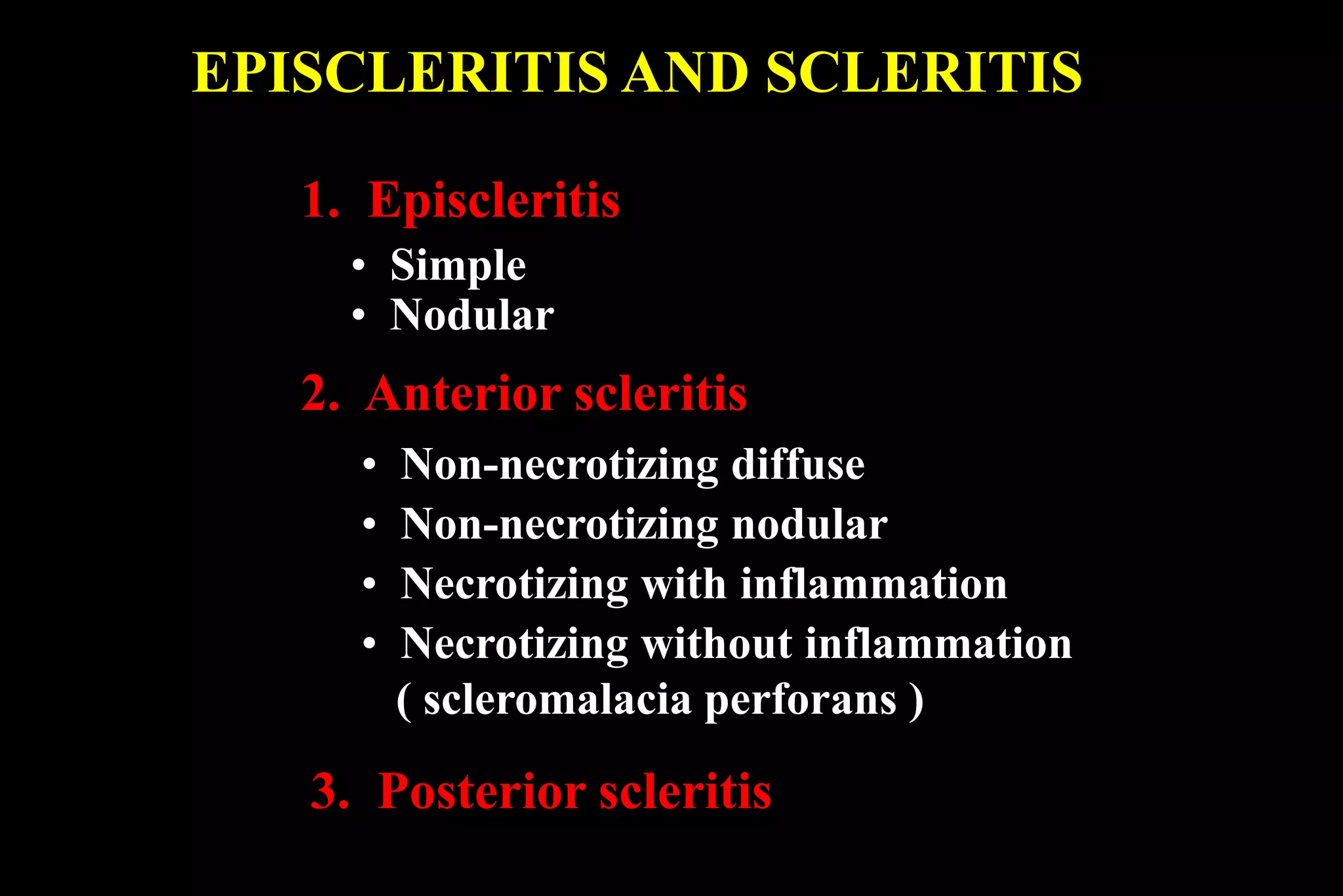
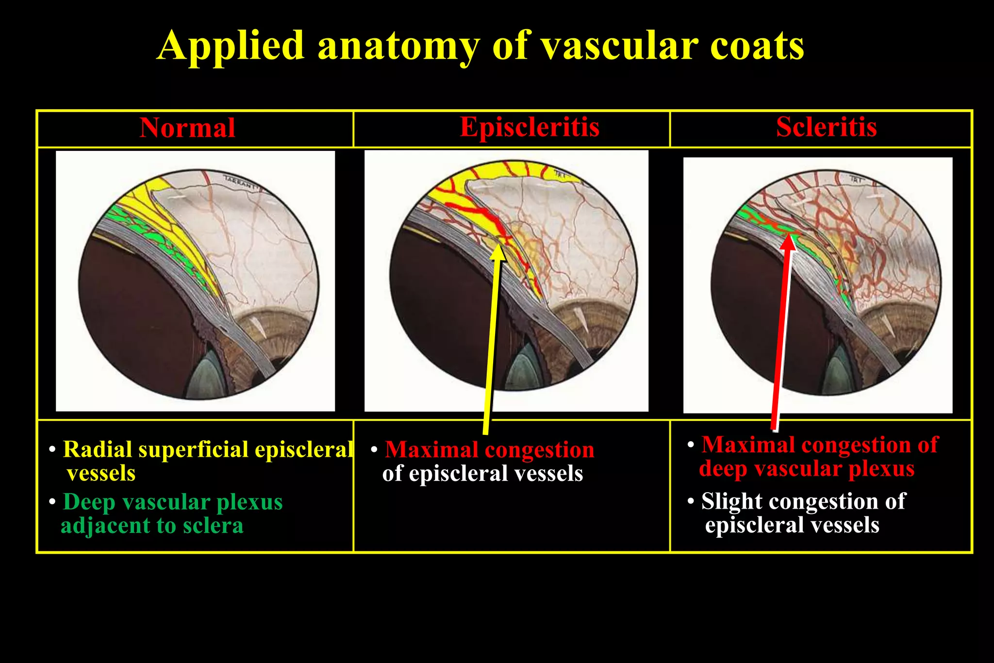
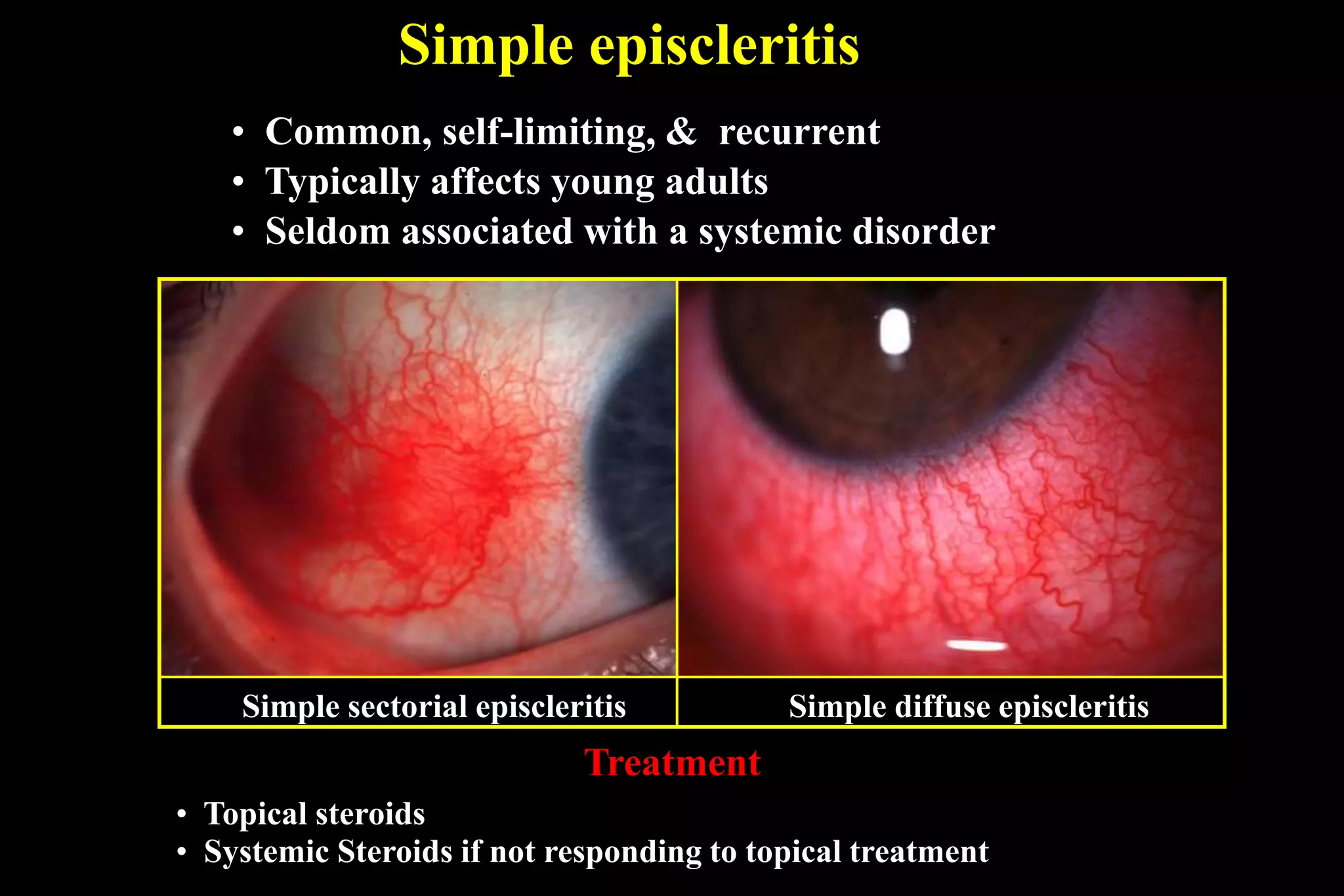
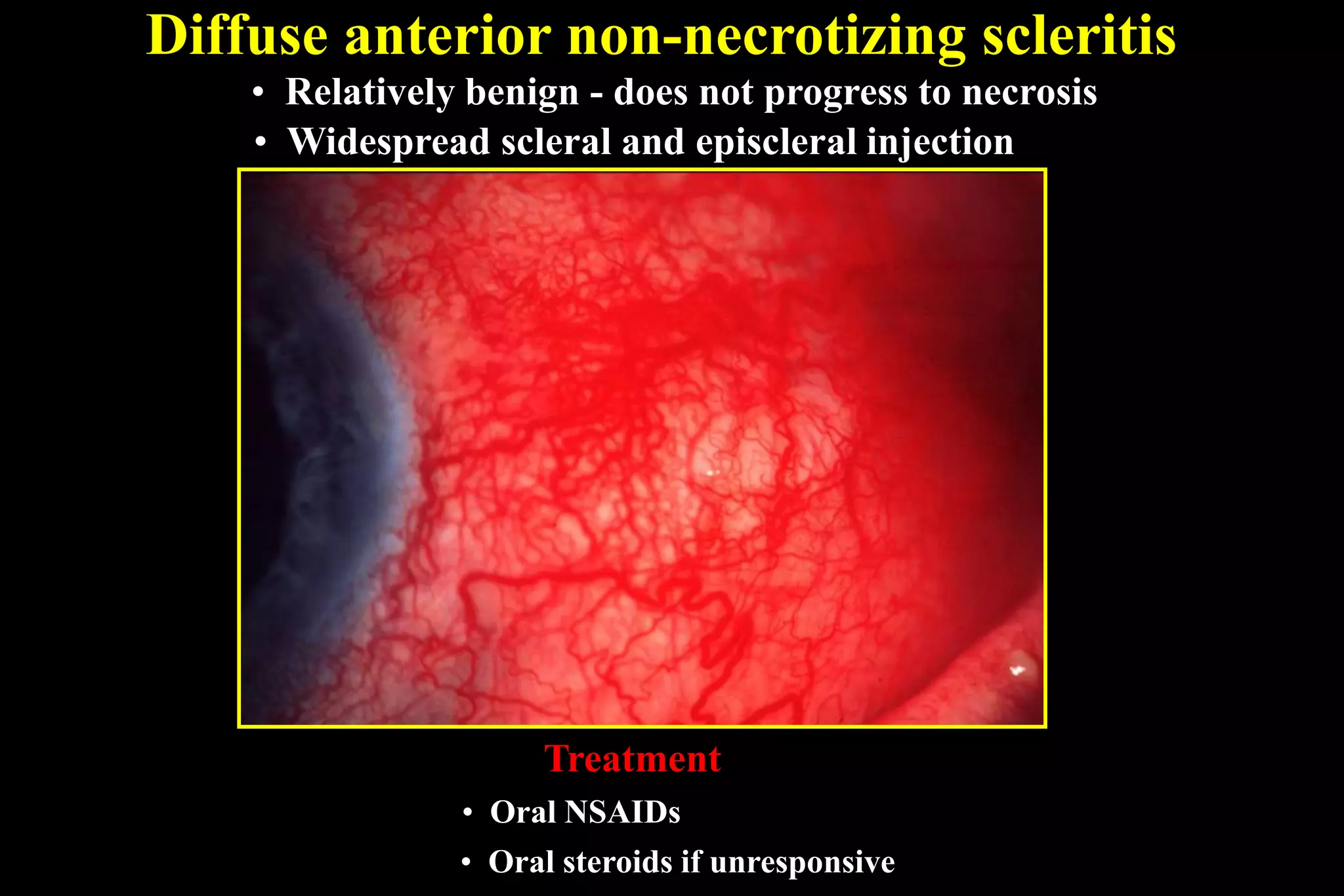
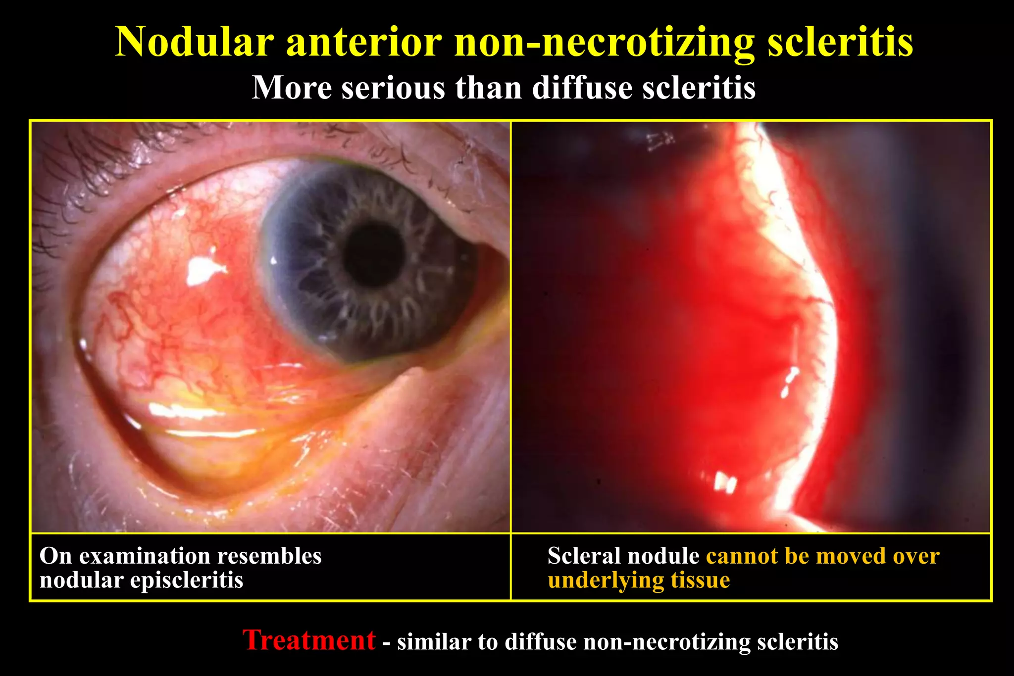
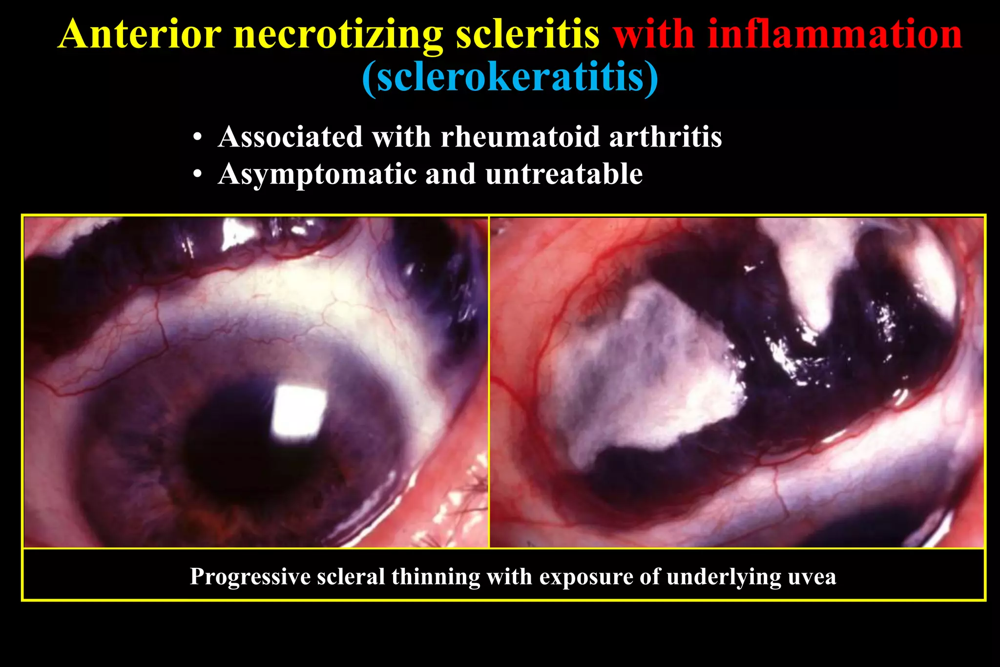
Sclera
Abdelmonem Hamed
Professor of Ophthalmology
Benha University
Special features of Sclera
• Avascular
• Dense fibrous tissue
• Lack of reaction to
insult
• Two types of
inflammation -
episcleritis and
scleritis
EPISCLERITIS AND SCLERITIS
1. Episcleritis
• Simple
• Nodular
2. Anterior scleritis
• Non-necrotizing diffuse
• Non-necrotizing nodular
• Necrotizing with inflammation
• Necrotizing without inflammation
( scleromalacia perforans )
3. Posterior scleritis
Applied anatomy of vascular coats
Scleritis
• Maximal congestion of
deep vascular plexus
• Slight congestion of
episcleral vessels
• Maximal congestion
of episcleral vessels
EpiscleritisNormal
• Radial superficial episcleral
vessels
• Deep vascular plexus
adjacent to sclera
Simple episcleritis
• Common, self-limiting, & recurrent
• Typically affects young adults
Treatment
• Seldom associated with a systemic disorder
Simple sectorial episcleritis Simple diffuse episcleritis
• Topical steroids
• Systemic Steroids if not responding to topical treatment
Nodular episcleritis
• Less common than simple episcleritis
• May take longer to resolve
• Treatment - similar to simple episcleritis
Localized nodule which can be moved over sclera
Causes of Scleritis:
1. Rheumatoid arthritis
• Wegener granulomatosis
• Polyteritis nodosa
• Systemic lupus erythematosus
2. Connective tissue disorders
3. Miscellaneous
• Relapsing polychondritis
• Herpes zoster ophthalmicus
• Surgically induced
Diffuse anterior non-necrotizing scleritis
• Widespread scleral and episcleral injection
• Relatively benign - does not progress to necrosis
• Oral steroids if unresponsive
Treatment
• Oral NSAIDs
Nodular anterior non-necrotizing scleritis
Scleral nodule cannot be moved over
underlying tissue
More serious than diffuse scleritis
On examination resembles
nodular episcleritis
Treatment - similar to diffuse non-necrotizing scleritis
Anterior necrotizing scleritis with inflammation
Progression
• Painful and most severe type
• Complications - uveitis, keratitis, cataract and glaucoma
Treatment
• Oral steroids
• Immunosuppressive agents (cyclosporin)
• Combined intravenous steroids and cyclophosphamide if unresponsive
Scleral necrosis and
visibility of uvea
Spread and coalescence
of necrosis
Avascular patches
• Asymptomatic and untreatable
• Associated with rheumatoid arthritis
Progressive scleral thinning with exposure of underlying uvea
Anterior necrotizing scleritis with inflammation
(sclerokeratitis)
Anterior necrotizing scleritis:
without inflammation
(Scleromalacia perforans)
 Asymptomatic
 Mainly in females with
longstanding RhA
 Large areas of uvea eventually
exposed
 Spontaneous perforation rare
 No effective treatment
Posterior scleritis
Signs
• About 20% of all cases of scleritis
• About 30% of patients have systemic disease
Choroidal folds Subretinal exudation
Proptosis and
ophthalmoplegia
Disc swelling Exudative retinal
detachment
Ring choroidal
detachment
• Decreased vision
• With or without pain
Differential Diagnoses
1. Allergic Conjunctivitis
2. Atopic Keratoconjunctivitis
3. Bacterial Conjunctivitis
4. Bacterial Keratitis
5. Epidemic Keratoconjunctivitis
6. Fungal Keratitis
7. Giant Papillary Conjunctivitis
8. Acute Angle-Closure Glaucoma
9. Herpes Simplex Virus Keratitis
10. Interstitial Keratitis
11. Keratoconjunctivitis Sicca
12. Superior Limbic Keratoconjunctivitis
13. Toxoplasmosis
14. Viral Conjunctivitis
Imaging in posterior scleritis
Ultrasound
a - Thickening of posterior scler
(flat tire sign)
b -Fluid in Tenon space (‘T’ sign)
Axial CT
Posterior scleral thickening
a
b
a
Treatment of necrotizing scleritis
 Oral prednisone 1mg/kg/day Or Pulsed IV
Methylprednisolone (solu-medrol 1 gm)
 Monitor pain in first 2-3 days
 Taper dose of steroids to response
 Immunosuppressives
 cyclosporine in steroid resistant cases
 Young people systemic NSAI.
Staphyloma
• Staphyloma is a clinical
condition characterised
by an ectasia of the
outer coats (cornea,or
sclera or both) with an
incarceration of uveal
tissue
TYPES
• Anterior
• Intercalary
• Ciliary
• Equatorial
• Posterior
Anterior staphyloma
• Partial or total
• Mostly after sloughed
cornea
• AC becomes flat with
secondary glaucoma
• Iris is incarcerated in
anterior staphyloma
Intercalary staphyloma
• Limbus
• Root of iris and anterior most part of
ciliary body
• Externally from limbus to 2mm behind
• Caused by:
1. perforating injury at peripheral cornea
involving limbus,
2. marginal corneal ulcers,
3. anterior scleritis,
4. scleromalacia perforans,
5. complicated cataract surgery with
wound dehisence, secondary glaucoma
Ciliary staphyloma
• Affects ciliary zone - upto 8 mm
behind the limbus
• Scleral ectasia with
incarceration of ciliary body
• Caused by:
1. developmental glaucoma,
2. end stage of primary or sec
glaucoma,
3. scleritis,
4. trauma to ciliary region of eye
Equatorial staphyloma
• Equatorial region of eye with
incarceration of choriod
• 14 mm behind the limbus weak
area due to passage of venae
vorticosae
• Caused by:
1. scleritis ,
2. chronic uncontrolled
glaucoma,
3. degenerative myopia
Posterior staphyloma
• Posterior pole of eye lined by
choroid
• Degenerative high myopia
• Detected by fundoscopy and
B- scan ultrasonography
Treatment
• Treat the cause
• Small –local excision with
corneo-scleral graft
(tectonic graft)
• Large unsightly blind
eyes are enucleated and
replaced with implant
BLUE SCLERA
1. In children.
2. In high myopia.
3. In Buphthalmos.
4. Over a staphyloma.
5. Osteogenesis imperfecta
a syndrome of :
a) Deafness (otosclerosis).
b) Fragility of bones.
c) Blue sclera.
Sclera

Sclera

  • 1.
    Sclera Abdelmonem Hamed Professor ofOphthalmology Benha University
  • 2.
    Special features ofSclera • Avascular • Dense fibrous tissue • Lack of reaction to insult • Two types of inflammation - episcleritis and scleritis
  • 3.
    EPISCLERITIS AND SCLERITIS 1.Episcleritis • Simple • Nodular 2. Anterior scleritis • Non-necrotizing diffuse • Non-necrotizing nodular • Necrotizing with inflammation • Necrotizing without inflammation ( scleromalacia perforans ) 3. Posterior scleritis
  • 4.
    Applied anatomy ofvascular coats Scleritis • Maximal congestion of deep vascular plexus • Slight congestion of episcleral vessels • Maximal congestion of episcleral vessels EpiscleritisNormal • Radial superficial episcleral vessels • Deep vascular plexus adjacent to sclera
  • 5.
    Simple episcleritis • Common,self-limiting, & recurrent • Typically affects young adults Treatment • Seldom associated with a systemic disorder Simple sectorial episcleritis Simple diffuse episcleritis • Topical steroids • Systemic Steroids if not responding to topical treatment
  • 6.
    Nodular episcleritis • Lesscommon than simple episcleritis • May take longer to resolve • Treatment - similar to simple episcleritis Localized nodule which can be moved over sclera
  • 7.
    Causes of Scleritis: 1.Rheumatoid arthritis • Wegener granulomatosis • Polyteritis nodosa • Systemic lupus erythematosus 2. Connective tissue disorders 3. Miscellaneous • Relapsing polychondritis • Herpes zoster ophthalmicus • Surgically induced
  • 8.
    Diffuse anterior non-necrotizingscleritis • Widespread scleral and episcleral injection • Relatively benign - does not progress to necrosis • Oral steroids if unresponsive Treatment • Oral NSAIDs
  • 9.
    Nodular anterior non-necrotizingscleritis Scleral nodule cannot be moved over underlying tissue More serious than diffuse scleritis On examination resembles nodular episcleritis Treatment - similar to diffuse non-necrotizing scleritis
  • 10.
    Anterior necrotizing scleritiswith inflammation Progression • Painful and most severe type • Complications - uveitis, keratitis, cataract and glaucoma Treatment • Oral steroids • Immunosuppressive agents (cyclosporin) • Combined intravenous steroids and cyclophosphamide if unresponsive Scleral necrosis and visibility of uvea Spread and coalescence of necrosis Avascular patches
  • 11.
    • Asymptomatic anduntreatable • Associated with rheumatoid arthritis Progressive scleral thinning with exposure of underlying uvea Anterior necrotizing scleritis with inflammation (sclerokeratitis)
  • 12.
    Anterior necrotizing scleritis: withoutinflammation (Scleromalacia perforans)  Asymptomatic  Mainly in females with longstanding RhA  Large areas of uvea eventually exposed  Spontaneous perforation rare  No effective treatment
  • 13.
    Posterior scleritis Signs • About20% of all cases of scleritis • About 30% of patients have systemic disease Choroidal folds Subretinal exudation Proptosis and ophthalmoplegia Disc swelling Exudative retinal detachment Ring choroidal detachment • Decreased vision • With or without pain
  • 14.
    Differential Diagnoses 1. AllergicConjunctivitis 2. Atopic Keratoconjunctivitis 3. Bacterial Conjunctivitis 4. Bacterial Keratitis 5. Epidemic Keratoconjunctivitis 6. Fungal Keratitis 7. Giant Papillary Conjunctivitis 8. Acute Angle-Closure Glaucoma 9. Herpes Simplex Virus Keratitis 10. Interstitial Keratitis 11. Keratoconjunctivitis Sicca 12. Superior Limbic Keratoconjunctivitis 13. Toxoplasmosis 14. Viral Conjunctivitis
  • 15.
    Imaging in posteriorscleritis Ultrasound a - Thickening of posterior scler (flat tire sign) b -Fluid in Tenon space (‘T’ sign) Axial CT Posterior scleral thickening a b a
  • 16.
    Treatment of necrotizingscleritis  Oral prednisone 1mg/kg/day Or Pulsed IV Methylprednisolone (solu-medrol 1 gm)  Monitor pain in first 2-3 days  Taper dose of steroids to response  Immunosuppressives  cyclosporine in steroid resistant cases  Young people systemic NSAI.
  • 17.
    Staphyloma • Staphyloma isa clinical condition characterised by an ectasia of the outer coats (cornea,or sclera or both) with an incarceration of uveal tissue
  • 18.
    TYPES • Anterior • Intercalary •Ciliary • Equatorial • Posterior
  • 19.
    Anterior staphyloma • Partialor total • Mostly after sloughed cornea • AC becomes flat with secondary glaucoma • Iris is incarcerated in anterior staphyloma
  • 20.
    Intercalary staphyloma • Limbus •Root of iris and anterior most part of ciliary body • Externally from limbus to 2mm behind • Caused by: 1. perforating injury at peripheral cornea involving limbus, 2. marginal corneal ulcers, 3. anterior scleritis, 4. scleromalacia perforans, 5. complicated cataract surgery with wound dehisence, secondary glaucoma
  • 21.
    Ciliary staphyloma • Affectsciliary zone - upto 8 mm behind the limbus • Scleral ectasia with incarceration of ciliary body • Caused by: 1. developmental glaucoma, 2. end stage of primary or sec glaucoma, 3. scleritis, 4. trauma to ciliary region of eye
  • 22.
    Equatorial staphyloma • Equatorialregion of eye with incarceration of choriod • 14 mm behind the limbus weak area due to passage of venae vorticosae • Caused by: 1. scleritis , 2. chronic uncontrolled glaucoma, 3. degenerative myopia
  • 23.
    Posterior staphyloma • Posteriorpole of eye lined by choroid • Degenerative high myopia • Detected by fundoscopy and B- scan ultrasonography
  • 24.
    Treatment • Treat thecause • Small –local excision with corneo-scleral graft (tectonic graft) • Large unsightly blind eyes are enucleated and replaced with implant
  • 25.
    BLUE SCLERA 1. Inchildren. 2. In high myopia. 3. In Buphthalmos. 4. Over a staphyloma. 5. Osteogenesis imperfecta a syndrome of : a) Deafness (otosclerosis). b) Fragility of bones. c) Blue sclera.