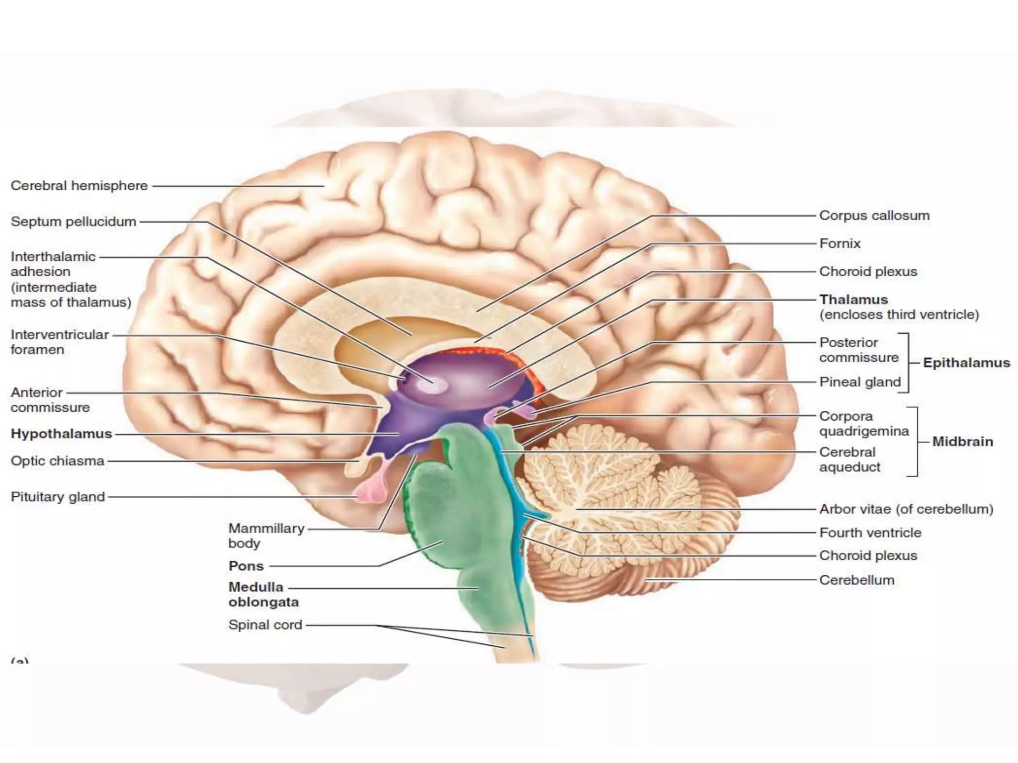
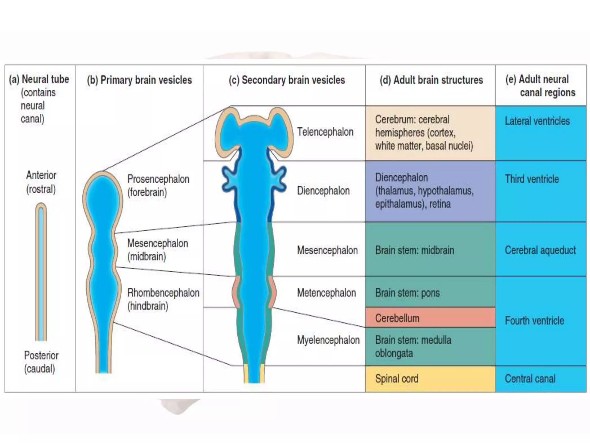
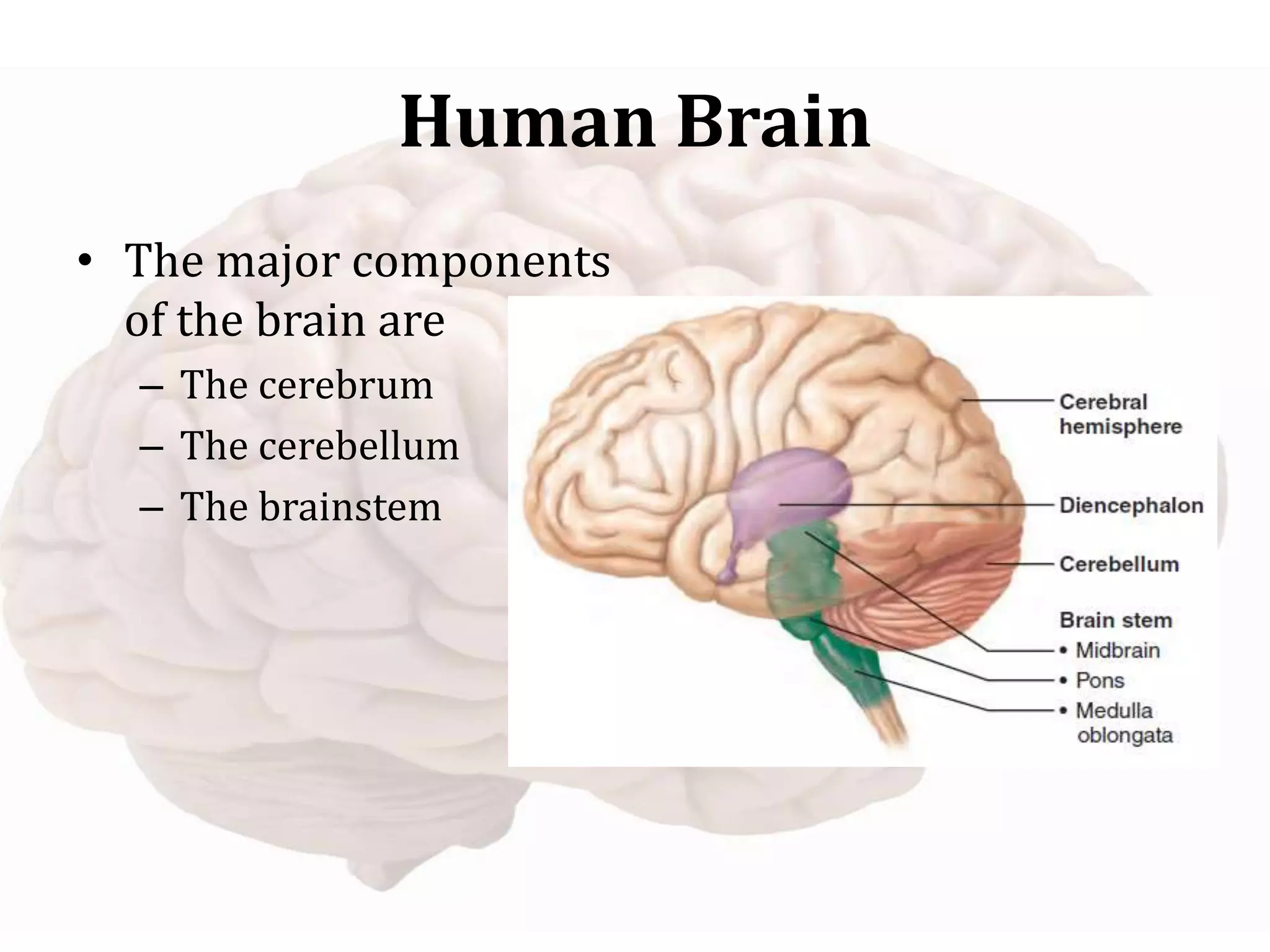
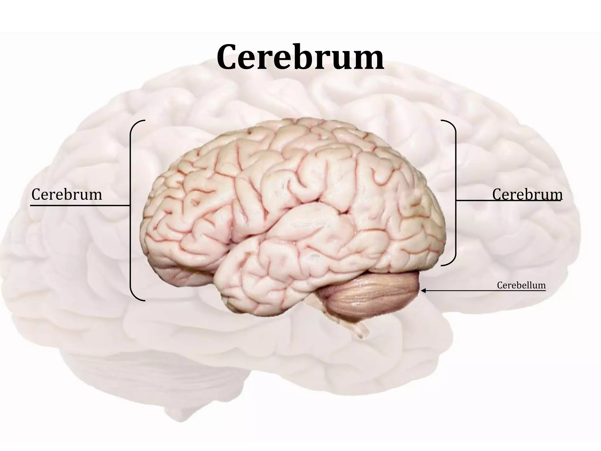
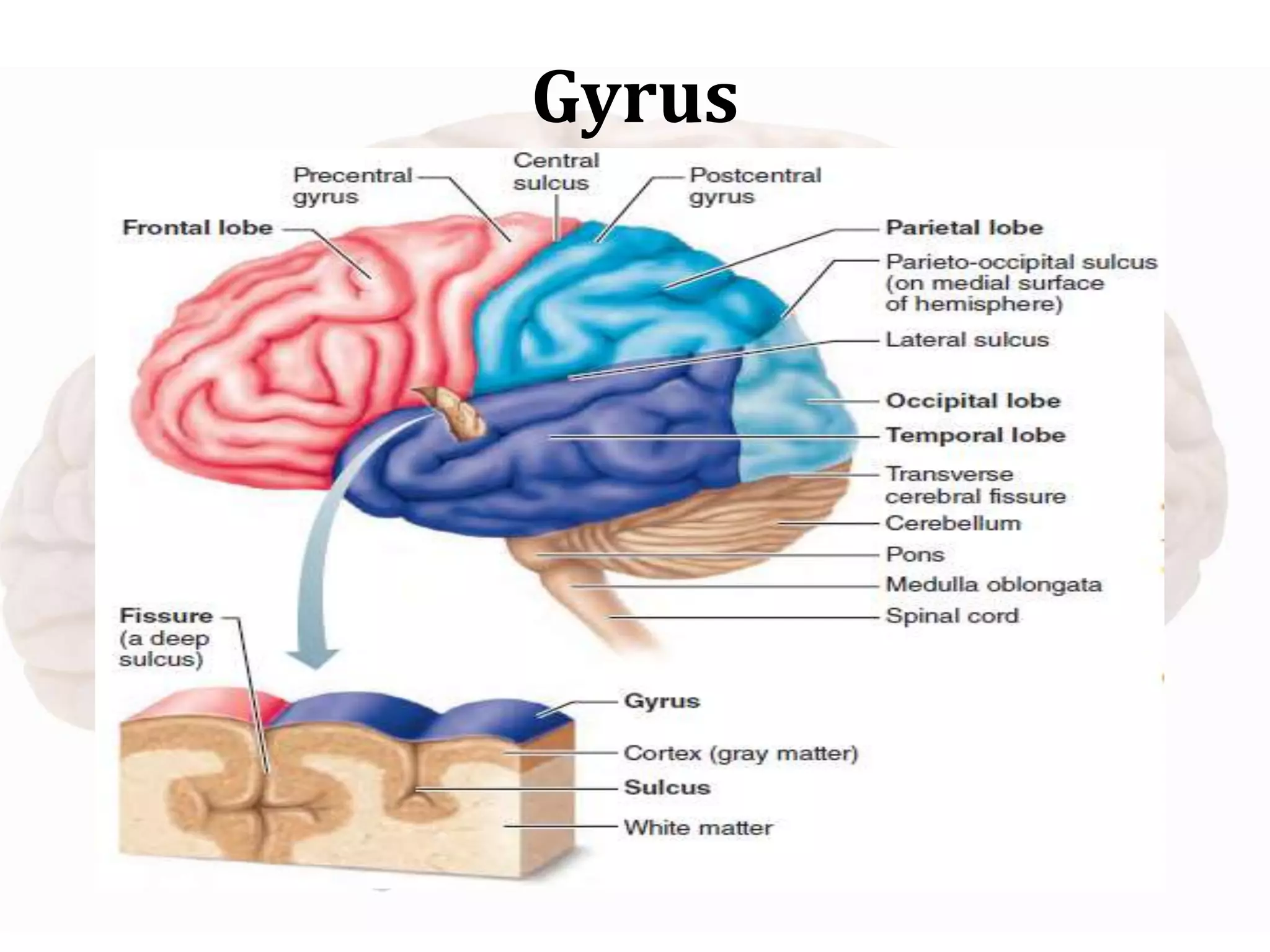
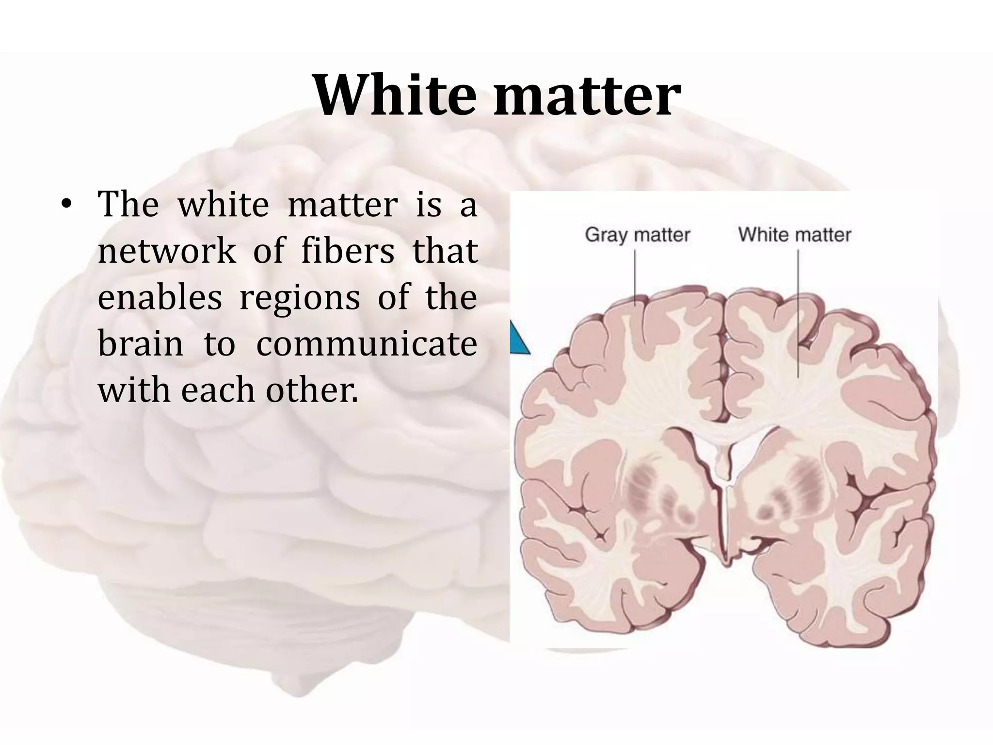
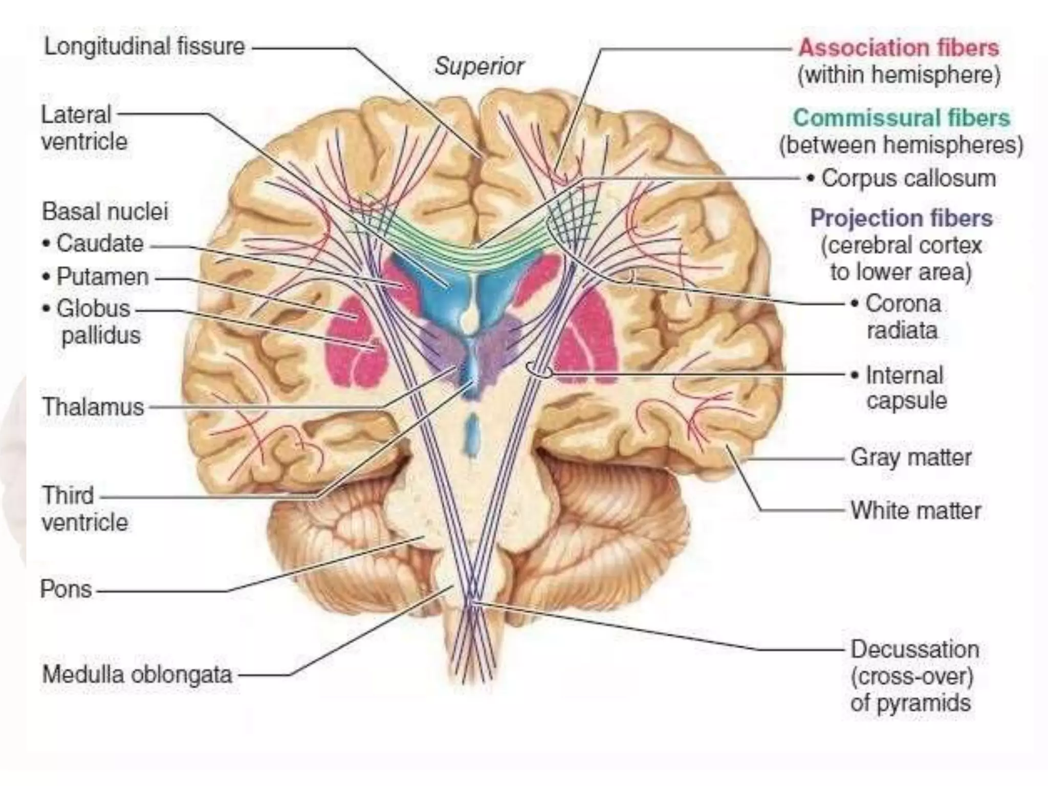
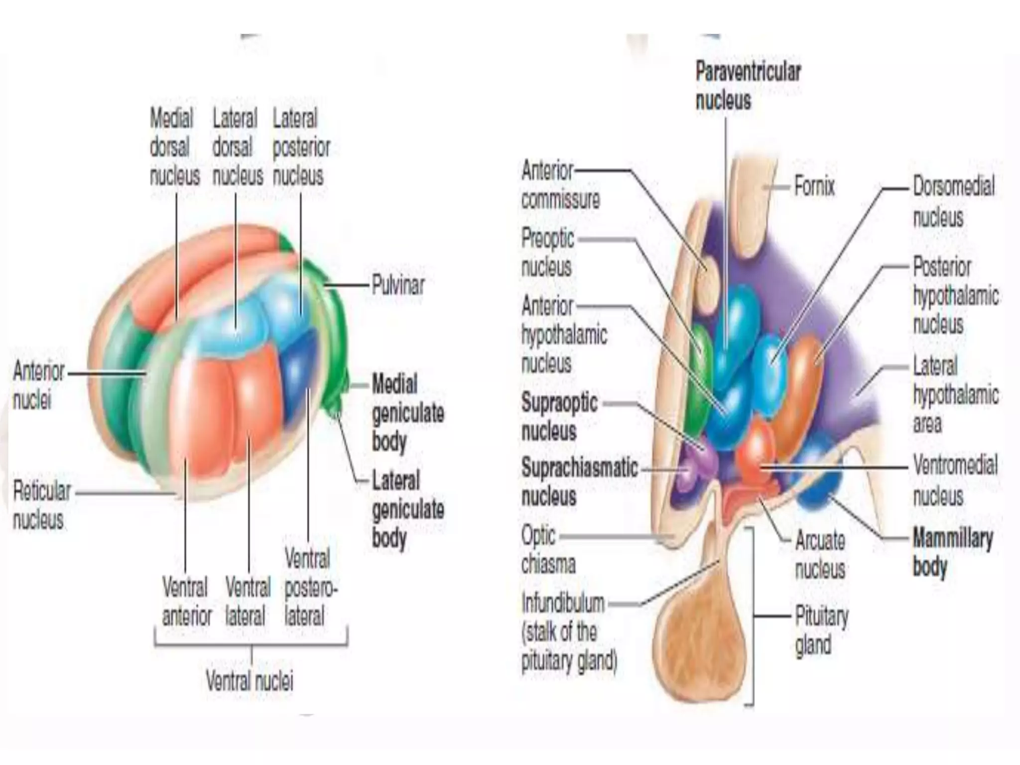
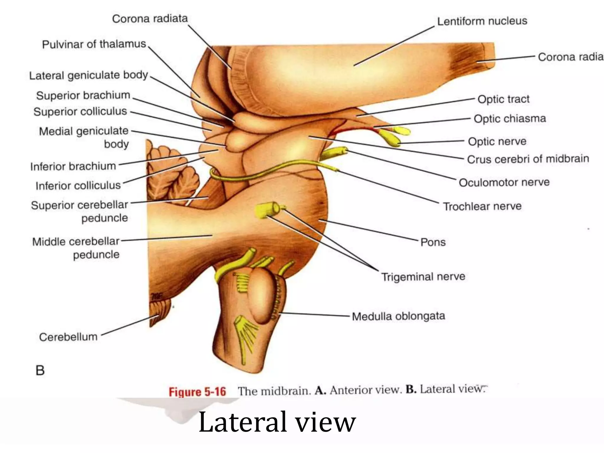
The brain is one of the most complex organs in the human body. It controls muscle movements, gland secretions, breathing, temperature regulation and processes sensory data. The brain and spinal cord begin as an embryonic structure called the neural tube which forms the three primary brain vesicles - forebrain, midbrain and hindbrain. These vesicles further differentiate into the adult brain structures. The major components of the adult brain are the cerebrum, cerebellum and brainstem. The cerebrum contains lobes, gyri, sulci and ventricles. It also contains white matter, basal ganglia and diencephalon structures like the thalamus.
cerebrum, sulci and gyri of cerebrum, lobes of cerebrum, frontal lobe , parietal lobe, temporal lobe and occipital lobe, sulci and gyri presnet in each lobes, and the functional areas , of cerebrum, brodmann areas of cerebrum, borders and surfaces of cerebrum, insula,
understanding spinal cord, its bransches, lesions, functions and anatomy.
hope to give you better knowledge of spinal cord by the end of it.
plese review ans comment for my future updates and corrections that iw ill be needing in this.
the all the content in this profile is completed by the teachers, students as well as other health care peoples.
thank you, all the respected peoples, for giving the information to complete this presentation.
this information is free to use by anyone.
control system in humans, neurons, types of neurons, nerves, human nervous system, CNS, PNS, ANS, Brain, parts of brain, spinal cord, functions of spinal cord, reflex arc, PNS, ANS,
CNS parts, Neuron, Synapse and its transmission, Grey and White matter of brain and spinal cord, 3 layers of meninges, Spinal cord structure and divisions, Brain parts and lobes with functions
New Directions in Targeted Therapeutic Approaches for Older Adults With Mantl...i3 Health
i3 Health is pleased to make the speaker slides from this activity available for use as a non-accredited self-study or teaching resource.
This slide deck presented by Dr. Kami Maddocks, Professor-Clinical in the Division of Hematology and
Associate Division Director for Ambulatory Operations
The Ohio State University Comprehensive Cancer Center, will provide insight into new directions in targeted therapeutic approaches for older adults with mantle cell lymphoma.
STATEMENT OF NEED
Mantle cell lymphoma (MCL) is a rare, aggressive B-cell non-Hodgkin lymphoma (NHL) accounting for 5% to 7% of all lymphomas. Its prognosis ranges from indolent disease that does not require treatment for years to very aggressive disease, which is associated with poor survival (Silkenstedt et al, 2021). Typically, MCL is diagnosed at advanced stage and in older patients who cannot tolerate intensive therapy (NCCN, 2022). Although recent advances have slightly increased remission rates, recurrence and relapse remain very common, leading to a median overall survival between 3 and 6 years (LLS, 2021). Though there are several effective options, progress is still needed towards establishing an accepted frontline approach for MCL (Castellino et al, 2022). Treatment selection and management of MCL are complicated by the heterogeneity of prognosis, advanced age and comorbidities of patients, and lack of an established standard approach for treatment, making it vital that clinicians be familiar with the latest research and advances in this area. In this activity chaired by Michael Wang, MD, Professor in the Department of Lymphoma & Myeloma at MD Anderson Cancer Center, expert faculty will discuss prognostic factors informing treatment, the promising results of recent trials in new therapeutic approaches, and the implications of treatment resistance in therapeutic selection for MCL.
Target Audience
Hematology/oncology fellows, attending faculty, and other health care professionals involved in the treatment of patients with mantle cell lymphoma (MCL).
Learning Objectives
1.) Identify clinical and biological prognostic factors that can guide treatment decision making for older adults with MCL
2.) Evaluate emerging data on targeted therapeutic approaches for treatment-naive and relapsed/refractory MCL and their applicability to older adults
3.) Assess mechanisms of resistance to targeted therapies for MCL and their implications for treatment selection
Flu Vaccine Alert in Bangalore Karnatakaaddon Scans
As flu season approaches, health officials in Bangalore, Karnataka, are urging residents to get their flu vaccinations. The seasonal flu, while common, can lead to severe health complications, particularly for vulnerable populations such as young children, the elderly, and those with underlying health conditions.
Dr. Vidisha Kumari, a leading epidemiologist in Bangalore, emphasizes the importance of getting vaccinated. "The flu vaccine is our best defense against the influenza virus. It not only protects individuals but also helps prevent the spread of the virus in our communities," he says.
This year, the flu season is expected to coincide with a potential increase in other respiratory illnesses. The Karnataka Health Department has launched an awareness campaign highlighting the significance of flu vaccinations. They have set up multiple vaccination centers across Bangalore, making it convenient for residents to receive their shots.
To encourage widespread vaccination, the government is also collaborating with local schools, workplaces, and community centers to facilitate vaccination drives. Special attention is being given to ensuring that the vaccine is accessible to all, including marginalized communities who may have limited access to healthcare.
Residents are reminded that the flu vaccine is safe and effective. Common side effects are mild and may include soreness at the injection site, mild fever, or muscle aches. These side effects are generally short-lived and far less severe than the flu itself.
Healthcare providers are also stressing the importance of continuing COVID-19 precautions. Wearing masks, practicing good hand hygiene, and maintaining social distancing are still crucial, especially in crowded places.
Protect yourself and your loved ones by getting vaccinated. Together, we can help keep Bangalore healthy and safe this flu season. For more information on vaccination centers and schedules, residents can visit the Karnataka Health Department’s official website or follow their social media pages.
Stay informed, stay safe, and get your flu shot today!
Pulmonary Thromboembolism - etilogy, types, medical- Surgical and nursing man...VarunMahajani
Disruption of blood supply to lung alveoli due to blockage of one or more pulmonary blood vessels is called as Pulmonary thromboembolism. In this presentation we will discuss its causes, types and its management in depth.
MANAGEMENT OF ATRIOVENTRICULAR CONDUCTION BLOCK.pdfJim Jacob Roy
Cardiac conduction defects can occur due to various causes.
Atrioventricular conduction blocks ( AV blocks ) are classified into 3 types.
This document describes the acute management of AV block.
Explore natural remedies for syphilis treatment in Singapore. Discover alternative therapies, herbal remedies, and lifestyle changes that may complement conventional treatments. Learn about holistic approaches to managing syphilis symptoms and supporting overall health.
ARTIFICIAL INTELLIGENCE IN HEALTHCARE.pdfAnujkumaranit
Artificial intelligence (AI) refers to the simulation of human intelligence processes by machines, especially computer systems. It encompasses tasks such as learning, reasoning, problem-solving, perception, and language understanding. AI technologies are revolutionizing various fields, from healthcare to finance, by enabling machines to perform tasks that typically require human intelligence.
Factory Supply Best Quality Pmk Oil CAS 28578–16–7 PMK Powder in Stockrebeccabio
Factory Supply Best Quality Pmk Oil CAS 28578–16–7 PMK Powder in Stock
Telegram: bmksupplier
signal: +85264872720
threema: TUD4A6YC
You can contact me on Telegram or Threema
Communicate promptly and reply
Free of customs clearance, Double Clearance 100% pass delivery to USA, Canada, Spain, Germany, Netherland, Poland, Italy, Sweden, UK, Czech Republic, Australia, Mexico, Russia, Ukraine, Kazakhstan.Door to door service
Hot Selling Organic intermediates
Tom Selleck Health: A Comprehensive Look at the Iconic Actor’s Wellness Journeygreendigital
Tom Selleck, an enduring figure in Hollywood. has captivated audiences for decades with his rugged charm, iconic moustache. and memorable roles in television and film. From his breakout role as Thomas Magnum in Magnum P.I. to his current portrayal of Frank Reagan in Blue Bloods. Selleck's career has spanned over 50 years. But beyond his professional achievements. fans have often been curious about Tom Selleck Health. especially as he has aged in the public eye.
Follow us on: Pinterest
Introduction
Many have been interested in Tom Selleck health. not only because of his enduring presence on screen but also because of the challenges. and lifestyle choices he has faced and made over the years. This article delves into the various aspects of Tom Selleck health. exploring his fitness regimen, diet, mental health. and the challenges he has encountered as he ages. We'll look at how he maintains his well-being. the health issues he has faced, and his approach to ageing .
Early Life and Career
Childhood and Athletic Beginnings
Tom Selleck was born on January 29, 1945, in Detroit, Michigan, and grew up in Sherman Oaks, California. From an early age, he was involved in sports, particularly basketball. which played a significant role in his physical development. His athletic pursuits continued into college. where he attended the University of Southern California (USC) on a basketball scholarship. This early involvement in sports laid a strong foundation for his physical health and disciplined lifestyle.
Transition to Acting
Selleck's transition from an athlete to an actor came with its physical demands. His first significant role in "Magnum P.I." required him to perform various stunts and maintain a fit appearance. This role, which he played from 1980 to 1988. necessitated a rigorous fitness routine to meet the show's demands. setting the stage for his long-term commitment to health and wellness.
Fitness Regimen
Workout Routine
Tom Selleck health and fitness regimen has evolved. adapting to his changing roles and age. During his "Magnum, P.I." days. Selleck's workouts were intense and focused on building and maintaining muscle mass. His routine included weightlifting, cardiovascular exercises. and specific training for the stunts he performed on the show.
Selleck adjusted his fitness routine as he aged to suit his body's needs. Today, his workouts focus on maintaining flexibility, strength, and cardiovascular health. He incorporates low-impact exercises such as swimming, walking, and light weightlifting. This balanced approach helps him stay fit without putting undue strain on his joints and muscles.
Importance of Flexibility and Mobility
In recent years, Selleck has emphasized the importance of flexibility and mobility in his fitness regimen. Understanding the natural decline in muscle mass and joint flexibility with age. he includes stretching and yoga in his routine. These practices help prevent injuries, improve posture, and maintain mobilit
Prix Galien International 2024 Forum ProgramLevi Shapiro
June 20, 2024, Prix Galien International and Jerusalem Ethics Forum in ROME. Detailed agenda including panels:
- ADVANCES IN CARDIOLOGY: A NEW PARADIGM IS COMING
- WOMEN’S HEALTH: FERTILITY PRESERVATION
- WHAT’S NEW IN THE TREATMENT OF INFECTIOUS,
ONCOLOGICAL AND INFLAMMATORY SKIN DISEASES?
- ARTIFICIAL INTELLIGENCE AND ETHICS
- GENE THERAPY
- BEYOND BORDERS: GLOBAL INITIATIVES FOR DEMOCRATIZING LIFE SCIENCE TECHNOLOGIES AND PROMOTING ACCESS TO HEALTHCARE
- ETHICAL CHALLENGES IN LIFE SCIENCES
- Prix Galien International Awards Ceremony
HOT NEW PRODUCT! BIG SALES FAST SHIPPING NOW FROM CHINA!! EU KU DB BK substit...GL Anaacs
Contact us if you are interested:
Email / Skype : kefaya1771@gmail.com
Threema: PXHY5PDH
New BATCH Ku !!! MUCH IN DEMAND FAST SALE EVERY BATCH HAPPY GOOD EFFECT BIG BATCH !
Contact me on Threema or skype to start big business!!
Hot-sale products:
NEW HOT EUTYLONE WHITE CRYSTAL!!
5cl-adba precursor (semi finished )
5cl-adba raw materials
ADBB precursor (semi finished )
ADBB raw materials
APVP powder
5fadb/4f-adb
Jwh018 / Jwh210
Eutylone crystal
Protonitazene (hydrochloride) CAS: 119276-01-6
Flubrotizolam CAS: 57801-95-3
Metonitazene CAS: 14680-51-4
Payment terms: Western Union,MoneyGram,Bitcoin or USDT.
Deliver Time: Usually 7-15days
Shipping method: FedEx, TNT, DHL,UPS etc.Our deliveries are 100% safe, fast, reliable and discreet.
Samples will be sent for your evaluation!If you are interested in, please contact me, let's talk details.
We specializes in exporting high quality Research chemical, medical intermediate, Pharmaceutical chemicals and so on. Products are exported to USA, Canada, France, Korea, Japan,Russia, Southeast Asia and other countries.
Title: Sense of Taste
Presenter: Dr. Faiza, Assistant Professor of Physiology
Qualifications:
MBBS (Best Graduate, AIMC Lahore)
FCPS Physiology
ICMT, CHPE, DHPE (STMU)
MPH (GC University, Faisalabad)
MBA (Virtual University of Pakistan)
Learning Objectives:
Describe the structure and function of taste buds.
Describe the relationship between the taste threshold and taste index of common substances.
Explain the chemical basis and signal transduction of taste perception for each type of primary taste sensation.
Recognize different abnormalities of taste perception and their causes.
Key Topics:
Significance of Taste Sensation:
Differentiation between pleasant and harmful food
Influence on behavior
Selection of food based on metabolic needs
Receptors of Taste:
Taste buds on the tongue
Influence of sense of smell, texture of food, and pain stimulation (e.g., by pepper)
Primary and Secondary Taste Sensations:
Primary taste sensations: Sweet, Sour, Salty, Bitter, Umami
Chemical basis and signal transduction mechanisms for each taste
Taste Threshold and Index:
Taste threshold values for Sweet (sucrose), Salty (NaCl), Sour (HCl), and Bitter (Quinine)
Taste index relationship: Inversely proportional to taste threshold
Taste Blindness:
Inability to taste certain substances, particularly thiourea compounds
Example: Phenylthiocarbamide
Structure and Function of Taste Buds:
Composition: Epithelial cells, Sustentacular/Supporting cells, Taste cells, Basal cells
Features: Taste pores, Taste hairs/microvilli, and Taste nerve fibers
Location of Taste Buds:
Found in papillae of the tongue (Fungiform, Circumvallate, Foliate)
Also present on the palate, tonsillar pillars, epiglottis, and proximal esophagus
Mechanism of Taste Stimulation:
Interaction of taste substances with receptors on microvilli
Signal transduction pathways for Umami, Sweet, Bitter, Sour, and Salty tastes
Taste Sensitivity and Adaptation:
Decrease in sensitivity with age
Rapid adaptation of taste sensation
Role of Saliva in Taste:
Dissolution of tastants to reach receptors
Washing away the stimulus
Taste Preferences and Aversions:
Mechanisms behind taste preference and aversion
Influence of receptors and neural pathways
Impact of Sensory Nerve Damage:
Degeneration of taste buds if the sensory nerve fiber is cut
Abnormalities of Taste Detection:
Conditions: Ageusia, Hypogeusia, Dysgeusia (parageusia)
Causes: Nerve damage, neurological disorders, infections, poor oral hygiene, adverse drug effects, deficiencies, aging, tobacco use, altered neurotransmitter levels
Neurotransmitters and Taste Threshold:
Effects of serotonin (5-HT) and norepinephrine (NE) on taste sensitivity
Supertasters:
25% of the population with heightened sensitivity to taste, especially bitterness
Increased number of fungiform papillae
1. THE BRAIN
Mr. Binu Babu
Asst. Professor
Mrs. Jincy Ealias
Asst. Professor
2. • The brain is one of the most complex and
magnificent organs in the human body.
Brain gives us awareness of ourselves and
our environment, processing a constant
stream of sensory data.
• It controls muscle movements, the
secretions of glands, and even breathing and
internal temperature.
3.
4. The Brain: Embryonic development
• Embryonic development of the human
brain.
– Formed by week 4, the neural tube quickly
subdivides into the primary brain vesicles,
which subsequently form the secondary brain
vesicles by week 5. These five vesicles
differentiate into the adult brain structures. The
adult structures derived from the neural canal.
5. • The brain and spinal cord begin as an embryonic
structure
• called the neural tube. The neural tube forms, its
anterior (rostral) end begins to expand and
constrictions
• appear that mark off the three primary brain
vesicles
– Prosencephalon or forebrain
– Mesencephalon or midbrain
– Rhombencephalon or hindbrain
• The remaining caudal (“toward the tail”), or
posterior, portion of the neural tube becomes the
spinal cord.
6.
7. Human Brain
• The major components
of the brain are
– The cerebrum
– The cerebellum
– The brainstem
8. Cerebrum
• The cerebrum is the largest and most
developmentally advanced part of the
human brain. It is responsible for several
higher functions, including higher
intellectual function, speech, emotion,
integration of sensory stimuli of all types,
initiation of the final common pathways for
movement, and fine control of movement.
10. Cerebral hemispheres
• The cerebrum is divided into a
right and a left hemispheres.
• The left hemisphere controls the
majority of functions on the right
side of the body, while the right
hemisphere controls most of
functions on the left side of the
body
• The crossing of nerve fibers takes
place in the brain stem. Thus,
injury to the left cerebral
hemisphere produces sensory and
motor deficits on the right side,
and vice versa.
11. Lobes of the Cerebrum
Fissures (deep
grooves) divide the
cerebrum into lobes
Cerebrum is
composed of pairs of
Frontal lobe
Parietal lobe
Occipital lobe
Temporal lobe
12. External features:
Gyri – Elevated ridges “winding” around the brain.
Sulci – Small grooves dividing the gyri
– Central Sulcus – Divides the frontal lobe from the parietal
lobe.
– Lateral sulcus - outlines the flaplike temporal lobe and
separates it from the parietal and frontal lobes.
Fissures – Deep grooves, generally dividing large
regions/lobes of the brain
• Transverse Fissure – Separates the cerebrum from the
cerebellum.
• Longitudinal Fissure – Divides the two cerebral
hemispheres
• Sylvian/Lateral Fissure – Divides the temporal lobe from
the frontal and parietal lobes
18. Regions of cerebral hemisphere
Cerebral hemisphere has three basic regions:
1. A superficial cerebral cortex of gray matter,
which looks gray in fresh brain tissue
2. Internal white matter
3. Basal nuclei, islands of gray matter situated
deep within the white matter
19. Grey matter
• It is a major component
of the central nervous
system consisting
of neuronal cell
bodies, neuropil
(dendrites and myelinat
ed as well as
unmyelinated axons)
glial cells (astrocytes
and oligodendrocytes)
synapses and capillaries.
20. Grey matter in the spinal cord
• Grey matter in the spinal cord is known as the grey
column which travels down the spinal cord
distributed in three grey columns that are presented
in an "H" shape.
• The forward-facing column is the anterior grey
column
• The rear-facing one is the posterior grey column
• The interlinking one is the lateral grey column.
• The grey matter on the left and right side is
connected by the grey commissure.
• The grey matter in the spinal cord consists
of interneurons, as well as cell bodies.
21. Cerebral cortex
• The cerebral cortex is the “executive suite” of the
nervous system.
• It enables us to be aware of ourselves and our
sensations, to communicate, remember, understand
and initiate voluntary movements.
• The cerebral cortex is composed of gray matter:
neuron cell bodies, dendrites, associated glia and
blood vessels, but no fiber tracts.
• It contains billions of neurons arranged in six layers.
• 2–4 mm (about 1/8 inch) thick
• It accounts for roughly 40% of total brain mass.
22.
23. • Based on the differences in lamination the
cerebral cortex can be classified into two
parts
– The large area of neocortex
– The smaller area of allocortex
24. Neocortex
• The neocortex (also known as the isocortex or
neopallium) is the part of the mature cerebral
cortex with six distinct layers.
27. Allocortex
• The allocortex is the part of the cerebral
cortex with less than six layers and has
three regions,
– The paleocortex with three cortical laminae
– The archicortex which has four or five cortical
laminae
– The periallocortex – a transitional zone
between the neocortex and the allocortex
28. White matter
• The white matter is a
network of fibers that
enables regions of the
brain to communicate
with each other.
29. • White matter consists largely of myelinated
fibers bundled into large tracts. These fibers
and tracts are classified according to the
direction in which they run as
– Association Fibers
– Projection Fibers
– Commissural Fibers
30.
31. • Association fibers: Connects different cortical
areas of the same hemisphere to one another.
– Eg:- superior longitudinal fasciculus
• Projection fibers: Connects cerebral cortex to
other parts of CNS like brain stem and spinal
cord.
– Eg: corona Radiata,internal capsule
• Commissural fibers: Connects the
corresponding parts of two hemispheres
32. White Matter of Cerebral
hemisphere
• Association tracts (fibers)- different regions
in cerebral cortex.
• Projection tracts (fibers)- cerebral cortex
other masses of gray matter.
• Commissures fibers - R L.
33. Internal capsule
• Internal capsule is
located between the
lentiform nucleus
laterally and
the thalamus and
caudate nucleus
medially.
• In a horizontal section of
cerebral hemisphere, the
internal capsule appears
as a V-shaped bundle of
fibers with the apex
directed medially.
34.
35. The internal capsule is divided into five parts.
– Anterior limb: part of the internal capsule that lies between the
anterior part of lentiform nucleus laterally and head of caudate
nucleus medially.
– Genu: It is the bend between the anterior and posterior limb of
internal capsule. The concavity of the bend faces laterally.
– Posterior limb: is that part of internal capsule which lies
between the thalamus medially and the posterior part of
lentiform nucleus laterally.
– Retrolentiform part: is located posterior to the lentiform
nucleus.
– Sublentiform part: is located inferior to the lentiform nucleus.
36.
37. Internal capsule contains both the motor
fibers and sensory fibers
Motor/Descending Fibers in Internal capsule
• Corticopontine :originate from all the lobes of cerebrum and relay
in ipsilateral pontine nuclei.
• Pyramidal fibers:
– Corticospinal: originate mainly in primary motor cortex and
relay in contralateral anterior horn of spinal cord.
– Corticonuclear: originate mainly in primary motor cortex
and relay usually in contralateral (some bilateral) motor
nuclei of cranial nerves.
• Extrapyramidal fibersCoticorubral, corticostriate, corticonigral
fibers etc.: these fibers originate in the motor and premotor areas of
cerebral cortex relay in subcortical nuclei viz. red nucleus, corpus
stiatum and substantia nigra respectively.
38. Internal Capsule -
Sensory/Ascending Fibers
• Thalmocortical and corticothalamic fibers: The
thalamocortical fibers are the 3rd order neurons of ascending
pathways.
• The four thalamic radiation bands are:
– Anterior thalamic radiation: Pass anteriorly to connect the
anterior and dorsomedial nucleus of thalamus to the frontal
lobe.
– Superior thalamic radiation: Pass superiorly to connect the
ventral tier of nucleus of thalamus to primary and premotor
areas of frontal lobe and sensory areas of parietal lobe.
– Posterior thalamic radiation (Optic radiation): pass
posteriorly to connect the lateral geniculate body to the
primary visual cortex.
– Inferior thalamic radiation (Auditory radiation): pass
inferiorly and laterally to connect the medial geniculate body
to the primary auditory area.
39.
40. Arrangement of Descending (Motor)
and Ascending (Sensory) Fibers
Anterior limb:
Motor
• Frontopontine fibers
Sensory
• Anterior thalamic radiation
Genu
Motor
• Frontopontine fibers,
• Corticonuclear
• Corticospinal fibers for head
and neck
Sensory
• Superior thalamic radiation
Retorlentiform part
Motor
• Parietopontine
• Occipitopontine fibers
Sensory
• Posterior thalamic ( Optic)
radiation
Sublentiform part
Motor
• Parietopontine
• Occipitopontine fibers
Sensory
• Inferior thalamic ( Auditory)
radiation
41. Basal Nuclei
• Deep within the cerebral white matter is the
third basic region of each hemisphere, a group
of subcortical nuclei called the basal nuclei or
basal ganglia.
• Basal nuclei include the
– Caudate nucleus
– Putamen
– Globus pallidus
• The comma-shaped caudate nucleus arches
superiorly over the diencephalon. Together
with the putamen, it forms the striatum
42.
43. • The putamen and globus pallidus together
form a lens-shaped mass, sometimes called the
lentiform nucleus.
• The basal nuclei are functionally associated
with the subthalamic nuclei of the diencephalon
and the substantia nigra of the midbrain.
• The basal nuclei receive input from the entire
cerebral cortex, as well as from other
subcortical nuclei
44.
45. The substantia nigra
• It is a brain structure
located in the
midbrain, and forms
part of the basal
ganglia. It is the largest
nucleus in the
midbrain.
• The substantia nigra
also plays an
important role in
reward and addiction.
46. Amygdaloid nucleus
• A small oval structure in the temporal
lobe of the brain that is closely connected to
the hypothalamus, the hippocampus, and
the cingulate gyrus.
• The amygdaloid nucleus is part of the
olfactory and limbic systems and plays a
role in the sense of smell, motivation, and
emotional behavior.
47.
48.
49. Ventricles
• The ventricles are structures that
produce cerebrospinal fluid, and transport
it around the cranial cavity. They are lined
by ependymal cells, which form a structure
called the choroid plexus. It is within
the choroid plexus that CSF is produced.
• Embryologically, the ventricular system is
derived from the lumen of the neural tube.
• There are four ventricles; right and left lateral
ventricles, third ventricle and fourth ventricle.
50. Lateral Ventricles
• The left and right lateral ventricles are located
within their respective hemispheres of the
cerebrum. They have ‘horns’ which project into the
frontal, occipital and temporal lobes.
• The paired lateral ventricles, one deep within each
cerebral hemisphere, are large C-shaped chambers
that reflect the pattern of cerebral growth.
• Anteriorly, the lateral ventricles lie close together,
separated only by a thin median membrane called
the septum pellucidum.
• The volume of the lateral ventricles increases with
age.
51. Third Ventricle
• The lateral ventricles are connected to
the third ventricle by the foramen of Monro.
The third ventricle is situated in between
the right and the left thalamus. The anterior
surface of the ventricle contains two
protrusions:
– Supra-optic recess – located above the optic
chiasm.
– Infundibular recess – located above the optic
stalk.
52. Fourth Ventricle
• The fourth ventricle is the last in the system – it
receives CSF from the third ventricle via
the cerebral aqueduct. It lies within the
brainstem, at the junction between
the pons and medulla oblongata. From the 4th
ventricle, the fluid drains into two places:
– Central spinal canal – Baths the spinal cord
– Subarachnoid cisterns – Baths the brain, between
arachnoid mater and pia mater. Here the CSF is
reabsorbed back into the circulation.
53.
54. CORPUS CALLOSUM
• It is the largest collection
of commissural fibers of
the cerebrum.
• It located in the floor of
the median longitudinal
fissure and connects two
cerebral hemisphere
except lower and anterior
part of the temporal lobe.
• Corpus callosum closely
related to lateral ventricle.
55. PARTS OF CORPUS
CALLOSUM
• 4 parts
• Anterior end called genu
• Central part is the Trunk
• Posterior bulbous part
called Splenium
• Rostrum is the
prologation from the
upper part of the genu to
the upper end of the
lamina terminals
56. FIBERS OF CORPUS
CALLOSUM
• Rostrum connects orbital
surfaces of frontal lobe.
• Forceps minor is made up
of fibers of genu that
connects the 2 frontal lobes.
• Forceps major is made up of
fibers of splenium that
connects the 2 occipital
lobes.
• The tapetum formed by
some fibers of the trunk
and splenium of corpus
callosum
FUNCTION OF CORPUS
CALLOSUM
COORDINATES THE ACTIVITIES
OF THE TWO CEREBRAL
HEMISPHERES
57. Diencephalon
• The diencephalon consists largely of three
paired structures:- the thalamus,
hypothalamus, and epithalamus.
• These gray matter areas collectively enclose
the third ventricle
58. Thalamus
• The thalamus consists of bilateral egg-shaped nuclei,
which form the superolateral walls of the third
ventricle.
• The thalamus is the relay station for information
coming into the cerebral cortex.
• Large mass of grey matter, lies immediately lateral to
third ventricle
Two poles
Anterior pole(or end)
Lies behind the interventricular foramen
Posterior pole(or end also called pulvinar lies just
above and lateral to superior colliculus.
59. Two thalami on each side of slit like cavity of 3rd ventricle
3 cms length x 1.5 cms breadth Long axis 300 oblique to
midline.
Interthalamic adhesion
60. Superior surface
Superior surface
• The superior (dorsal)
surface of the thalamus
is covered by a thin layer
of white matter, the
stratum zonale
• It extends laterally and
forms the roof of the
third ventricle
• More laterally it forms
part of the floor of the
lateral ventricle.
• Related laterally to
caudate nucleus
61. Medial surface
The medial surface
• The medial surface of the
thalamus is the superior
(dorsal) part of the lateral
wall of the third ventricle.
• It is usually connected to the
contralateral thalamus by an
interthalamic adhesion
behind the interventricular
foramina.
• The thalamus is continuous
with the midbrain
tegmentum, the subthalamus
and the hypothalamus
62. Inferior surface of thalamus
• Inferior surface of the tegmentum is related
to hypothalamus anteriorly and to ventral
thalamus posteriorly.
• The ventral thalamus seperates the
thalamus from tegmentum of midbrain.
63.
64. Internal structure of the
thalamus
• Thalamus consists of mainly of grey matter
• Superior surface is covered by a thin layer of white
matter called stratum zonale
• Lateral surface is covered by a similar layer called
externalmedullary layer.
• Internally, the thalamus is divided into anterior, medial
and lateral nuclear groups by a vertical Y-shaped sheet
of white matter, the internal medullary lamina
• Nuclei of the anterior part.
• Anterior nucleus.
• Nuclei in the medial part
• Largest nuclei among them medial dorsal nucleus.
65. Blood supply of thalamus
• Perforating branches of the posterior cerebral
artery
• Posteromedial group (thalamo - perforating
arteries) supply medial and anterior part.
• Posterolateral group (thalamo-geniculate
branches) supply
• posterior and lateral part of thalamus.
• Also recieves branches from posterior
communicating anterior choroidal,posterior
choroidal and middle cerebral artery.
66. Hypothalamus
• Hypothalamus which lies below the thalamus. It
the floor and lower parts of the lateral walls of the
3rd ventricle.
• The hypothalamus caps the brain stem and forms
the inferolateral walls of the third ventricle.
• The hypothalamus extends from the optic chiasma
(crossover point of the optic nerves) to the posterior
margin of the mammillary bodies.
• The mammillary bodies paired pea like nuclei that
bulge anteriorly from the hypothalamus, are relay
stations in the olfactory pathways.
67. • The hypothalamus is the main visceral
control center of the body.
• Small in size weighing only4gm, forms only
0.3 % of total brain mass.
• Mainly acts through 3 systems.
a. Autonomous nervous system
b. Endocrine system
c. The limbic system
68. Boundaries
• Anteriory - lamina
terminalis
• Posteriorly -subthalamus
• Inferiorly - structures in
the floor of the 3rd
ventricle.ie, tuber
cinereum, infundibulam
and mammillary bodies.
• Superiorly – thalamus
• Lateral boundary -
internal capsule.
• Medially - bounded by the
cavity of 3rd ventricle.
69. Anteroposteriorly divided into
• Preoptic region - area adjoining
the lamina terminalis.
• Supra optic region - above the
optic chiasma
• Tubreal region includes the tuber
cinereum,infundibulam and area
around it.
• Mammillary region - includes the
mammillary bodies and area
around it.
70. Hypothalamic nuclei
• Hypothalamus is made up of numerous small nuclear
masses called hypothalamic nucleus.
• They are divided into groups according to their regional
distribution and they differ in their functions also.
71. The subthalamic nuclei
• The subthalamic nuclei are
small paired structures that
are part of the
functional basal ganglia.
• They are located ventral to
the thalamus, dorsal to
the substantia nigra and
medial to the internal
capsule.
• The subthalamic nucleus
receives its main input from
the lateral pallidum (external
segment of the globus
pallidus). Excitatory,
glutamatergic inputs come
from the cerebral cortex.
72.
73. Region Nucleus Functions
Preotic region Preoptic nucleus
Contains the sexually
dimorphic nucleus, which
releases GnRH
Supraoptic region
Supra optic nucleus Vassopressin release
Anterior nucleus Thermo regulation, sweating
Paraventricular nucleus
TRH, CRH Corticotropin &
Oxytocin release
Tuberal region
Arcuate (infundibular
nucleus)
GHRH, feeding and
Dopamine release
Ventromedial nucleus Satiety centre
Dorsomedial nucleus
Controls BP, HR and GI
stimulation
76. Autonomic control of body
• Hypothalamus controls autonomic functions of body
through sympathetic as well as parasympathetic
fibres including cardiac, and respiratory activity,
vasomotor tone of blood vessels ,digestive process .
77. Part of limbic system
The limbic system is a complex
set of structures that lies on both
sides of the thalamus, just under
the cerebrum. It includes the
hypothalamus, the hippocampus,
the amygdala, and several other
nearby areas. It appears to be
primarily responsible for our
emotional life, and has imp. Role
in formation of memories.
79. Regulation of Food & water intake
• The extreme lateral part of
the ventromedial nucleus of the
hypothalamus is responsible for
the control of food intake.
Stimulation of this area causes
increased food intake.
Bilateral lesion of this area
causes complete cessation of
food intake.
•
80. Temperature regulation
• The cold and heat sensors
located in the hypothalamus
respond appropriately to
maintain the body
temperature at optimum level
in diverse conditions.
• The ant. Portion of
hypothalamus prevents the
rise in body temperature
while,
• Posterior portion promotes
heat conservation and heat
production.
81. Circadian rhythm
• Hypothalamus regulates the
cyclic activities of the body
,viz sleeping and waking cycle
but itself affected by diurnal
rhythm.
• The circadian rhythm for
many body functions is of
about 24 hours.
82. Sexual dimorphism
• Several hypothalamic nuclei are sexually dimorphic i.e.
there are clear differences in both structure and function
between males and females.
83. Other functions
• EMOTIONAL EXPRESSION:_
Hypothalamus regulates the
expression of emotions through
its control over the ANS
(laughting,crying,sweating,cryi
ng etc.)
• Fear processing:_The medial
zone of hypothalamus is part of
a circuitry that controls
motivated behaviors, like
defensive behaviors.
84. APPLIED ANATOMY
• Lesions in the hypothalamus will cause one of the
following syndromes;
• Obesity
• Diabetes insipidus
• Sexual disturbance
• Disturbance in sleep: somnolence,narcolepsy
• Hyperglycemia and glycosuria
• Diencepahlic autonomic epilepsy : characterized by
flushing,sweating,salivation,lacrimation,tacycardia,r
etardation of respiartory rate,unconsciousness etc.
85. Epithalamus
• The most dorsal portion of the diencephalon
• The epithalamus forms the roof of the third
ventricle. Extending from its posterior border
and visible externally is the pineal gland or
body.
• The pineal gland secretes the hormone
melatonin and, along with hypothalamic nuclei,
helps regulate the sleep-wake cycle.
• The posterior commissure forms the caudal
border of the epithalamus.
86. The brain stem
• The brain stem regions are
– Midbrain
– Pons
– Medulla oblongata
87. Midbrain
• Connects the pons and cerebellum with
the forebrain.
• It is about 0.8 inch in length
• The midbrain is traversed by a narrow
channel called cerebral aqueduct filled with
CSF.
88.
89.
90. Anterior Surface
Anteriorly, it presents
two large bundles of
fibers, one on each side
of the midline, called
crus cerebri.
The oculomotor nerve
emerges from the
medial aspect of the crus
of the same side
The crus cerebri bounds
the interpeduncular
fossa on either side.
91. Posterior Surface
Posteriorly, the midbrain presents
four rounded swellings called
colliculi- One Superior and
inferior colliculi one on each side.
Each colliculus is laterally related to
a ridge called brachium.
Superior and inferior brachium
arises from respective colliculi
Superior brachium connects the
superior colliculus to lateral
geniculate body.
Inferior brachium connects the
inferior colluculus to medial
geniculate body
In the midline below the inferior
colliculus, the trochlear nerve
emerges which then winds round
the side of the midbrain to reach its
ventral aspect.
93. Structure of Midbrain
• The midbrain comprises two lateral halves called the
Cerebral peduncles;
• The Cerebral peduncles are separated in to two parts by a
pigmented band of gray matter called substantia nigra
– Anterior part - crus cerebri
– Posterior part - tegmentum
The central narrow cavity is called the cerebral aqueduct
or aqueduct of Sylvius, which connects the 3rd and 4th
ventricles.
The tectum is the part of the midbrain posterior to the
cerebral aqueduct; it has four small surface swellings
referred to previously; these are two superior and two
inferior colliculi.
95. Blood Supply
Artery –
• posterior cerebral
artery
• superior cerebellar
artery
• basilar artery
Veins –
• Drained by basal or
great cerebral veins
96. PONS
• It is situated anteriorly to the cerebellum
measuring about 1 inch in length.
• Connects the medulla oblongata to the
midbrain.
• Its also connects two cerebellar
hemispheres and its name comes from this
bridge like appearance.
97.
98. Anterior surface of Pons
Pons has a convex anterior surface marked by
transversely running fibers which laterally form a bundle
called middle cerebellar peduncle.
Main Features:
• The trigeminal nerve emerges from the anterior surface at
its junction with middle cerebellar peduncle.
• Presents a basilar sulcus in the midline which lodges
basilar artery
• In the groove between Pons and the medulla oblongata,
there emerge, from medial to lateral, abducent, facial and
vestibulocochlear nerves.
99.
100. Posterior Surface
• Posterior surface contains
median sulcus which is
bounded by medial eminence
on both sides.
• Inferior end of medial
eminence is slightly expanded
to form facial colliculus, which
is produced by facial nerve
root.
• The upper end of sulcus
limitans presents a bluish-gray
coloration called substantia
ferruginosa.
101. • Respiratory nuclei:
• Apneustic & pneumotaxic centers work with
the medulla to maintain respiratory rhythm
• Nuclei & tracts that process and relay info
to/from the cerebellum
• Ascending, descending, and transverse
tracts that interconnect other portions of
the CNS
102. Structure of Pons
Posterior Part
• Tegmentum
Anterior part
• Basis pontis traversed
by running fibers of
Trapezoid body.
104. APPLIED ANATOMY
• Unilateral lesions in the lower part of the
pons results in paralysis of facial nerve in
the same side and limbs in the opposite side.
105. Medulla Oblangata
• The medulla oblongata (medulla) is one of
the three regions that make up the
brainstem. It is the most inferior of the three
and is continuous above with the pons and
below with the spinal cord. The medulla
houses essential ascending and descending
nerve tracts as well as brainstem nuclei
106. • Relays ascending sensory pathway impulses
from skin and proprioceptors through nuclei
cuneatus and gracilis
– Contains visceral nuclei controlling heart rate,
blood vessel diameter, respiratory rate, vomiting,
coughing, etc.
– Relays sensory information to the cerebellum
through inferior olivary nuclei
– Contains nuclei of cranial nerves VIII–XII
– Contains projection fibers
– Site of decussation of pyramids
107.
108. Structures of the Medulla
Oblongata
• Flanking the midline on the medulla’s
ventral aspect are two longitudinal ridges
called pyramids, formed by the large
pyramidal (corticospinal) tracts descending
from the motor cortex most of these fibers
cross over to the osite side before
continuing into the spinal cord.
• This crossover point is called the
decussation of the pyramids
109. • The olives are oval swellings. These
swellings are caused mainly by the wavy
folds of gray matter of the underlying
inferior olivary nuclei. These nuclei relay
sensory information on the degree of
stretch in muscles and joints to the
cerebellum.
110. External Anatomy of the Medulla
• The superior margin of the medulla is located at the
junction between the medulla and pons
• The inferior margin is marked by the origin of the first pair
of cervical spinal nerves. This occurs just as the medulla
exits the skull through the foramen magnum.
• Length is about 3 cm, width is about 2 cm
• Surfaces shows series of fissures
– Anterior median fissure
– Posterior median fissure
112. Anterior Surface
• There are several structures visible on the anterior surface of
the medulla – namely the three fissures/sulci, the pyramids, the
olives, and five cranial nerves.
– Anterior median fissure, which is continuous along the
length of the spinal cord. Two sulci are visible –
the ventrolateral sulcus and the posterolateral sulcus.
– The pyramids are paired swellings found between the
anterior median fissure and the ventrolateral sulcus.
– The olives are another pair of swellings located laterally to
the pyramids – between the ventrolateral and posterolateral
sulci.
• Arising from the junction between the pons and medulla is the
abducens nerve (CN VI). Extending out of the ventrolateral
sulcus is the hypoglossal nerve (CN XII). In the posteriolateral
sulcus, three more cranial nerves join the medulla (CN IX, CN X,
and CN XI).
113.
114. Ventral surface of medulla oblongata contains
• Pyramid
• It is an elevation between anterior median and
anterolateral sulcus
• Formed due to decussation of corticospinal fibres.
Pyramid
Olive
Olive
• Oval swelling between anterolateral
posterolateral sulcus,half an inch
long
• Produced by large mass of gray
matter called inferior olivary
nucleus
115. The posterior surface
• The posterior surface has a midline structure –
the posterior median sulcus – which is
continuous below as the posterior median
sulcus of the spinal cord. Above, the sulcus
ends at the point in which the fourth ventricle
develops.
• As we move lateral from the midline,
the fasciculus gracilis and fasciculus
cuneatus are seen, separated by the posterior
intermediate sulcus.
116.
117. Internal Anatomy of the Medulla
• The internal structures of the medulla must
be viewed in cross section to understand the
layout. Three levels of the medulla are
typically discussed (inferior – superior):
– Level of decussation of the pyramids
– Level of decussation of the medial lemnisci
– Level of the olives
118. • The medulla contains gray matter, while
the outer portions consist
of white matter. The posterior white matter
contains the fasiculus gracilis and the
more lateral fasiculus cuneatus.
Corresponding portions of gray matter
extend to these regions and are the nucleus
gracilis and nucleus cuneatus associated
with a tract called the medial lemniscus.
119.
120. Functions of the Medulla
Oblongata
• The small size of the medulla belies its crucial role as an
autonomic reflex center involved in maintaining body
homeostasis. The medulla contains these important
functional groups of visceral motor nuclei:
Cardiovascular center. Adjusts the force and rate of heart
contraction to meet the body’s needs, and the vasomotor
center, which changes blood vessel diameter to regulate
blood pressure.
Respiratory centers. These generate the respiratory
rhythm and control the rate and depth of breathing.
Various other centers. Additional centers regulate such
activities as vomiting, hiccuping, swallowing, coughing, and
sneezing
121. The Limbic System
• The limbic system is a group of structures
located on the medial aspect of each
cerebral hemisphere and diencephalon.
• These functional system:
– Includes cerebral and diencephalon structures
(e.g., hypothalamus and anterior thalamic
nuclei)
– Mediates emotional response
– Involved in memory processing
122.
123. • The limbic system is our emotional, or affective
(feelings), brain.
• Amygdaloid body is critical for responding to
perceived threats such as angry or fearful facial
expressions with fear or aggression.
• The cingulate gyrus plays a role in expressing
our emotions through gestures and in resolving
mental conflicts when we are frustrated.
• Rhinencephalon helps the brain in responds to
odors.
124. Reticular Formation
• The reticular formation is a
set of interconnected nuclei
that are located throughout
the brainstem. The reticular
formation is not anatomically
well defined because it
includes neurons located in
diverse parts of the brain.
• A functional system:
– Maintains cerebral cortical
alertness (reticular activating
system)
– Filters out repetitive stimuli
– Helps regulate skeletal and
visceral muscle activity
125. Reticular Activating System
• The neurons of the part of the reticular
formation known as the reticular activating
system (RAS).
• Reticular activating system send a
continuous stream of impulses to the cerebral
cortex, keeping the cortex alert and conscious
and enhancing its excitability. Impulses from all
the great ascending sensory tracts synapse
with RAS neurons, keeping them active and
enhancing their arousing effect on the
cerebrum.
126. • The reticular formation is divided into three
columns:
– Raphe nuclei (median),
– Magnocellular red nucleus (medial zone),
– Parvocellular reticular nucleus(lateral zone).
127. • The raphe nuclei is the place of synthesis of
the neurotransmitter serotonin, which plays
an important role in mood regulation.
• The magnocellular red nucleus is involved in
motor coordination.
• The parvocellular nucleus
regulates exhalation.