Downloaded 144 times


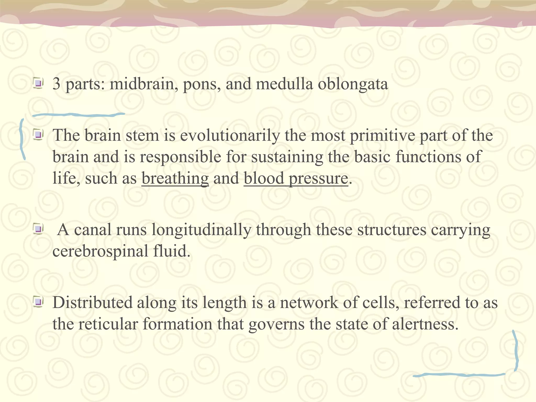


The nervous system has three main components - the brain, nerves, and spinal cord. The brain is the control center located in the cranial cavity and is made up of the cerebrum, brainstem, and cerebellum. The cerebrum is divided into four lobes and is responsible for functions like memory, thinking, language, and movement. The brainstem consists of the midbrain, pons, and medulla oblongata and controls vital functions. The cerebellum, located in the back of the brain, coordinates movement and balance. The nervous system allows communication and coordination throughout the body to control all functions.
Overview of the nervous system presented by students: Abalos, Ailyn; Juatas, Kenneth; Llorin, Patricia; Mansibang, Rania; Sunio, Leslie.
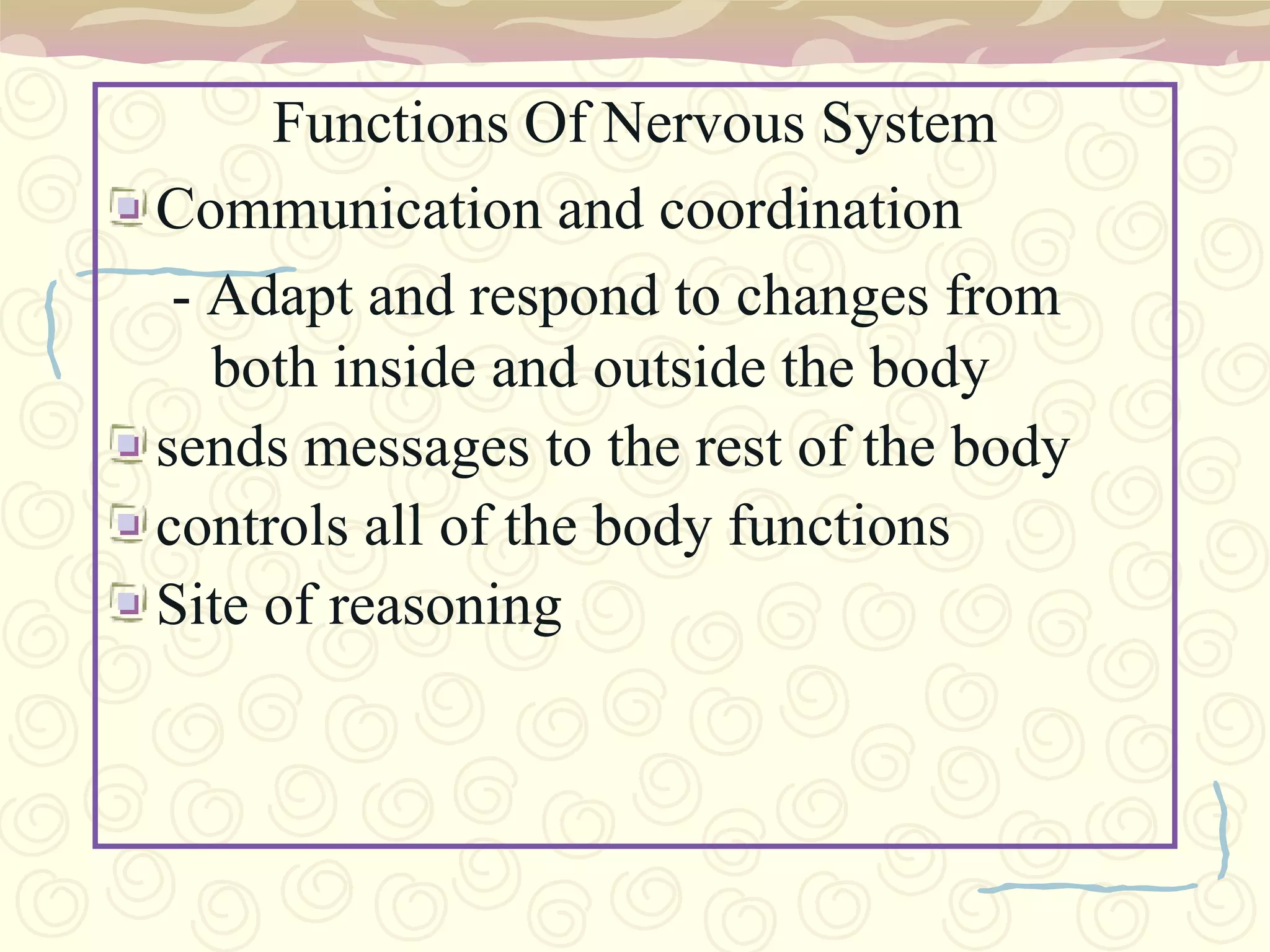
Communication, coordination, adaptability, control of functions, and reasoning.
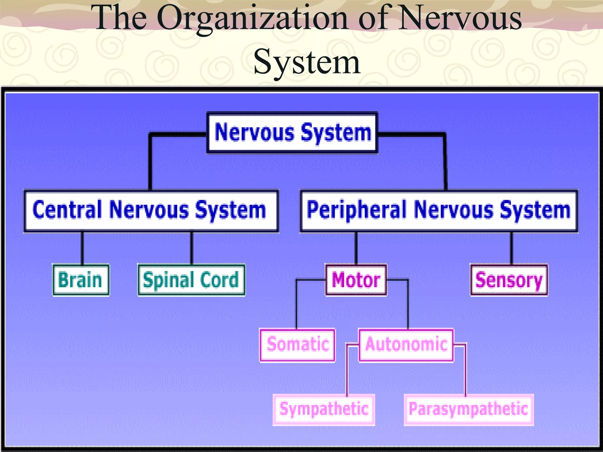
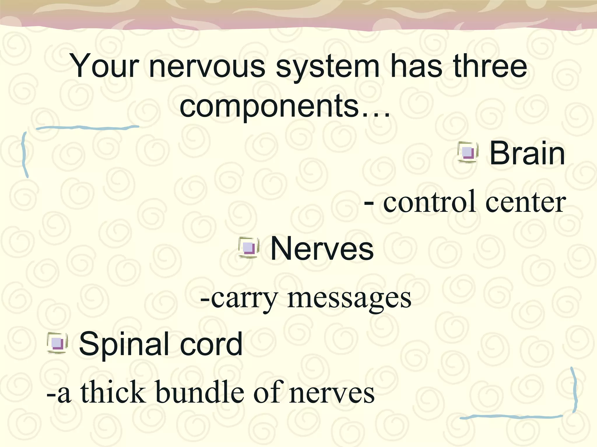
Components include the Brain (control center), Nerves (messaging), and Spinal Cord (nerve bundle).
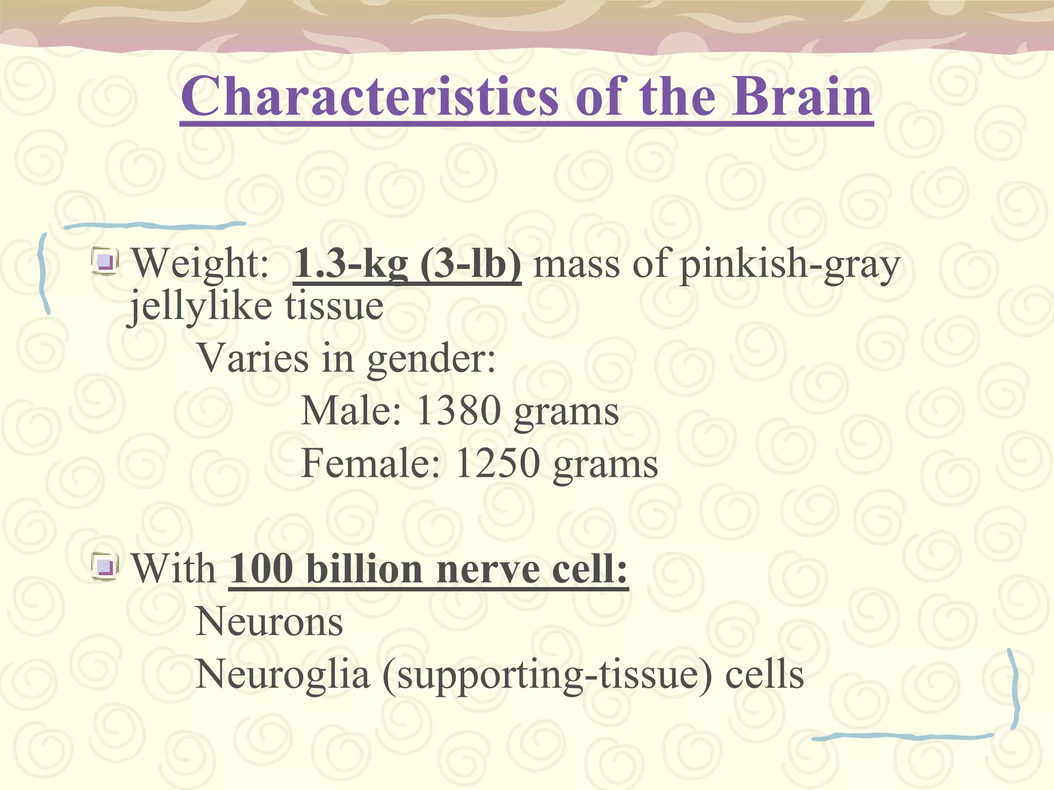
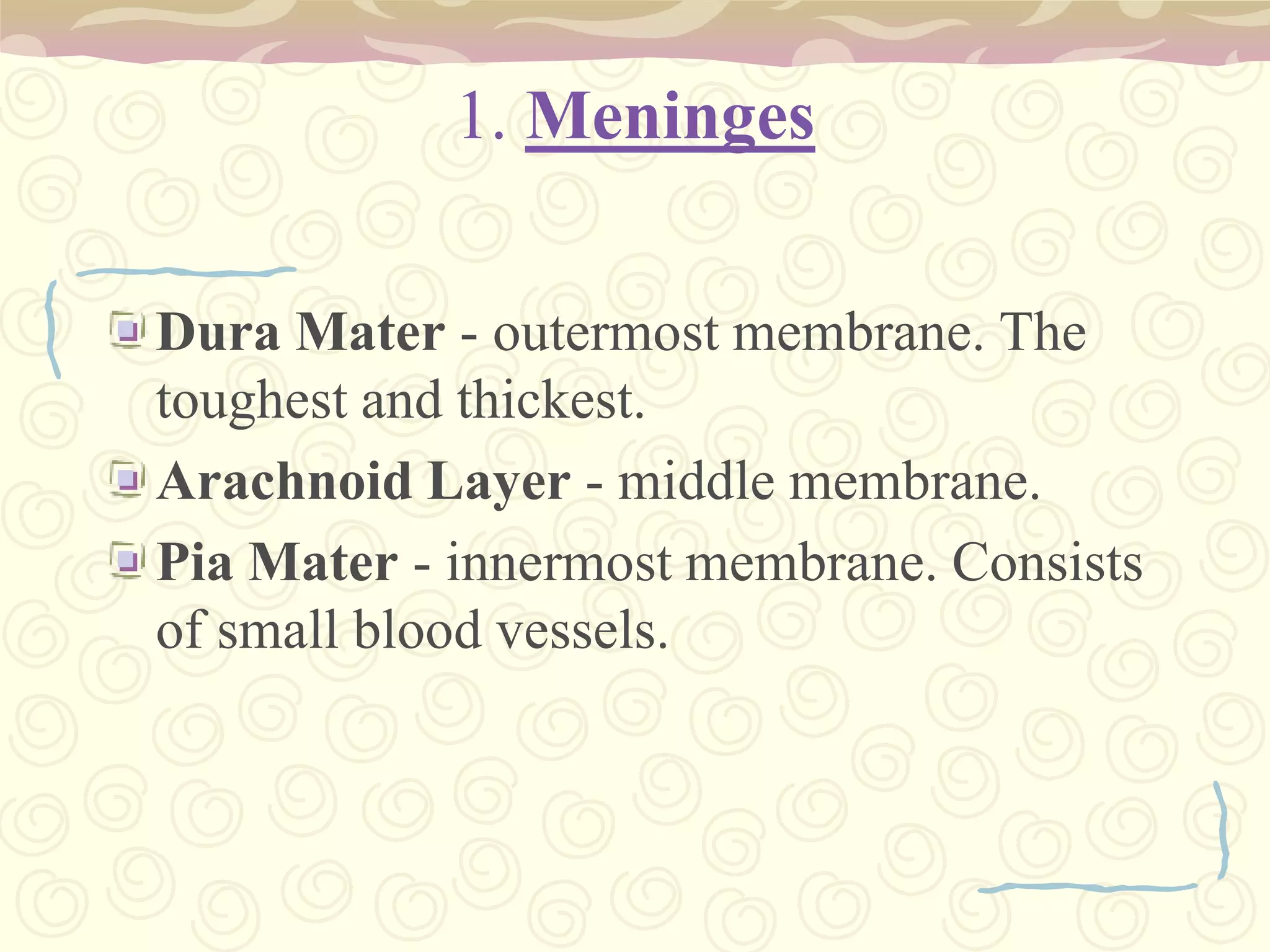
Brain's Attributes: 1.3 kg weight, 100 billion neurons, 4 types of memory, meninges, cerebrospinal fluid.
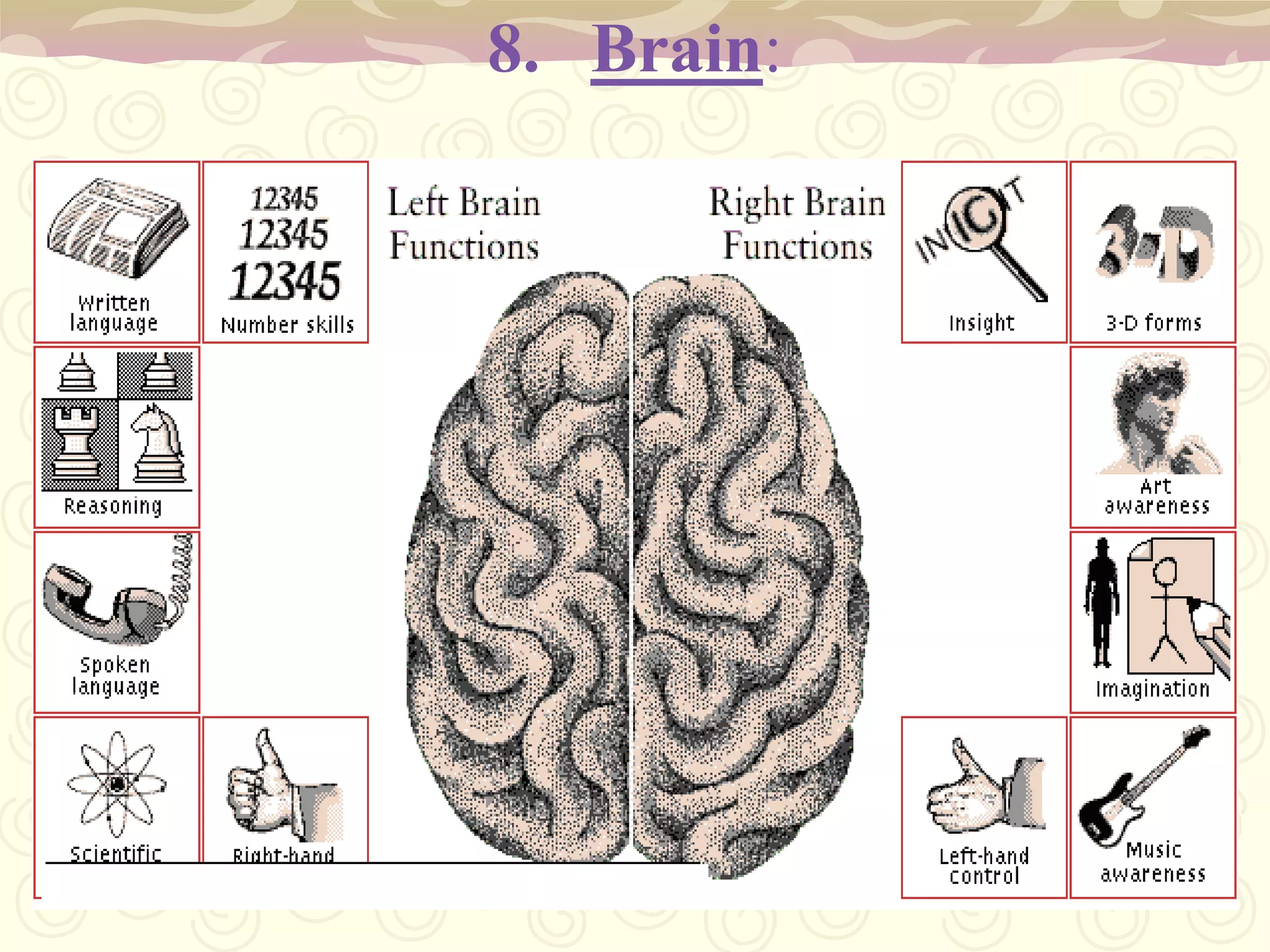
Right brain (artistic ability) vs Left brain (logical, analytical thinking).
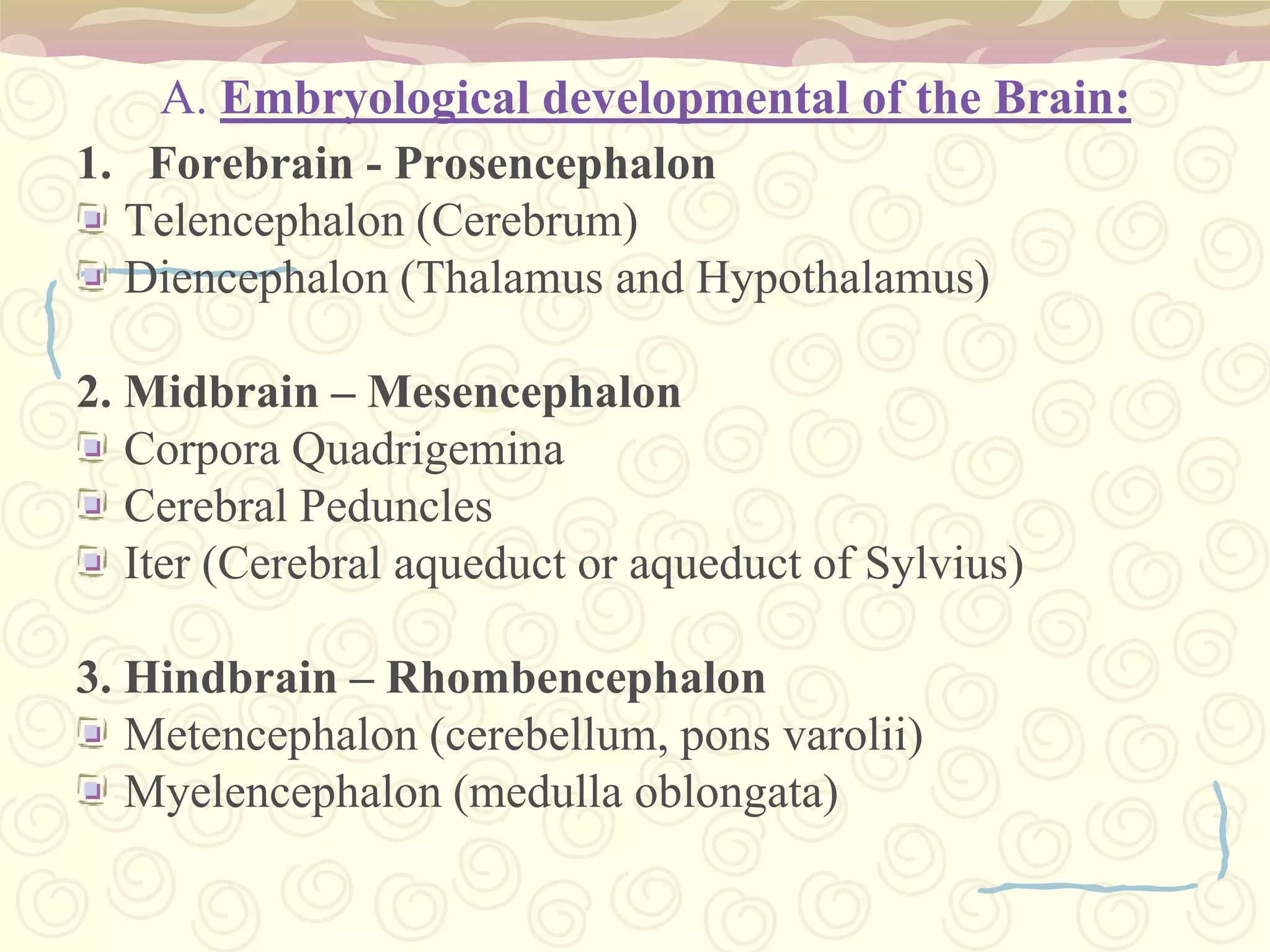
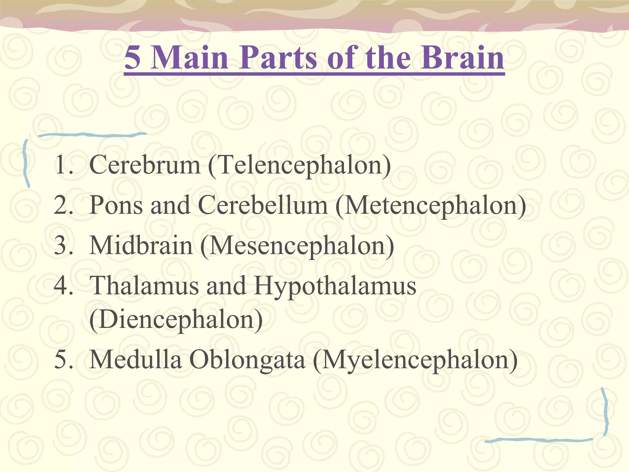
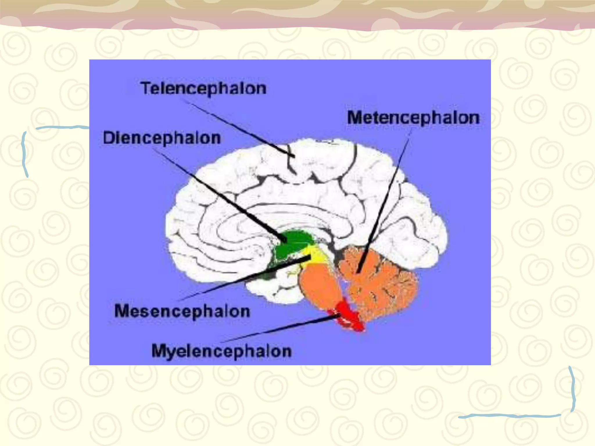
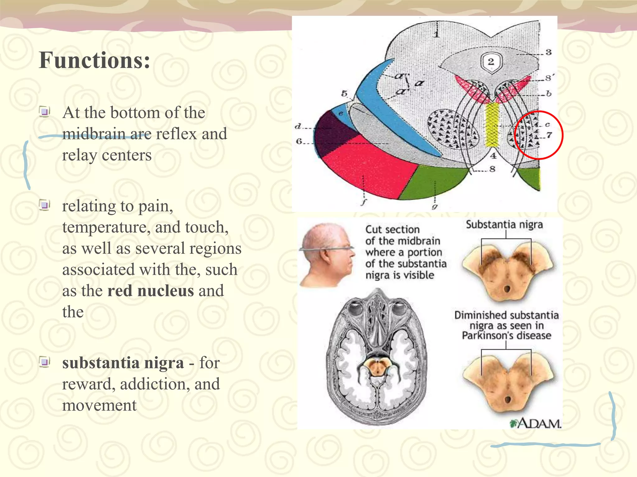
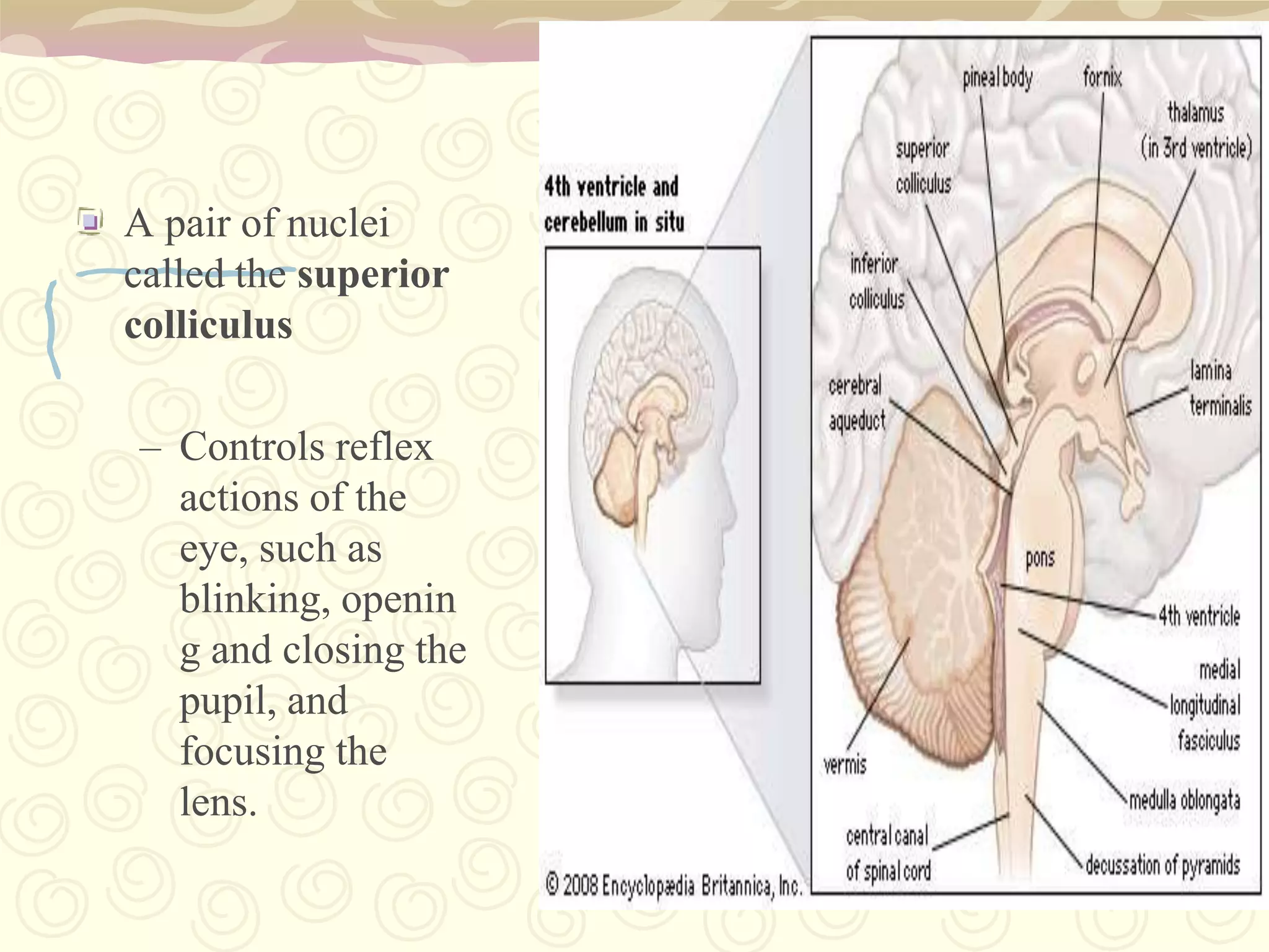
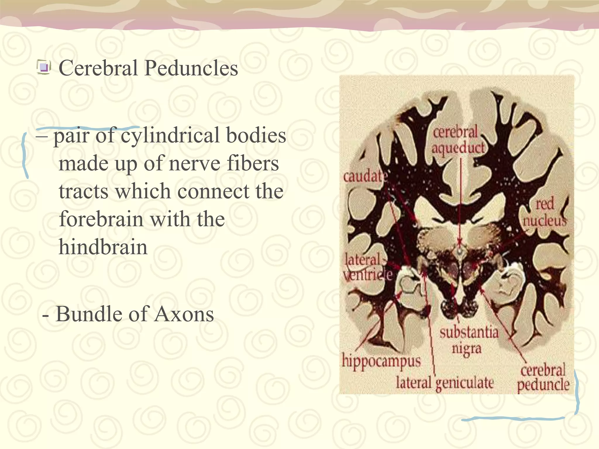
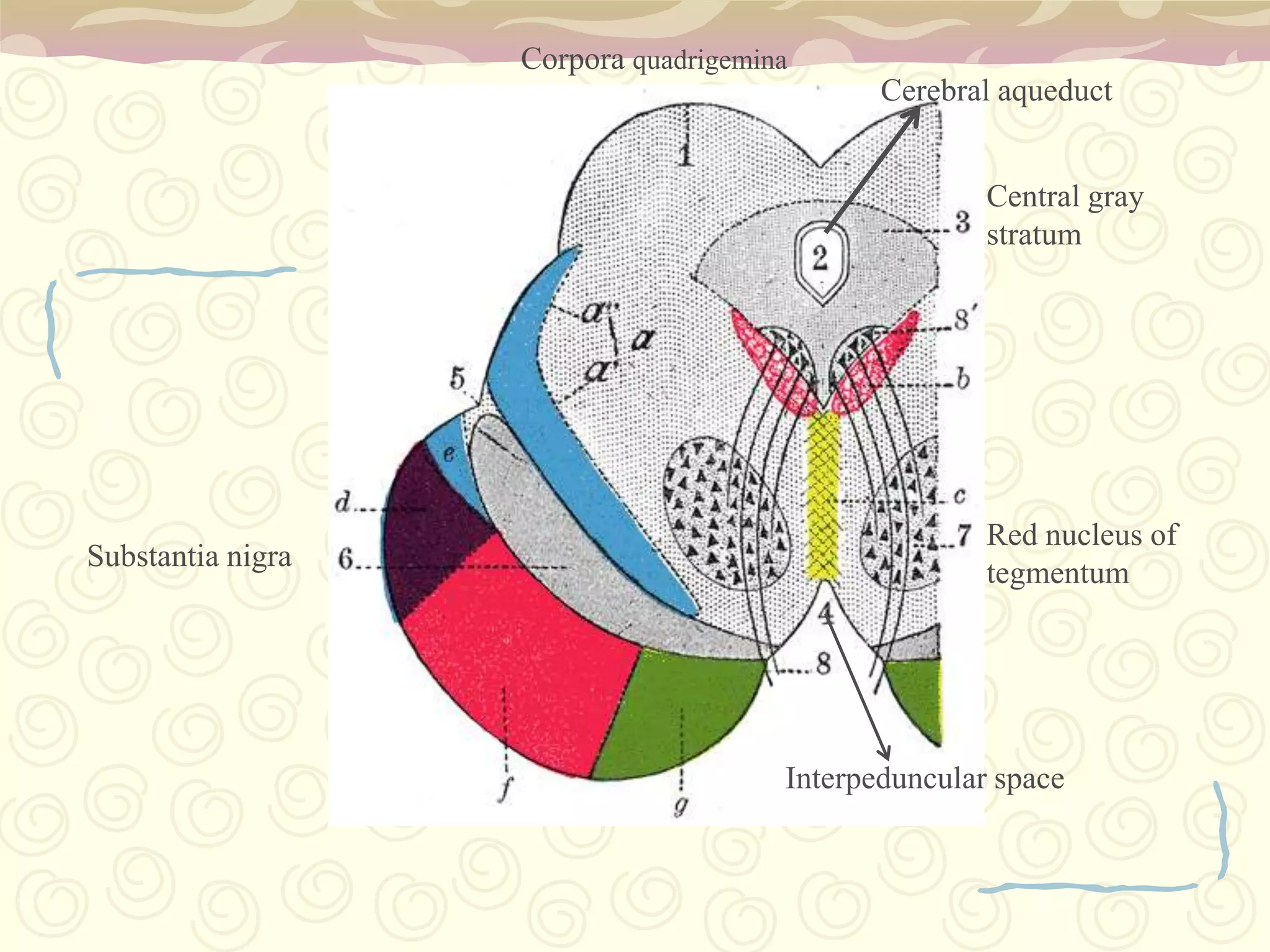
Embryological development of brain: Forebrain, Midbrain, Hindbrain; parts include Cerebrum, Pons, Thalamus.
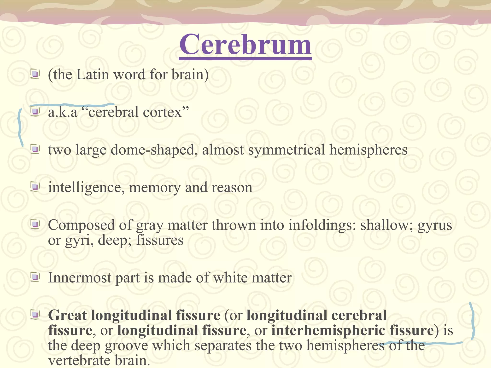
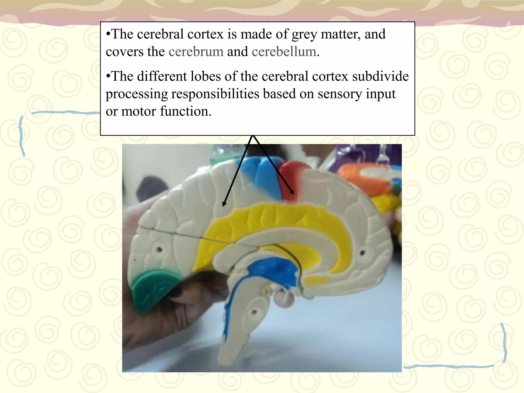
Cerebrum features: intelligence, memory, consisting of grey and white matter, along with cortex functions.
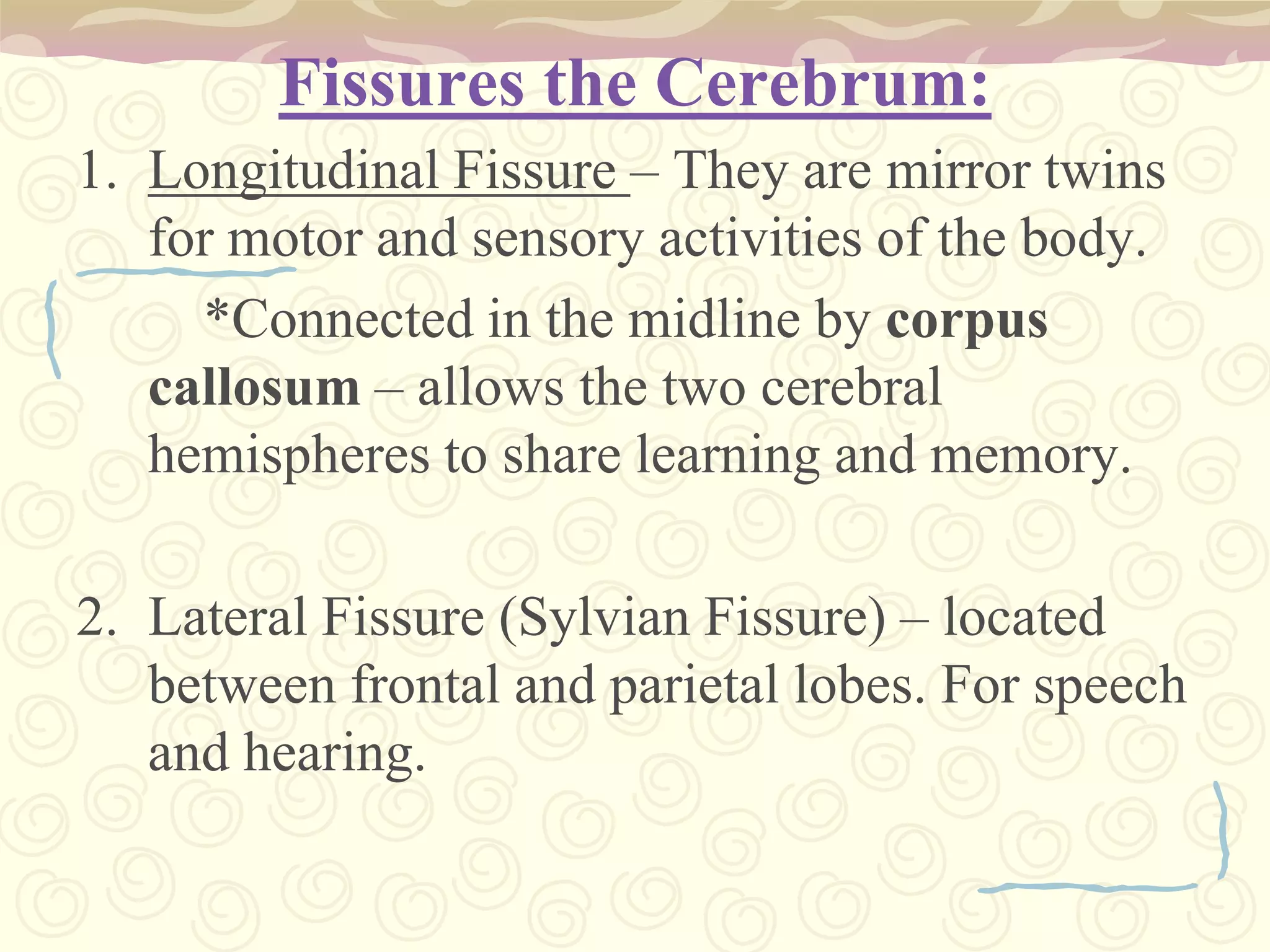
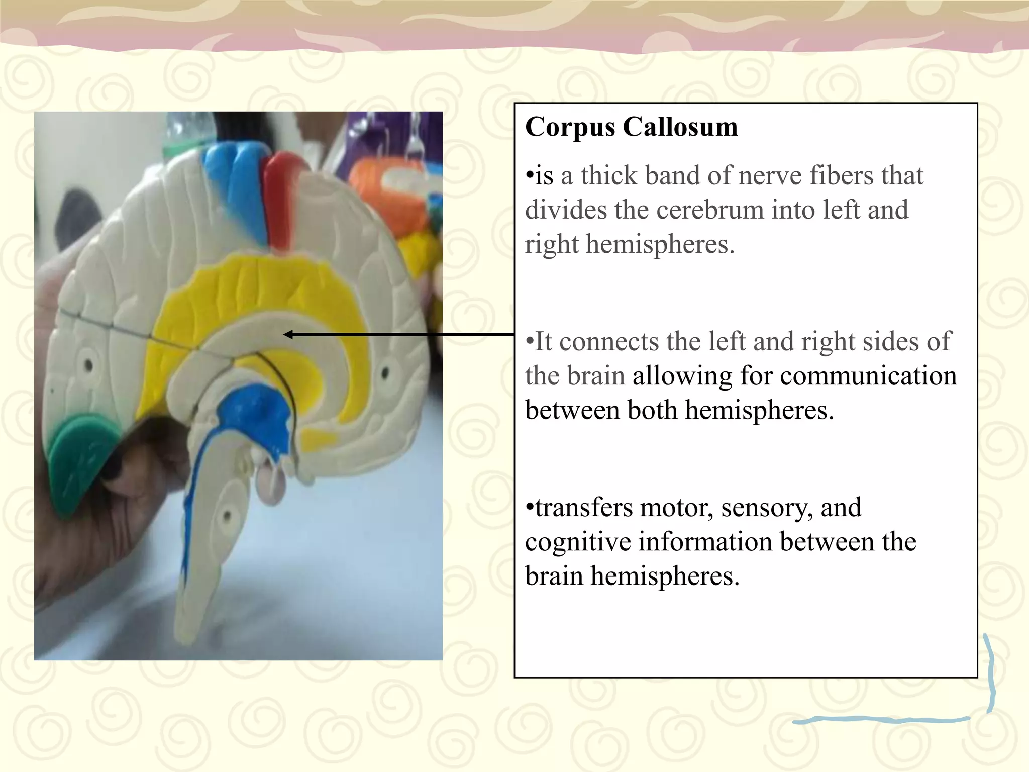
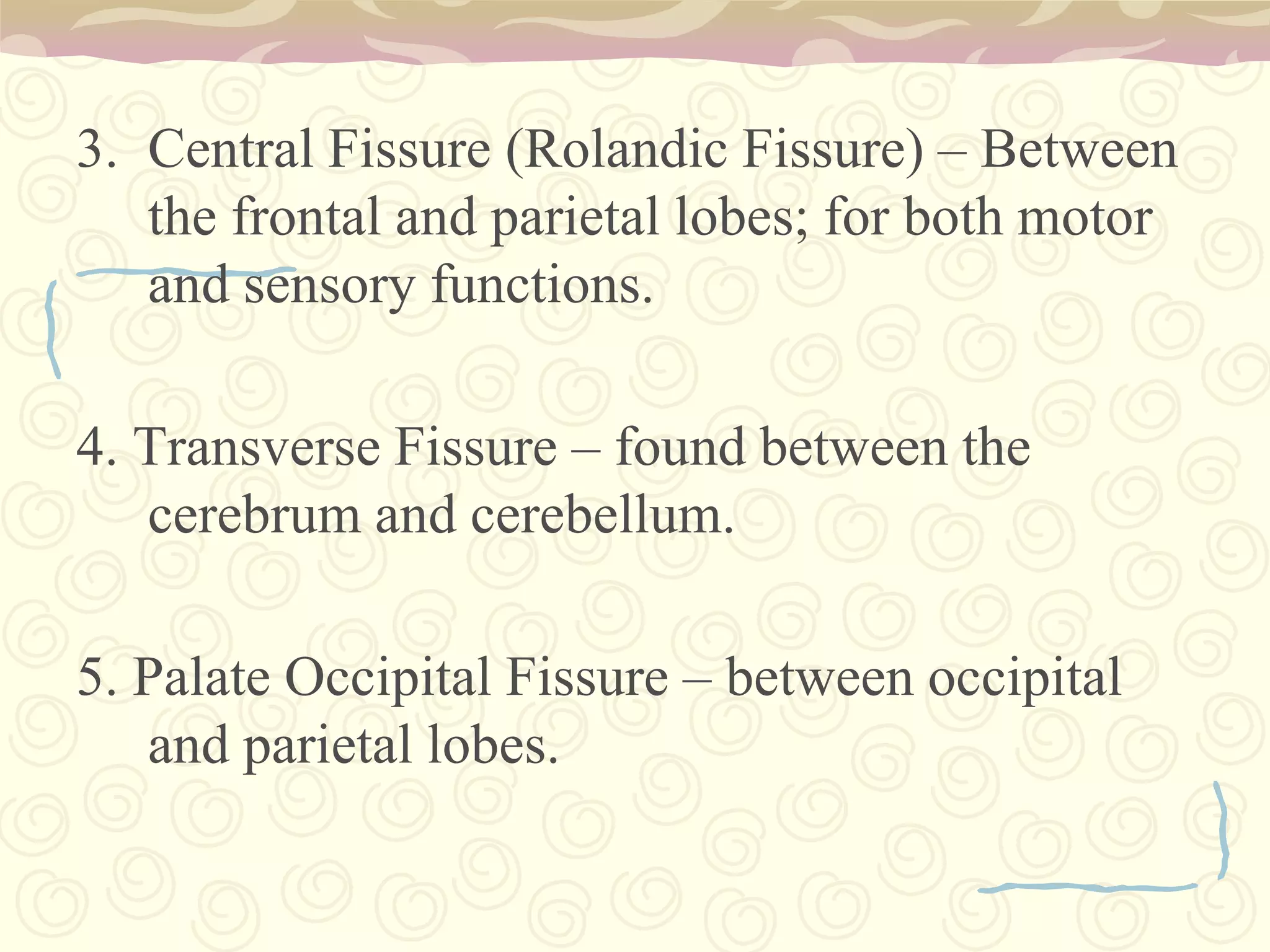
Fissures allow communication between hemispheres; central, lateral, and longitudinal fissures structured for functionality.
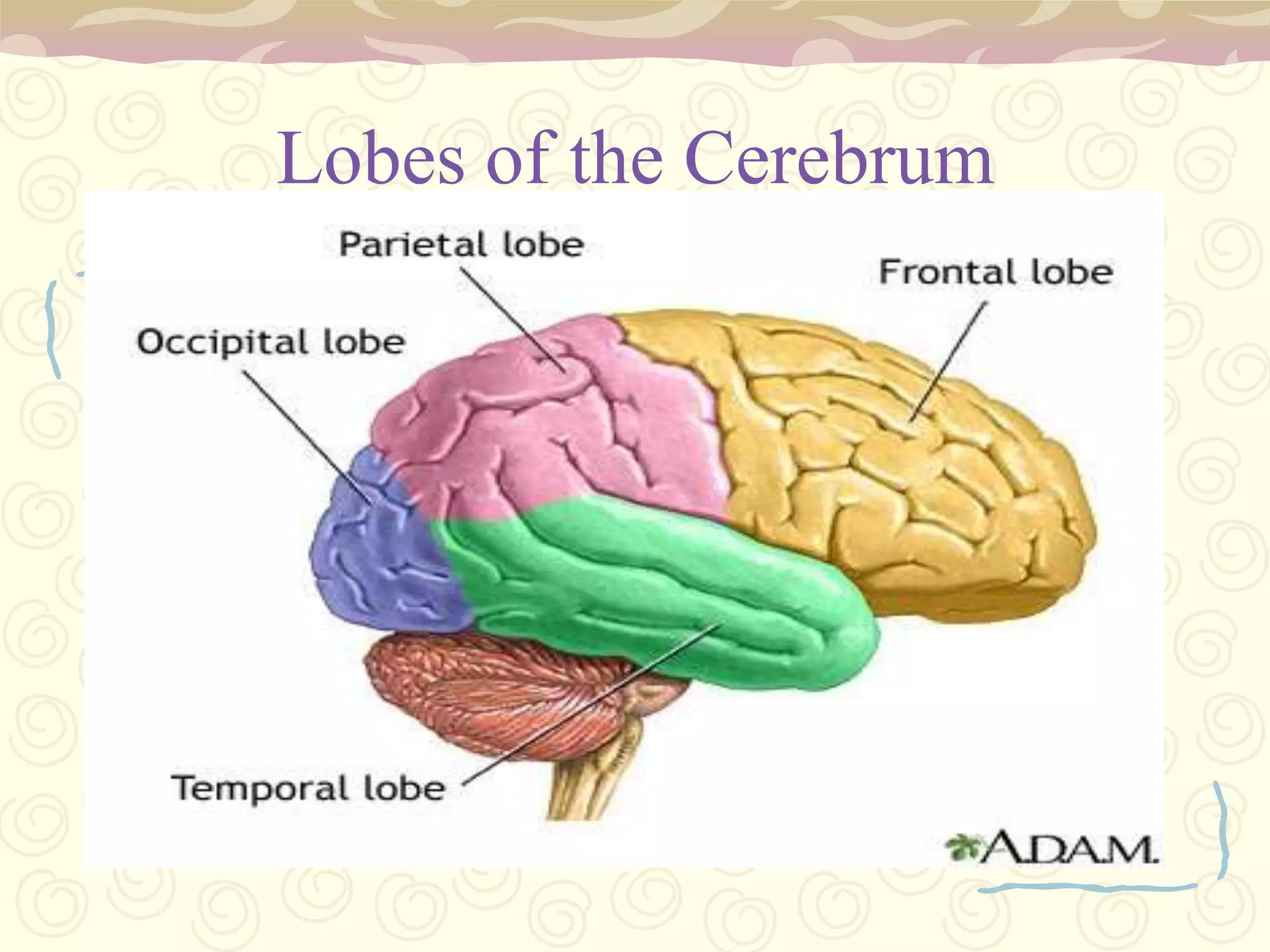
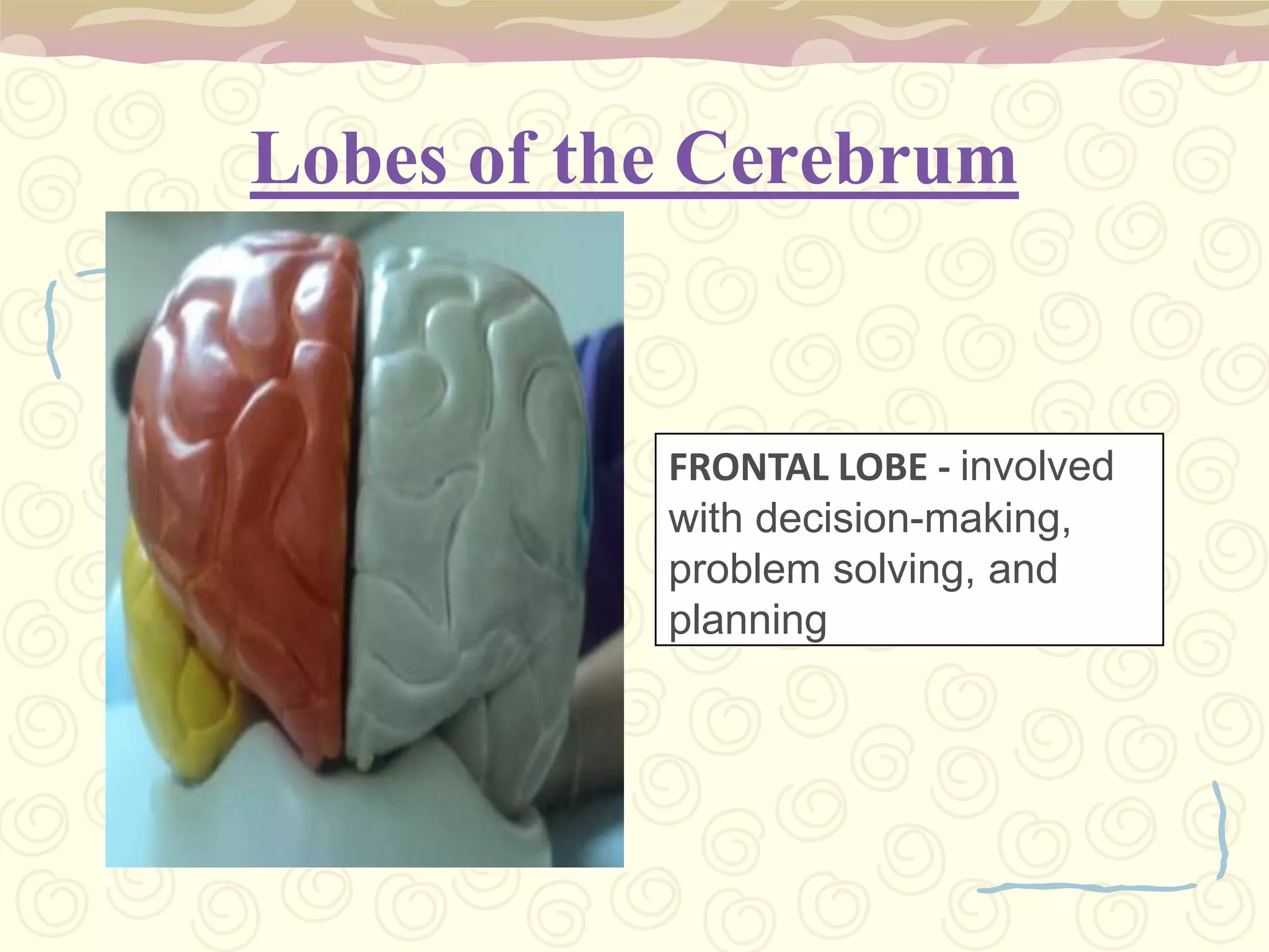
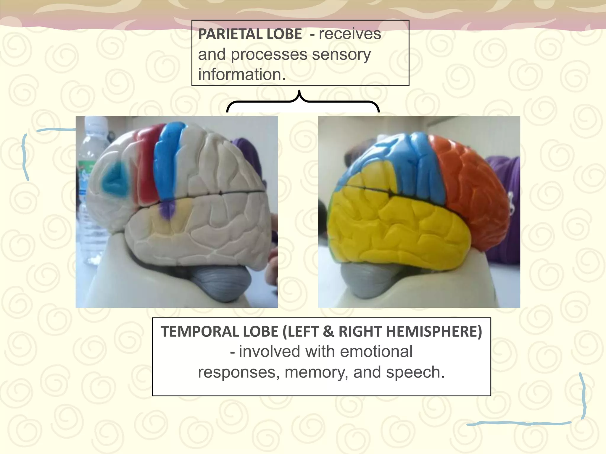
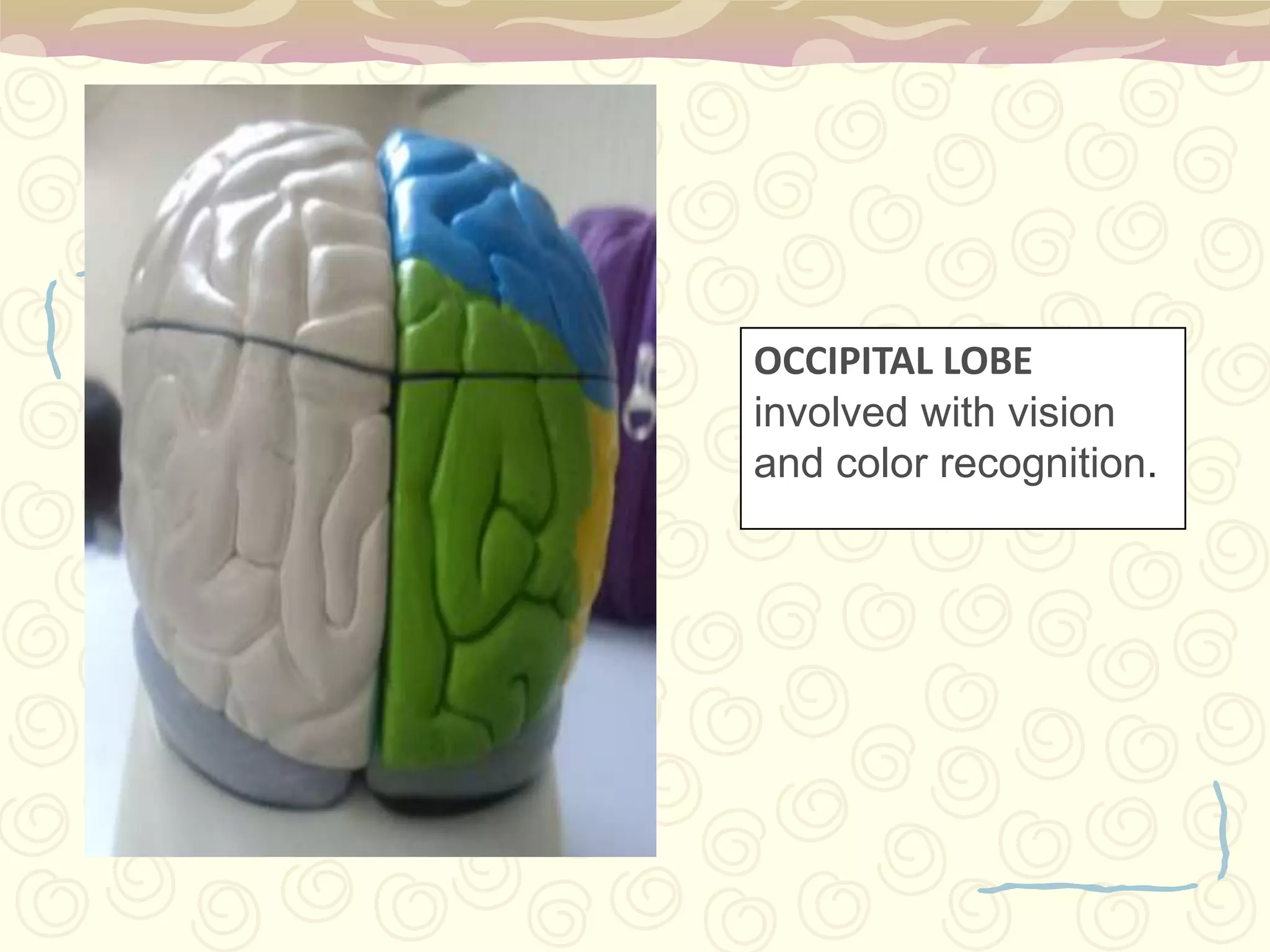
Frontal (decision-making), Parietal (sensory processing), Temporal (memory and emotion), Occipital (vision).
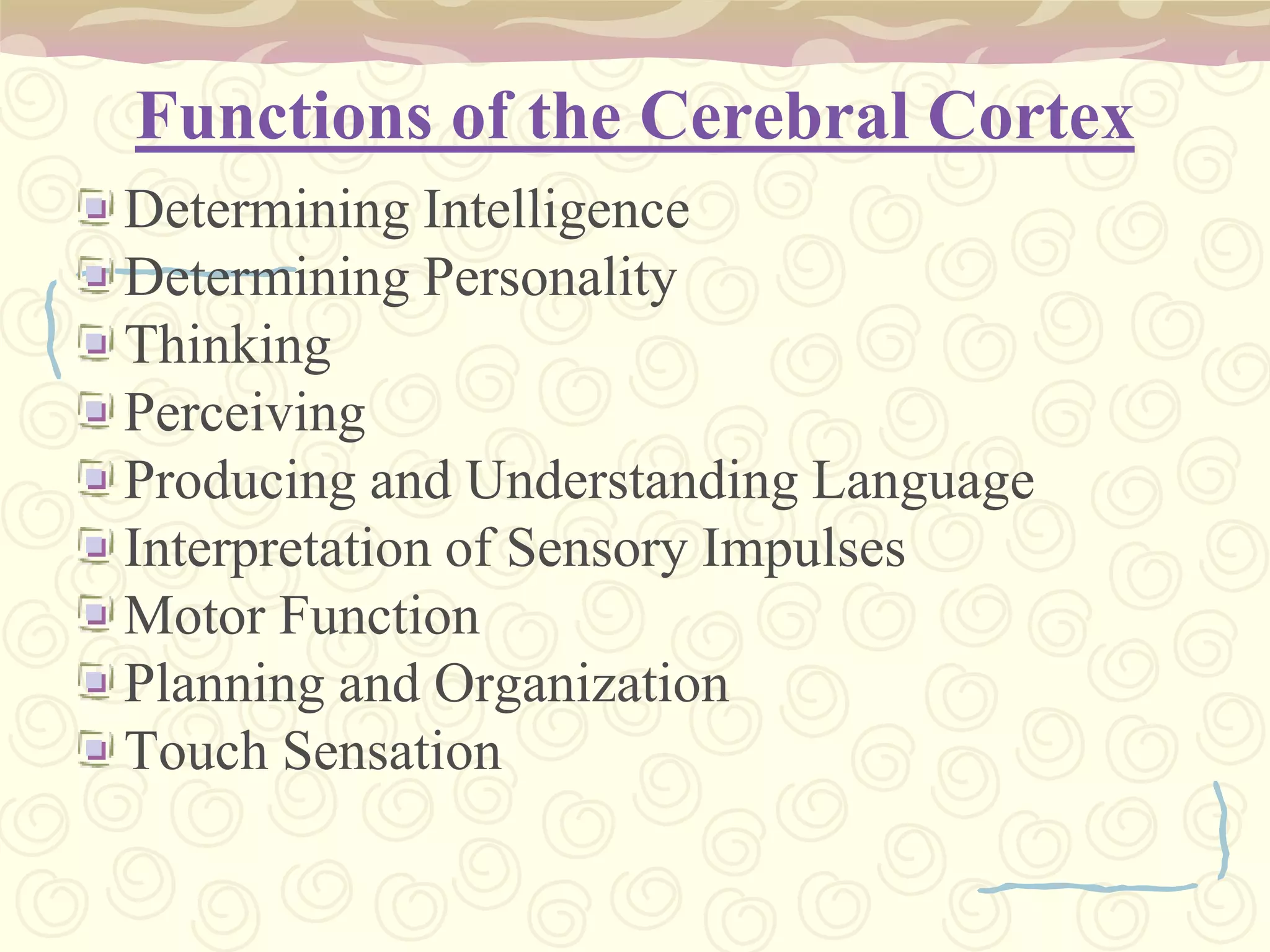
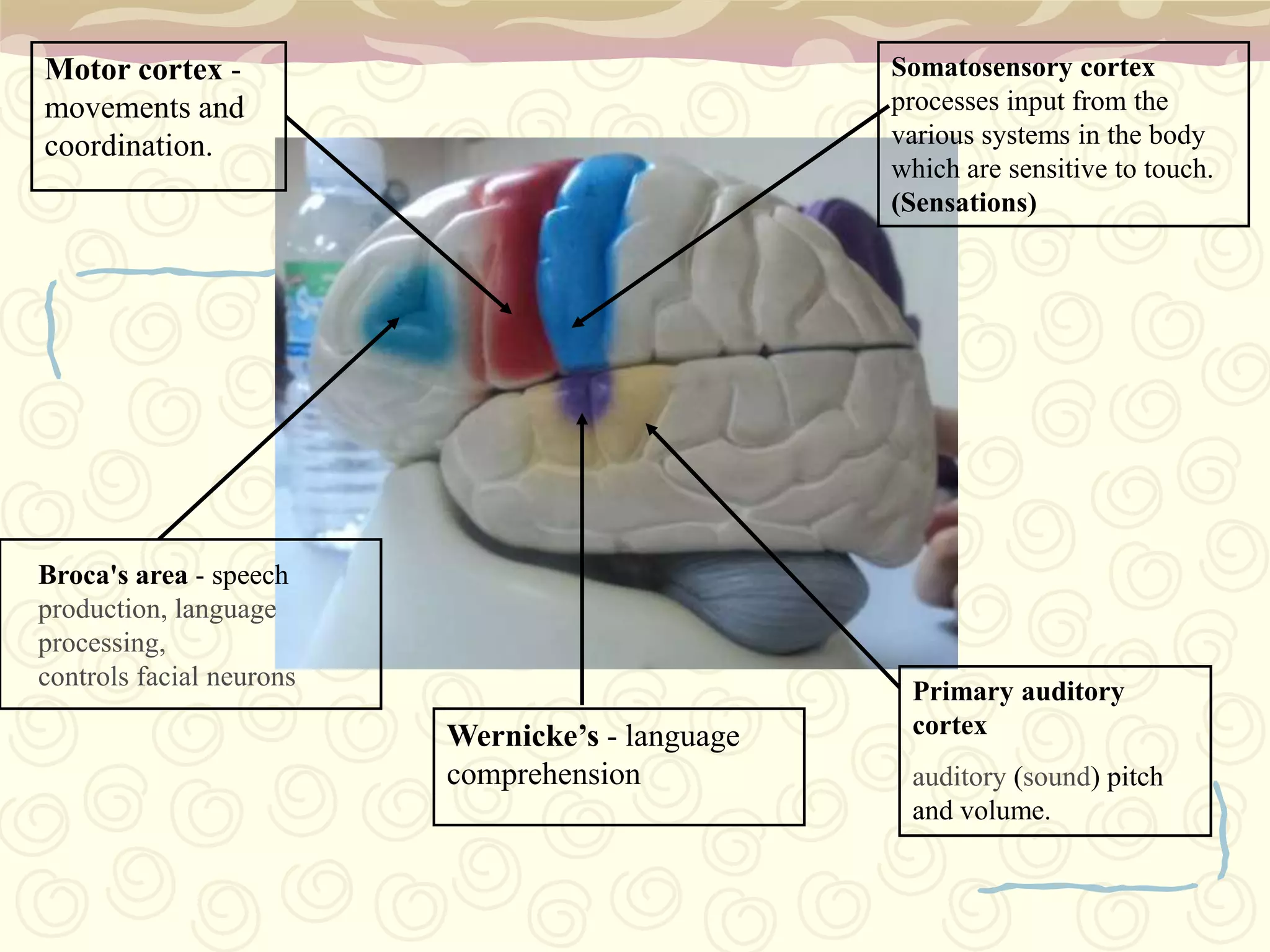
Responsibilities: intelligence, personality, sensory perception, motor functions.
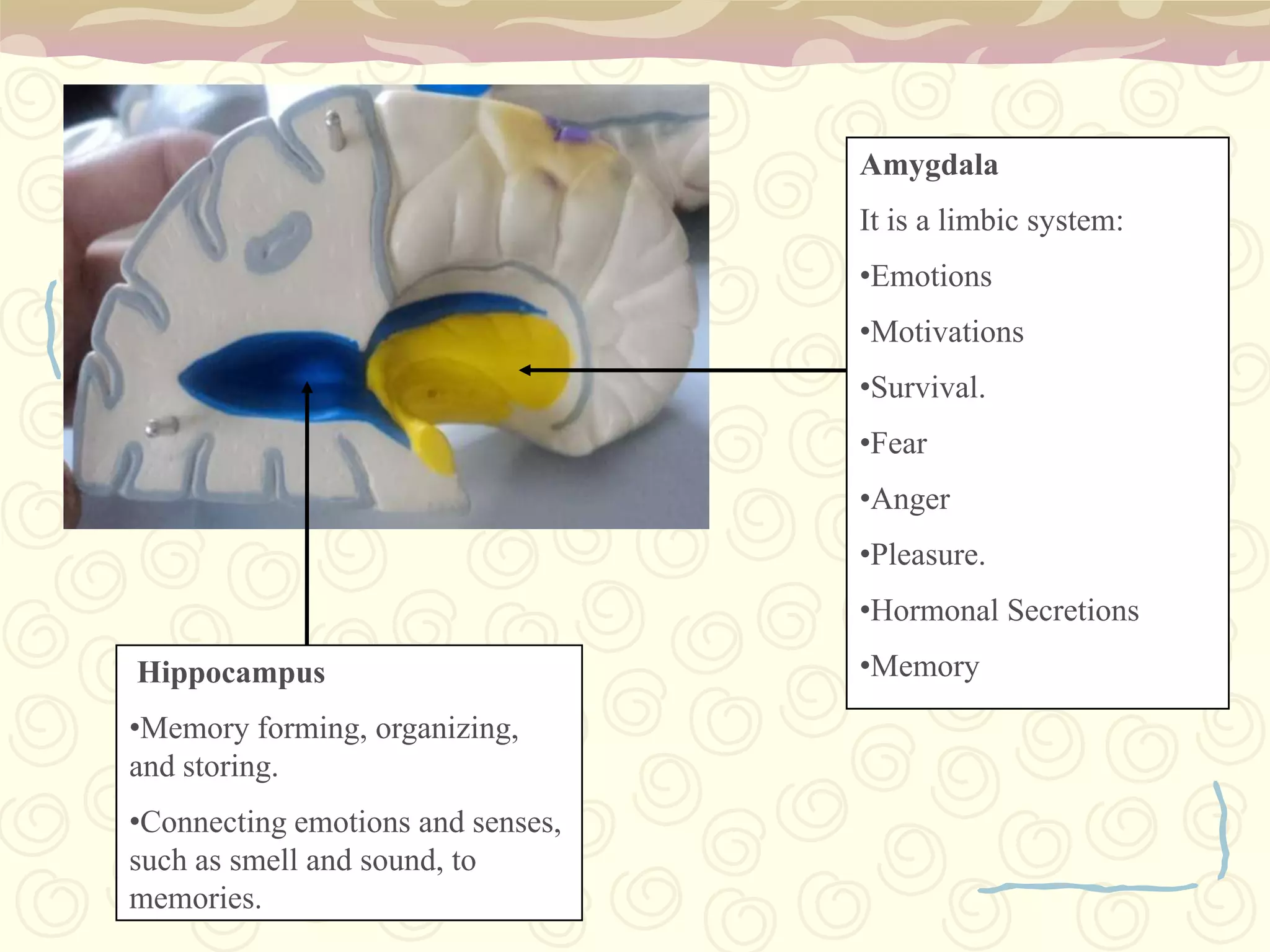
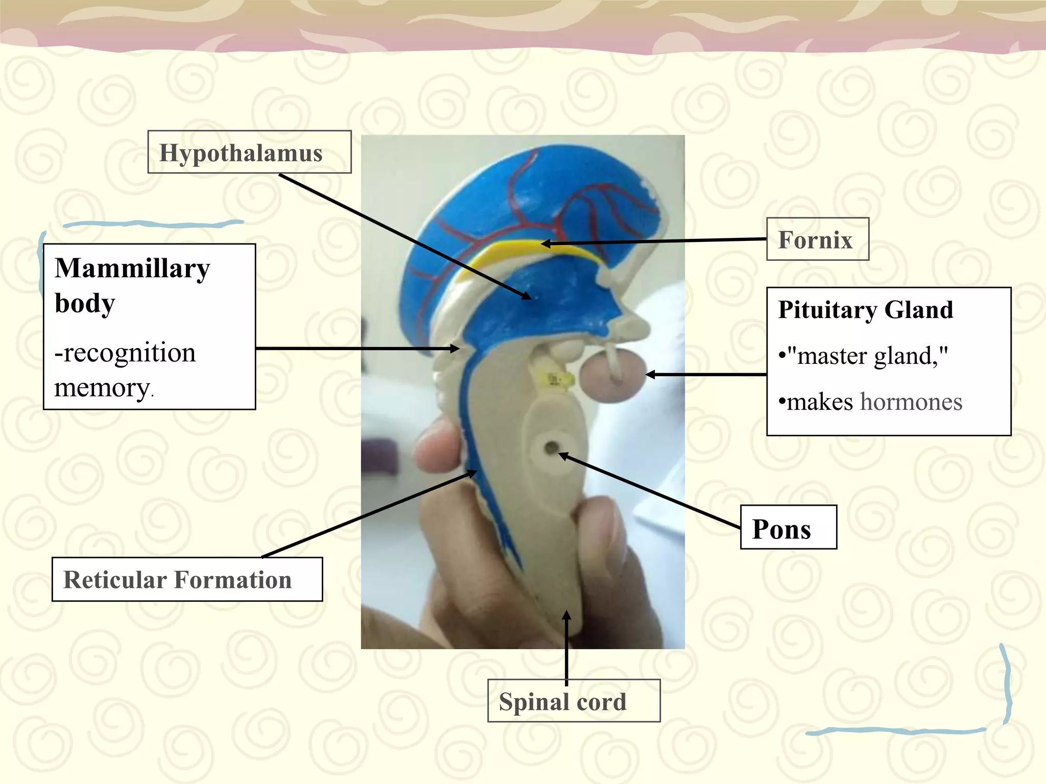
Amygdala (emotions) and Hippocampus (memory organization, sensory connection).
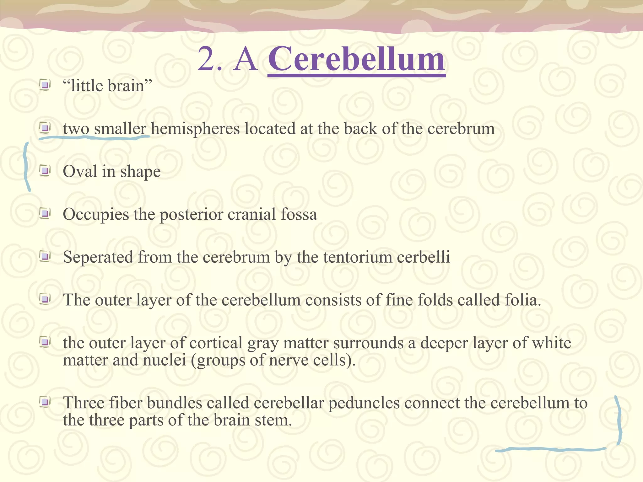
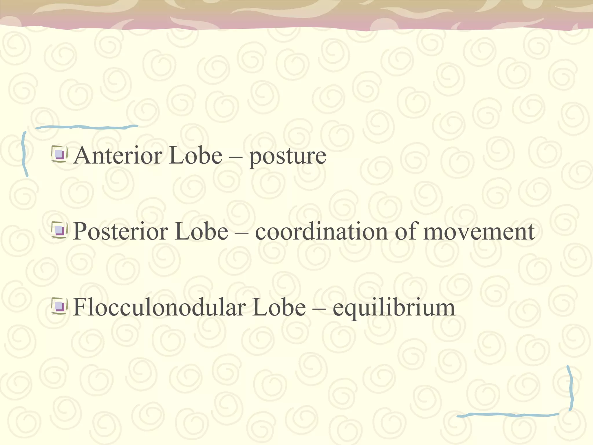
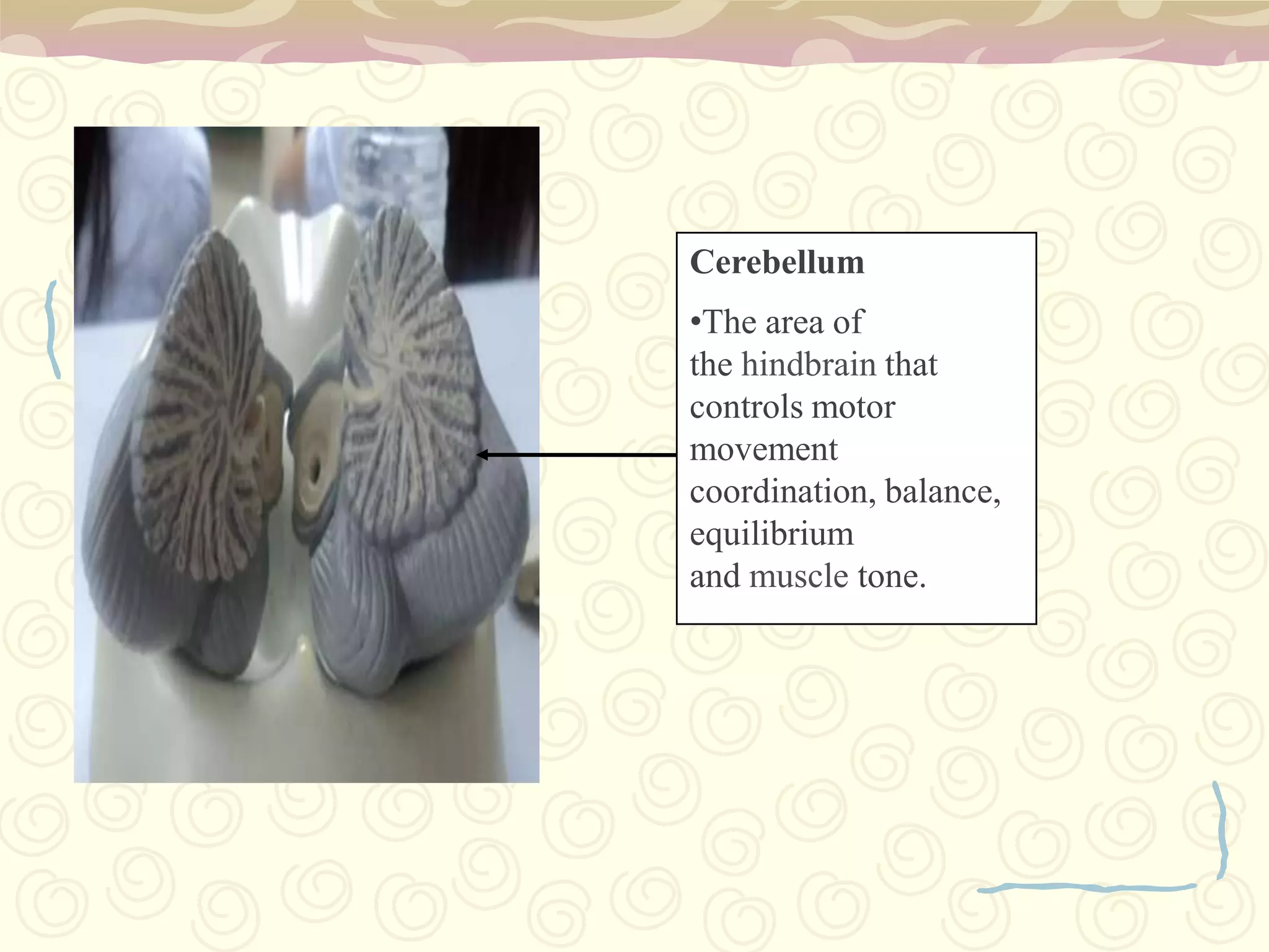
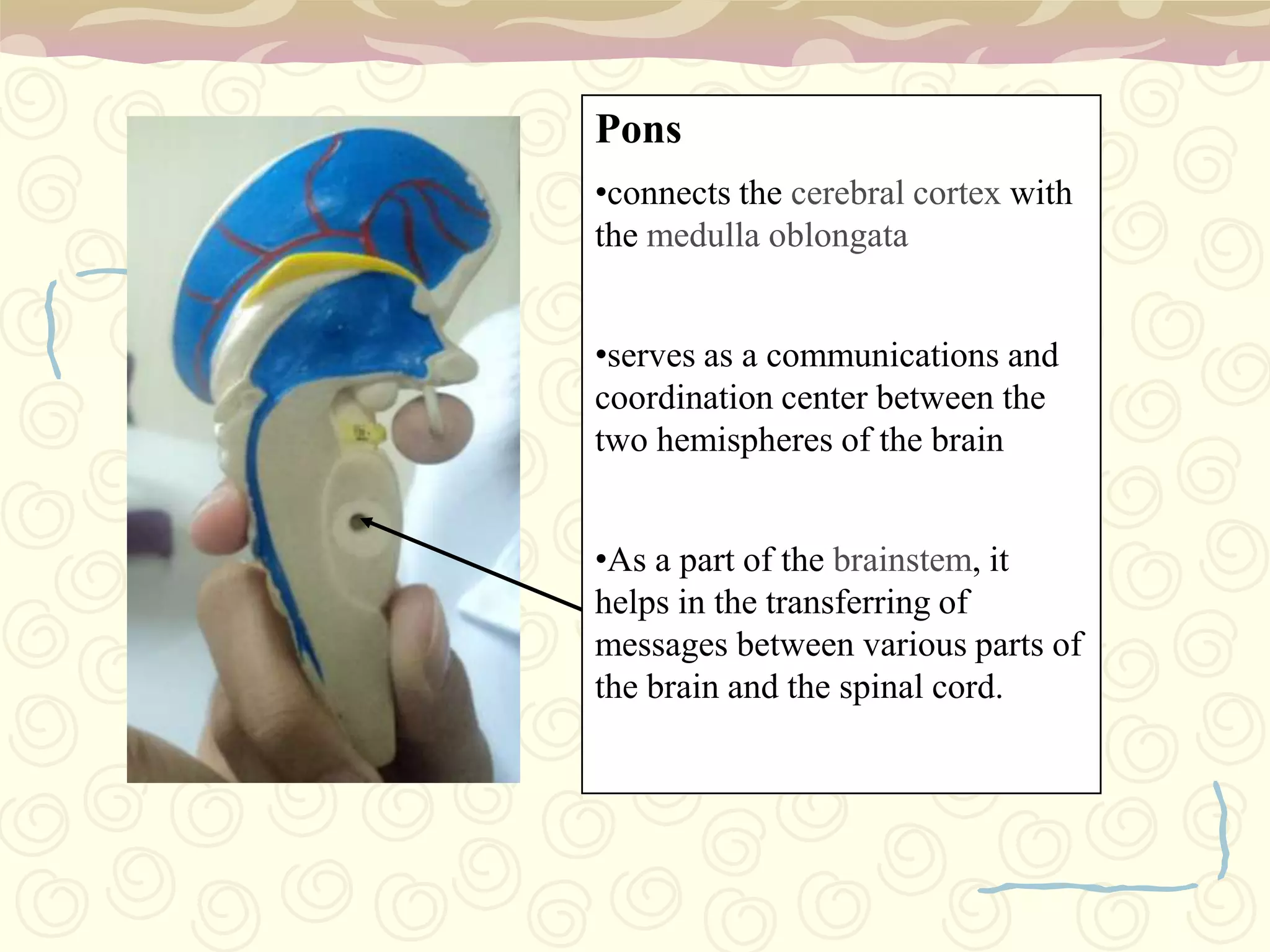
Functions: coordination, balance, muscle tone (cerebellum) and communication (pons).
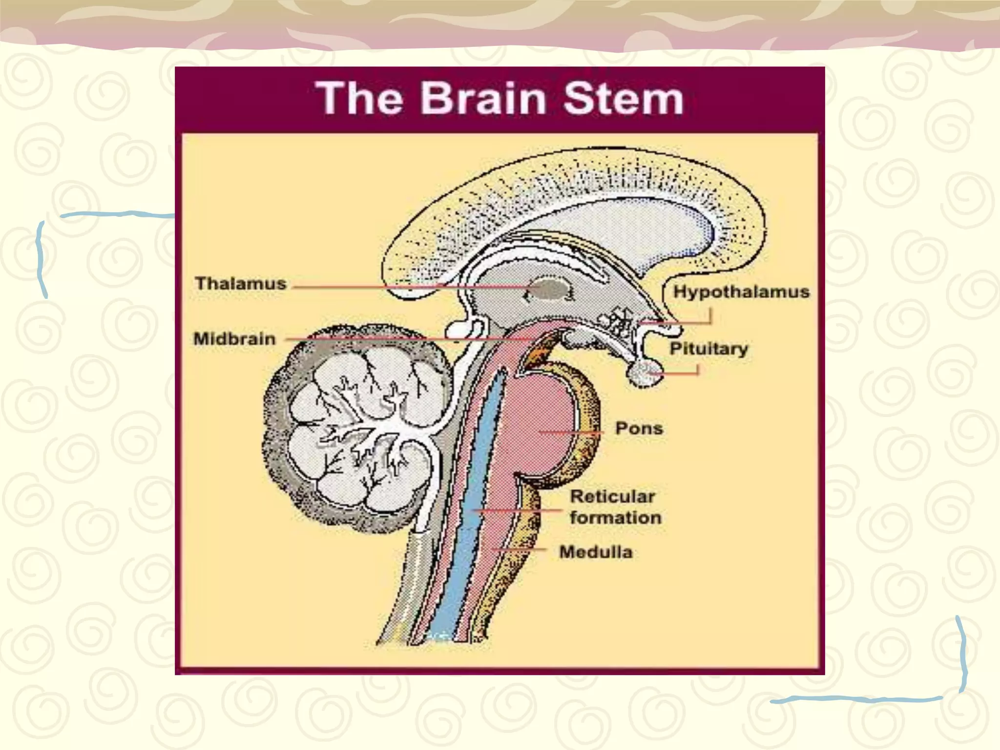
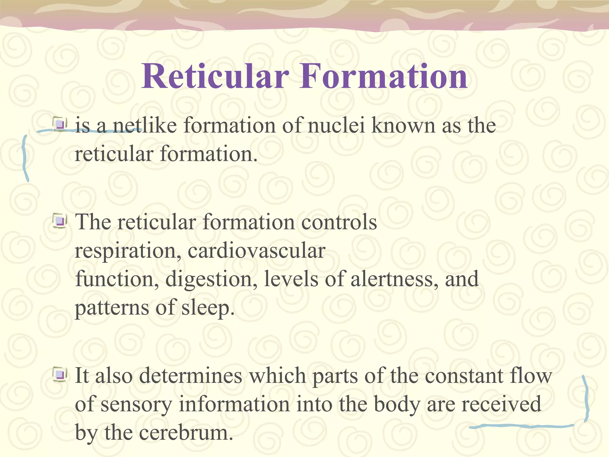
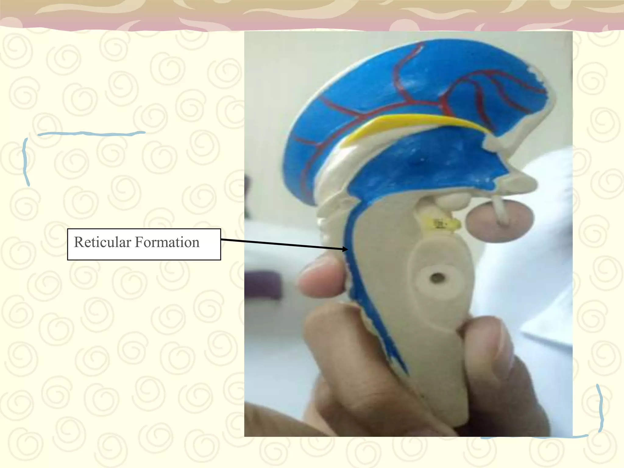
Brainstem's roles: sustains life functions, spinal cord communication, and reticular formation effects.
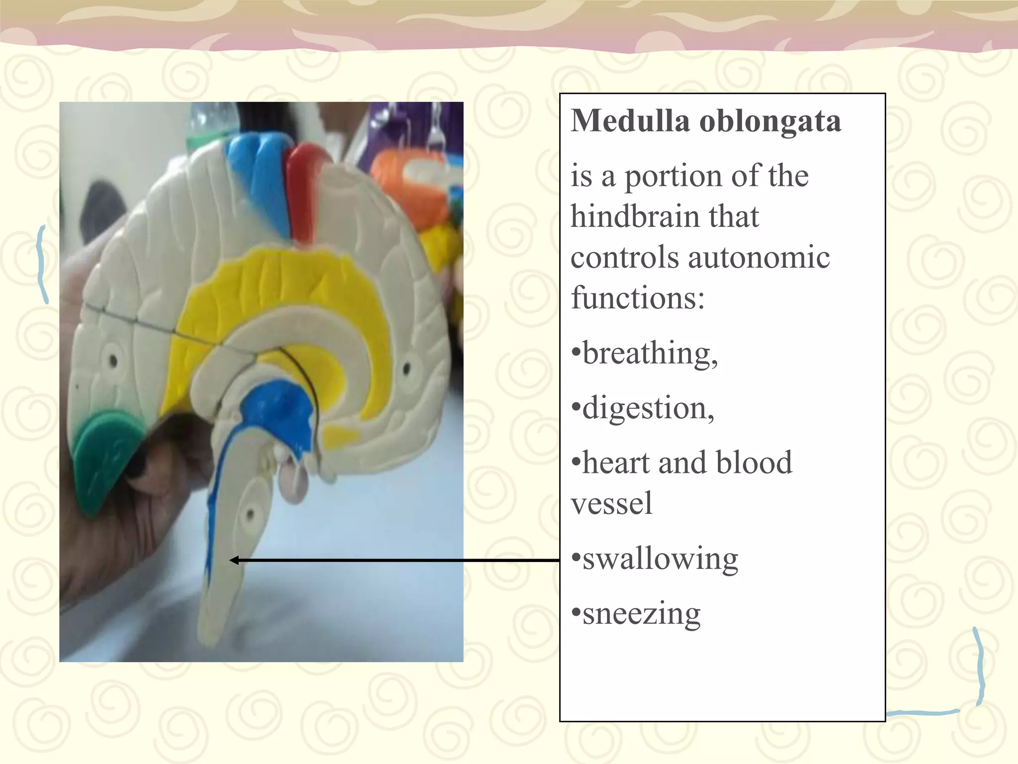
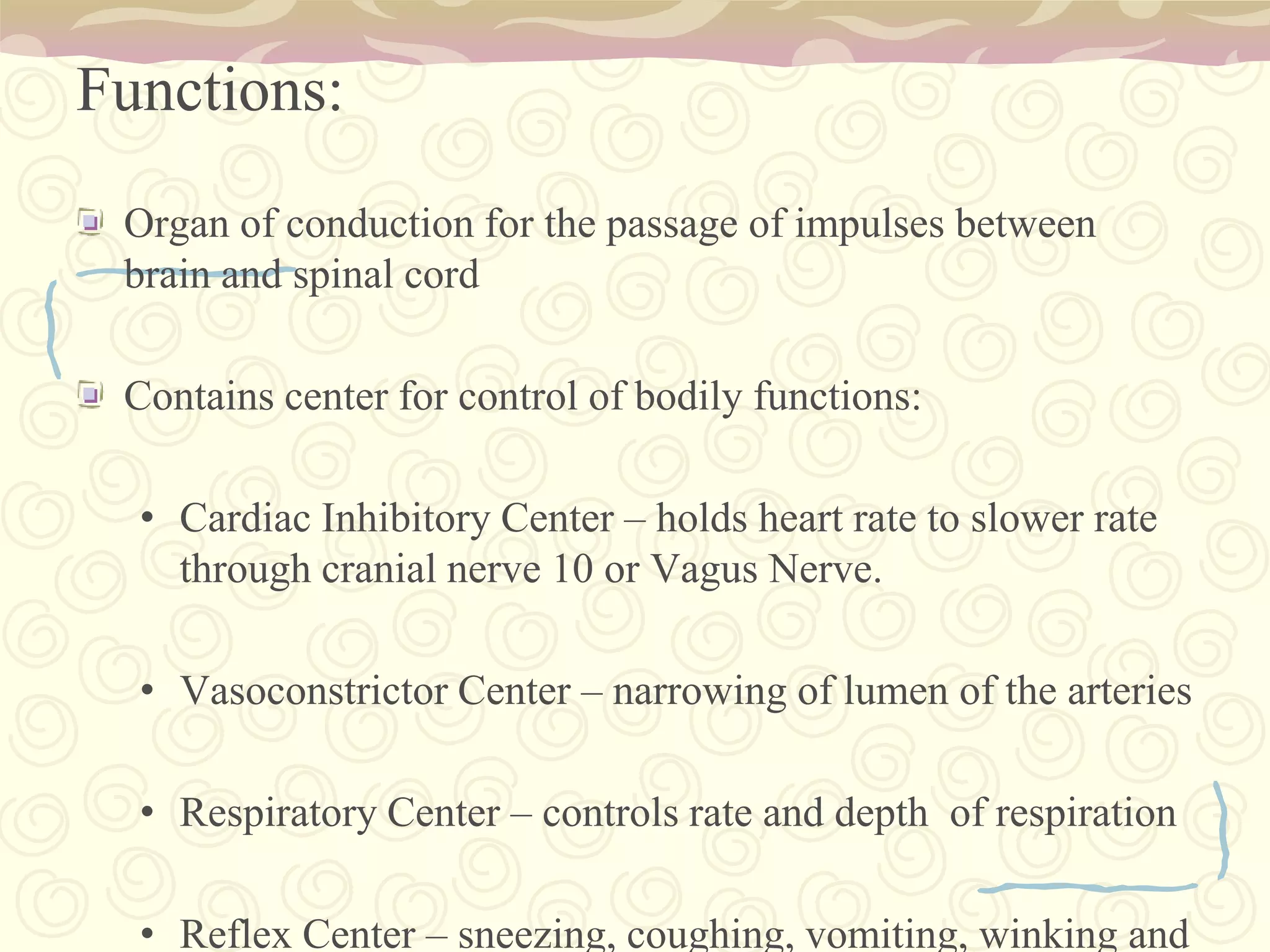
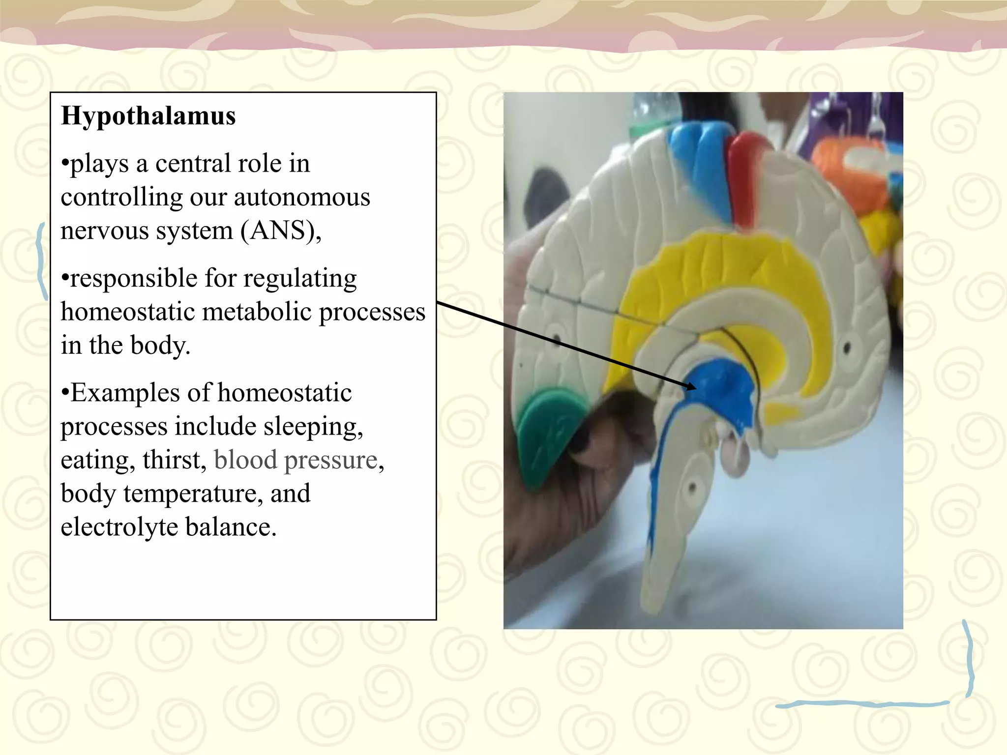
Lowest brainstem part and autonomic function control: heart rate, breathing, digestion.Thalamus as a relay station; Hypothalamus regulates hunger, thirst, and homeostasis.
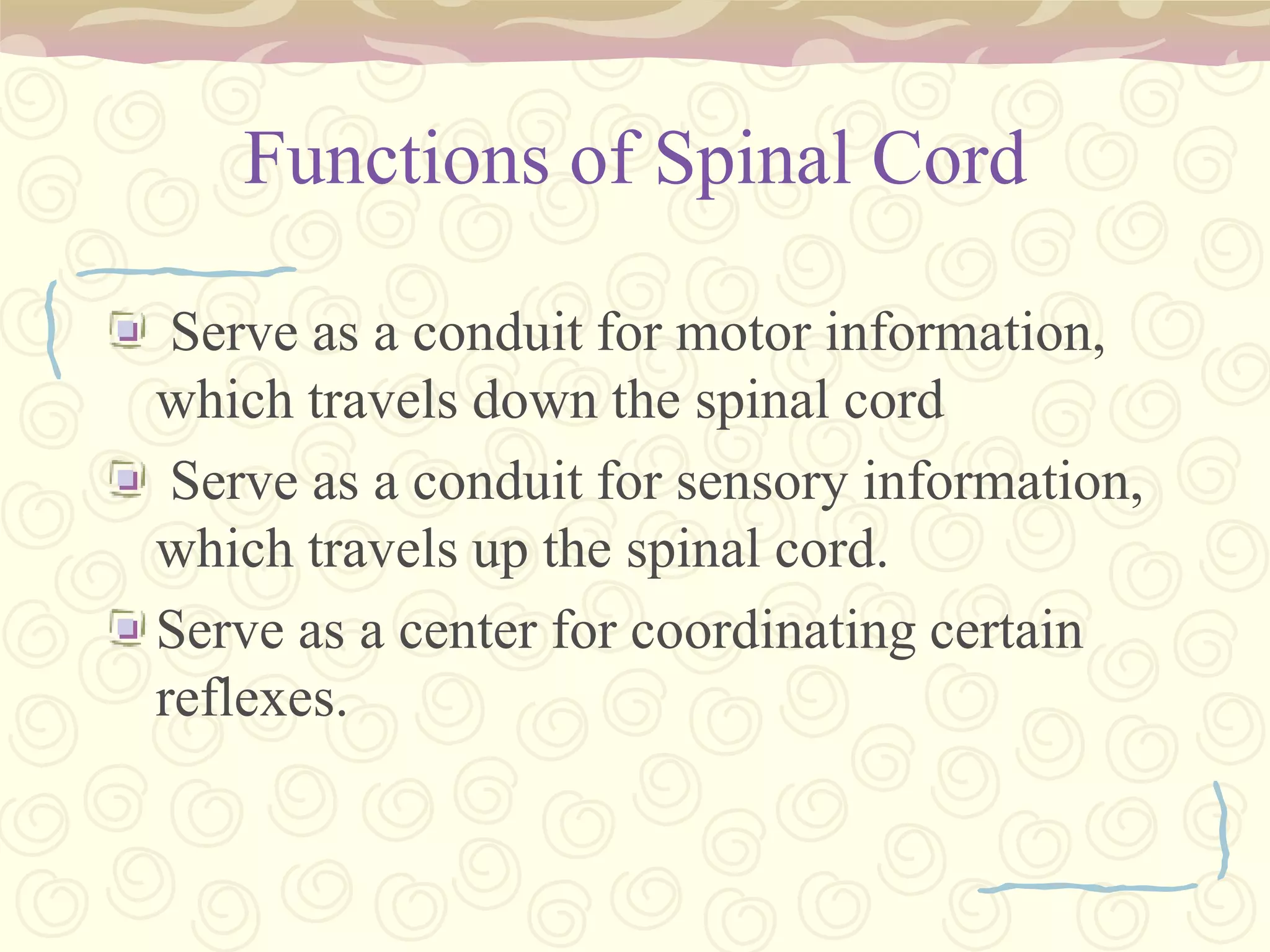
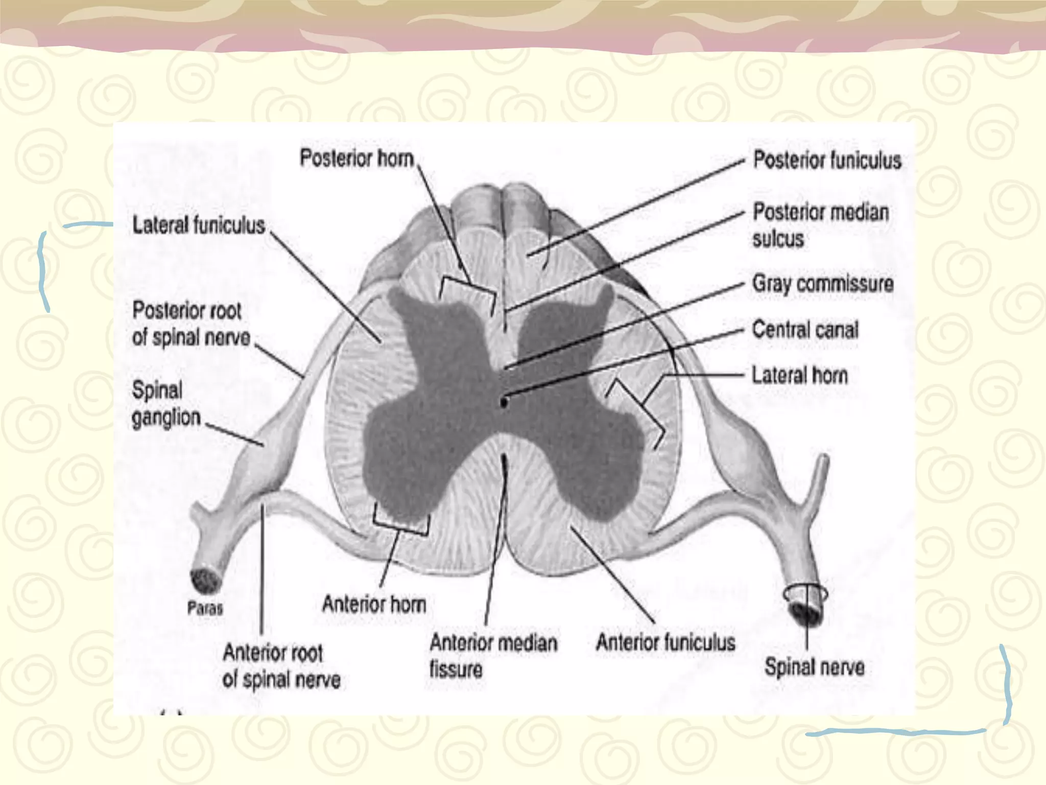
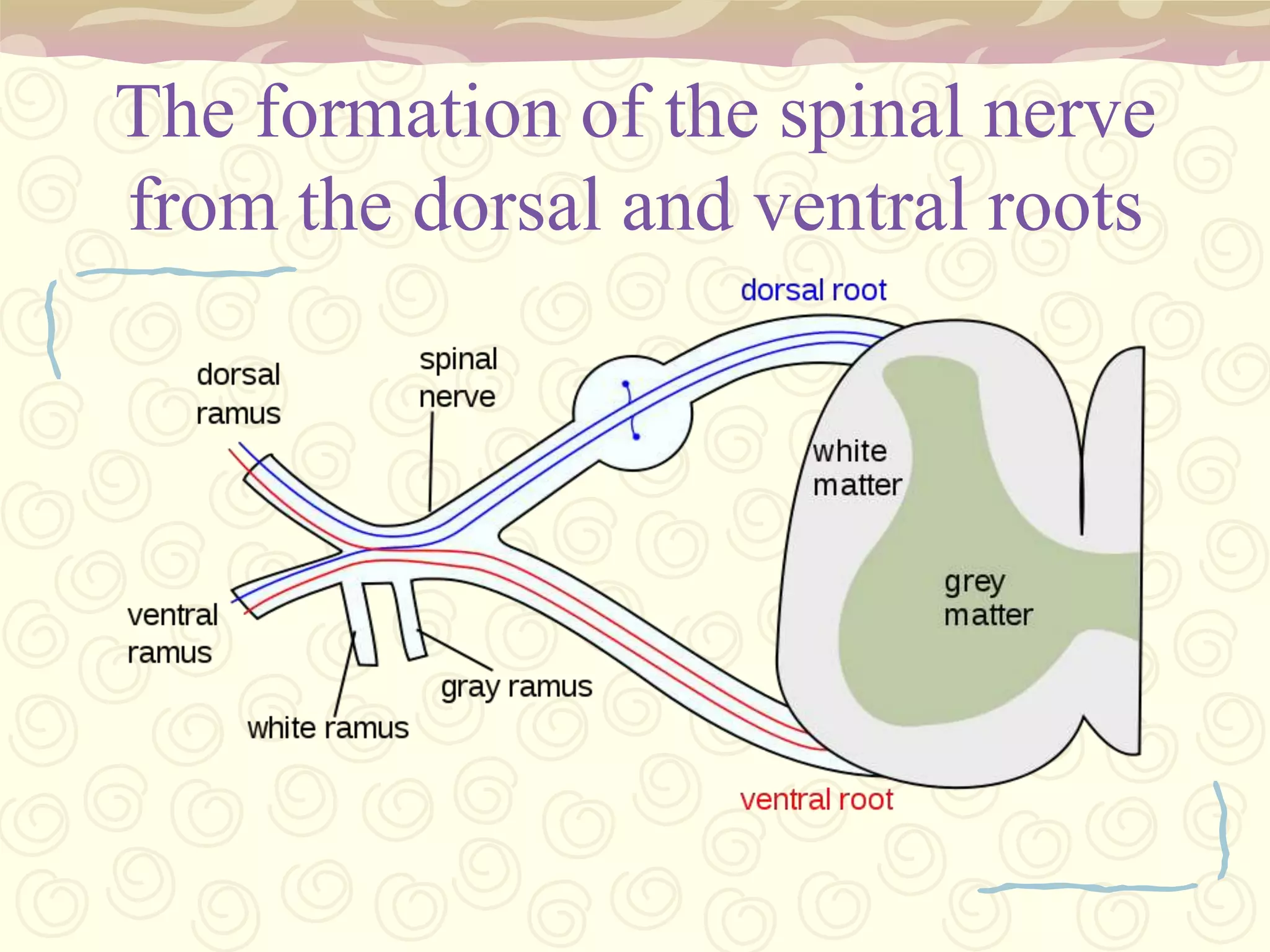
Structure and functions: conduit for motor/sensory info, coordination of reflexes.
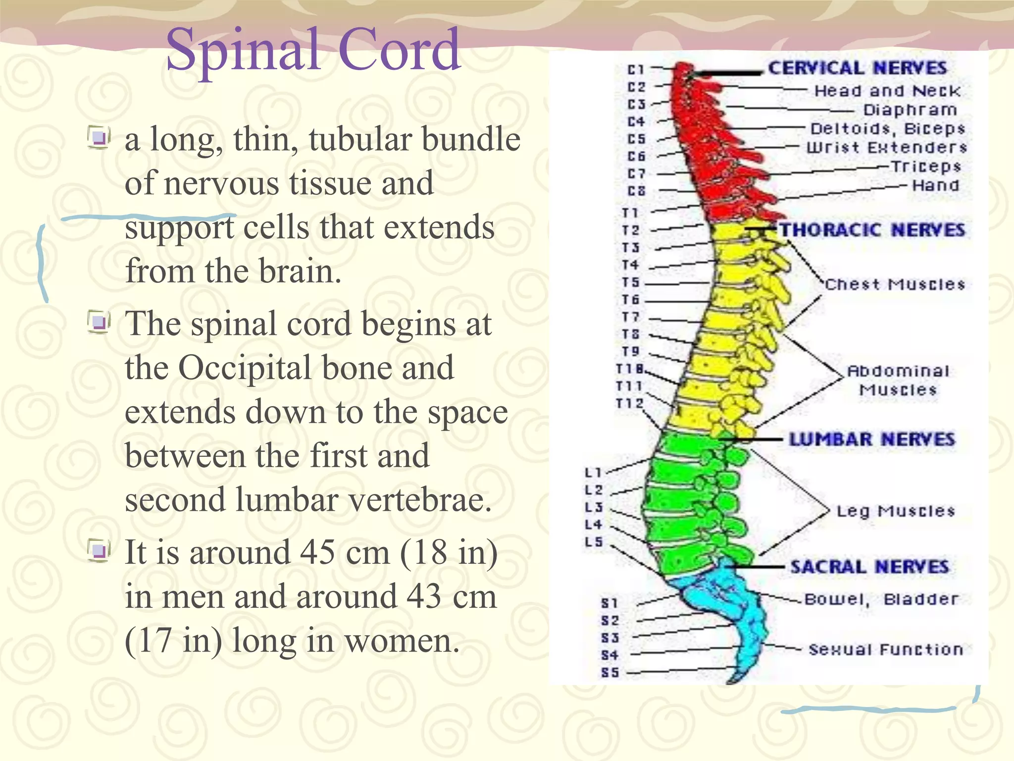
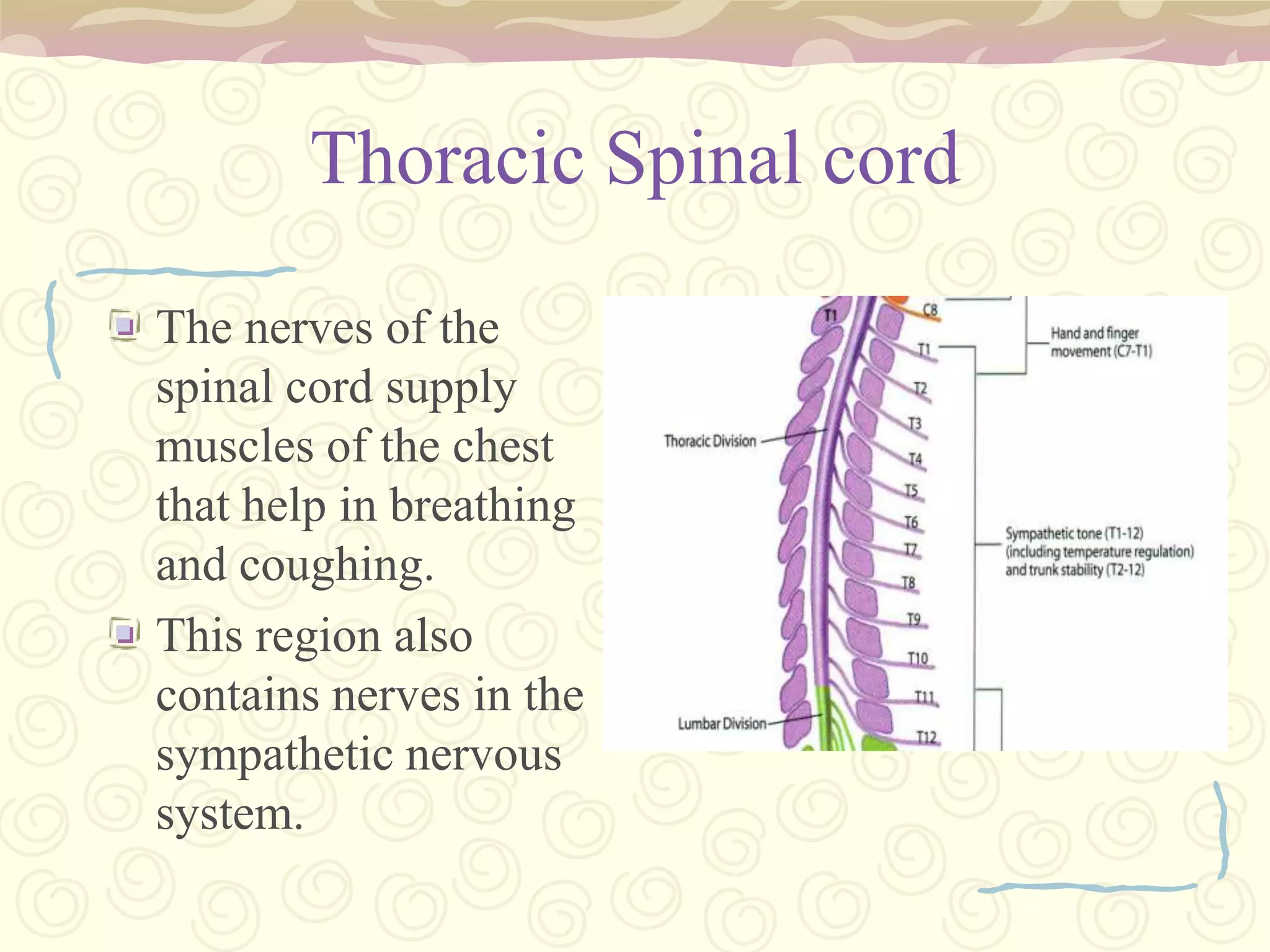
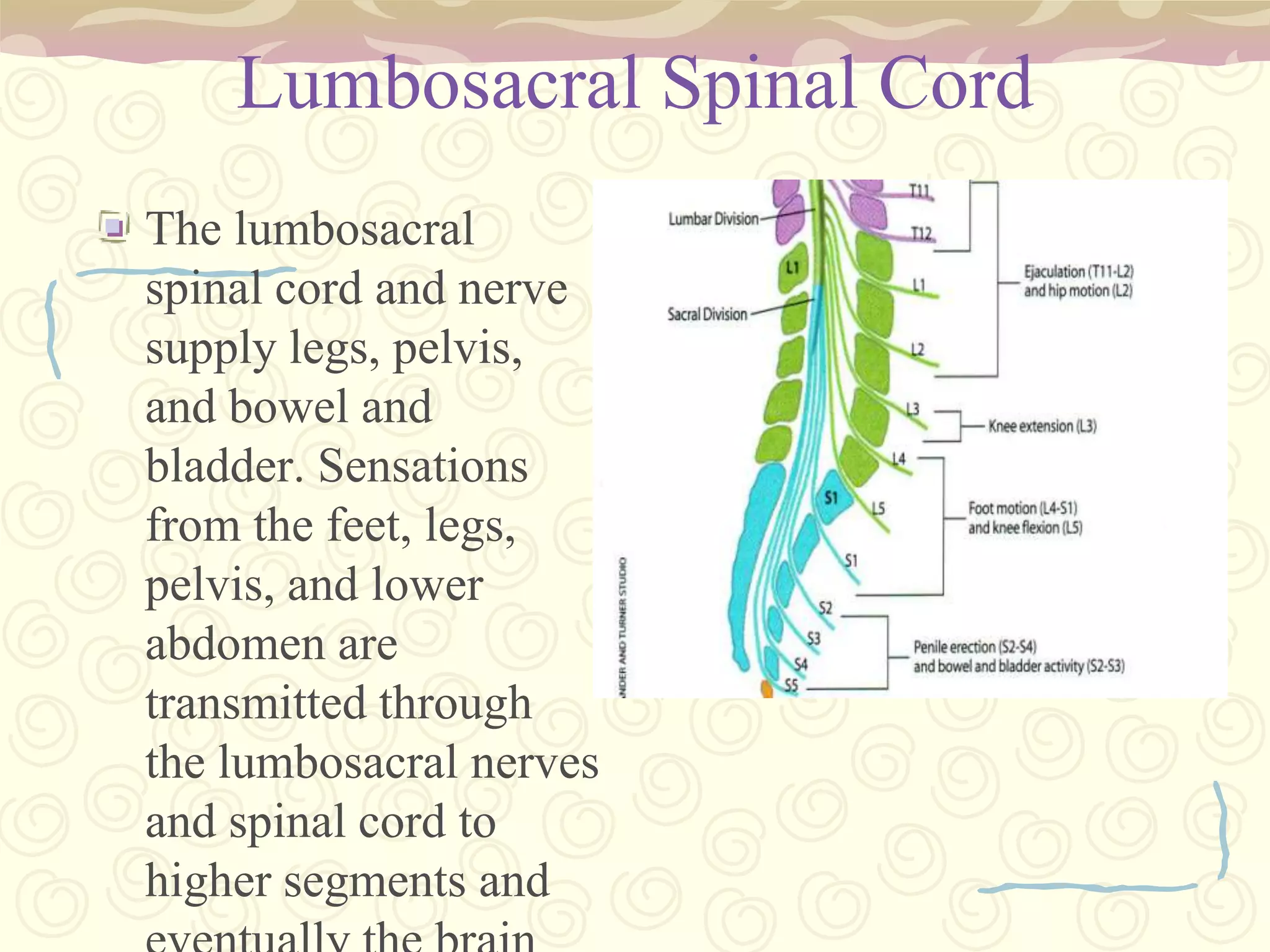
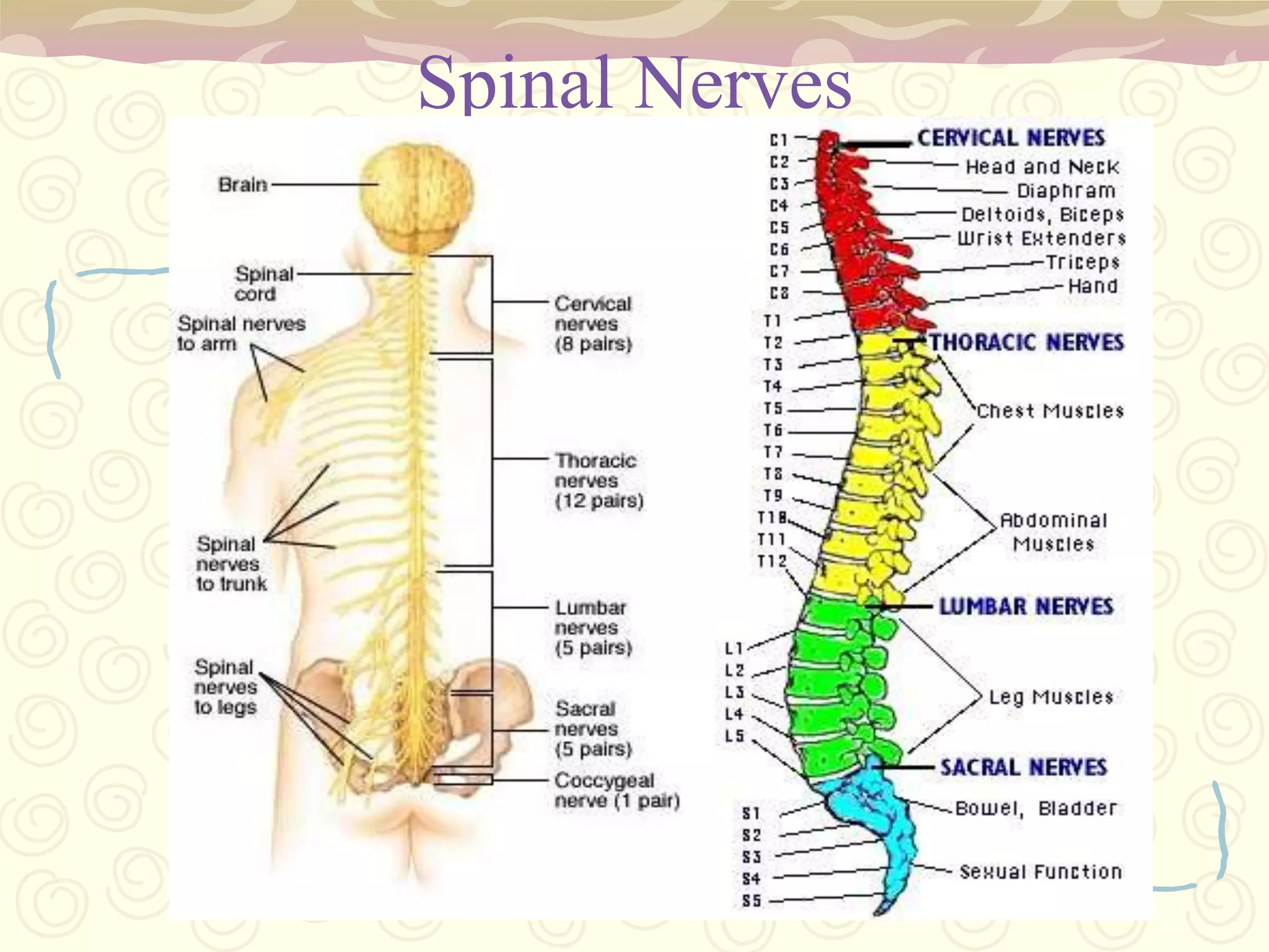
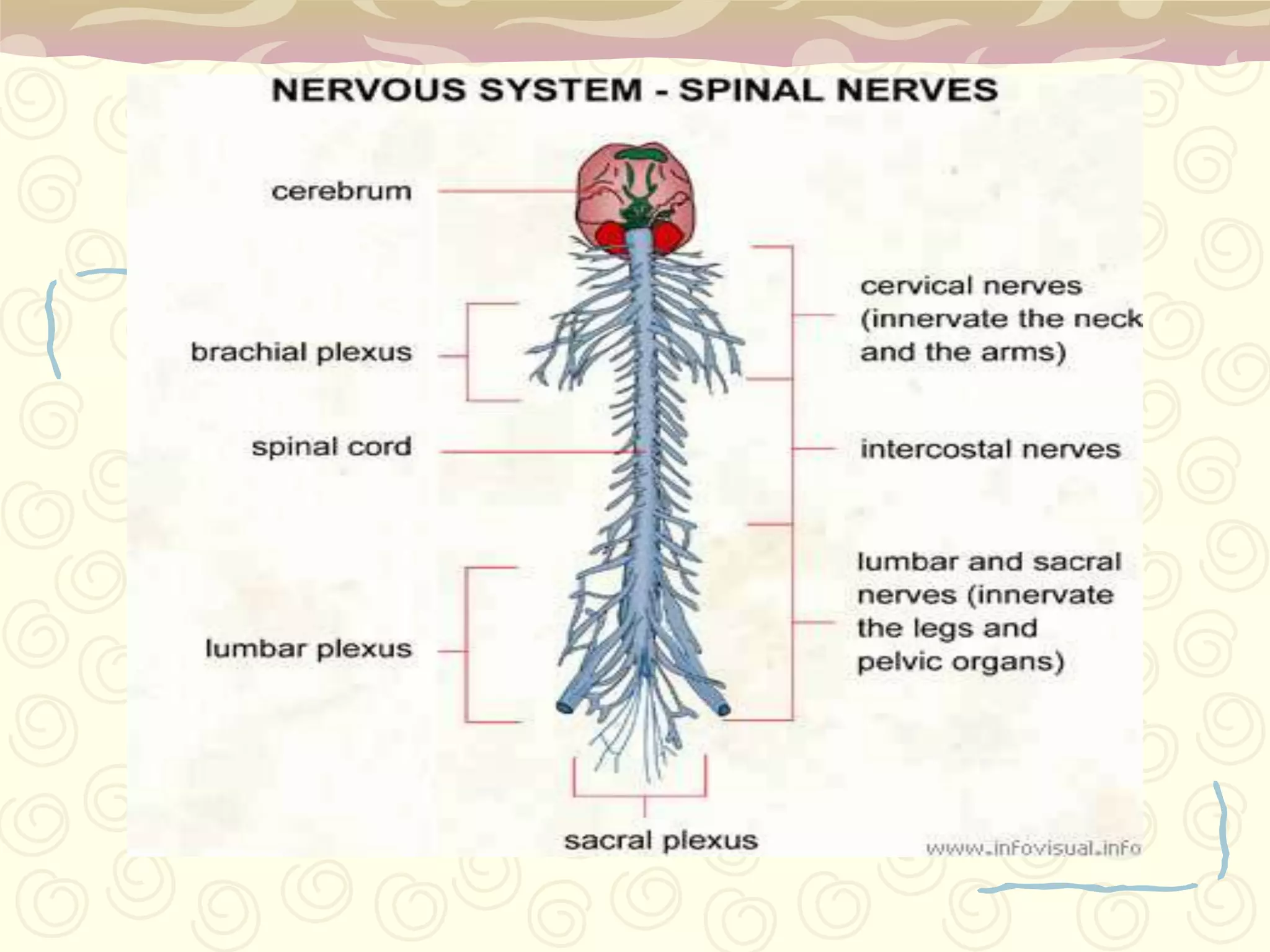
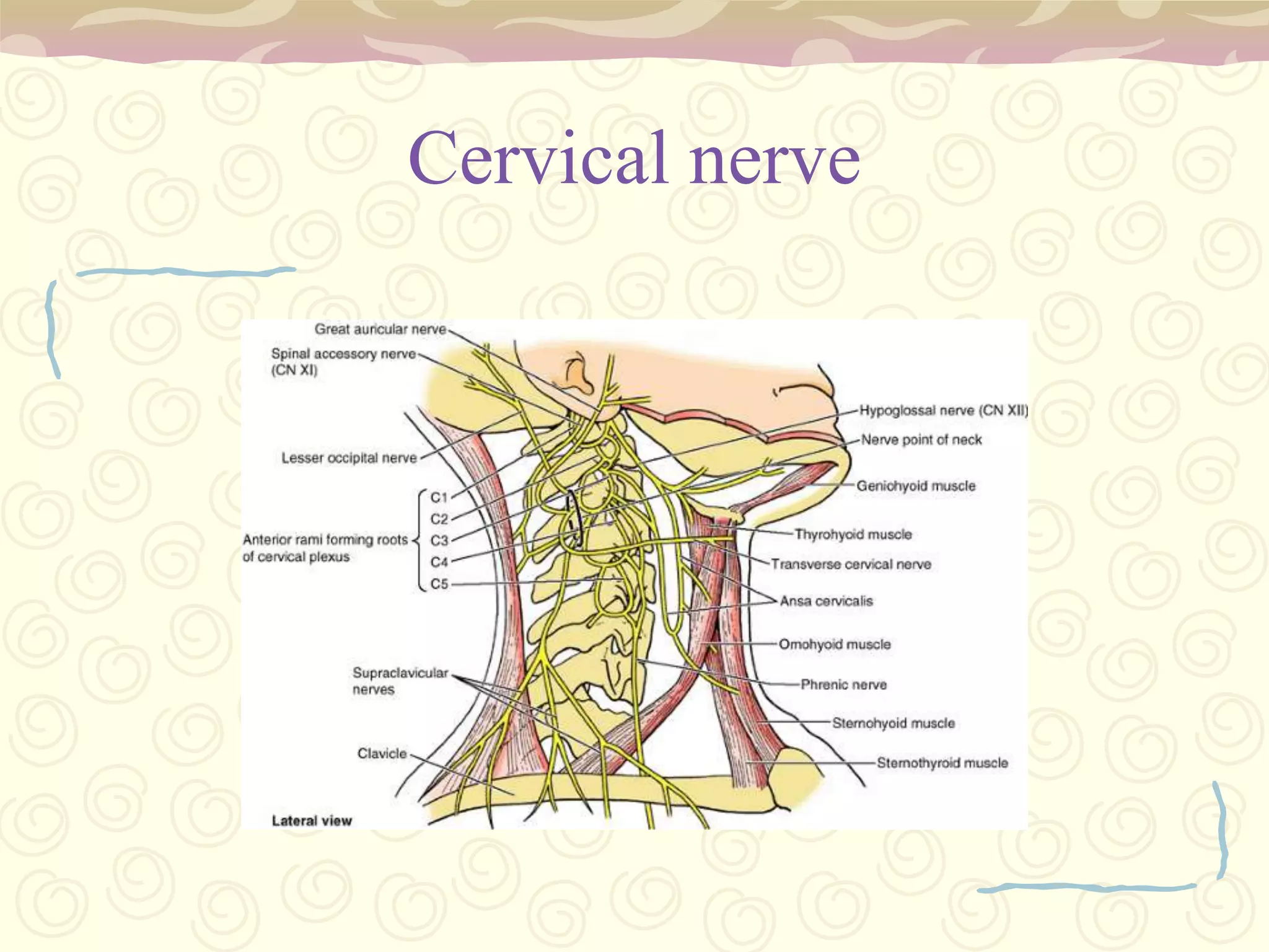
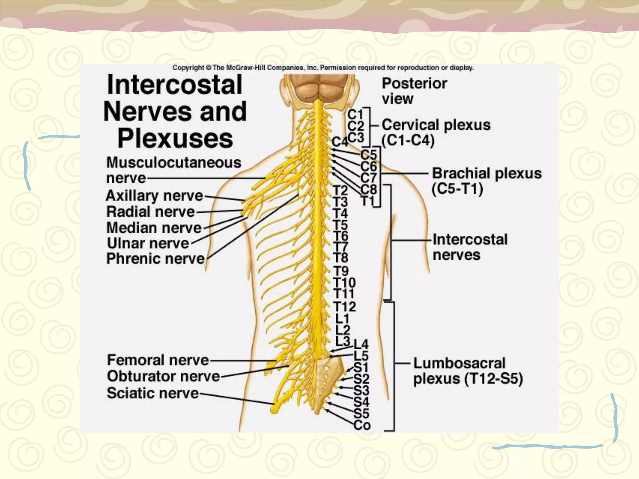
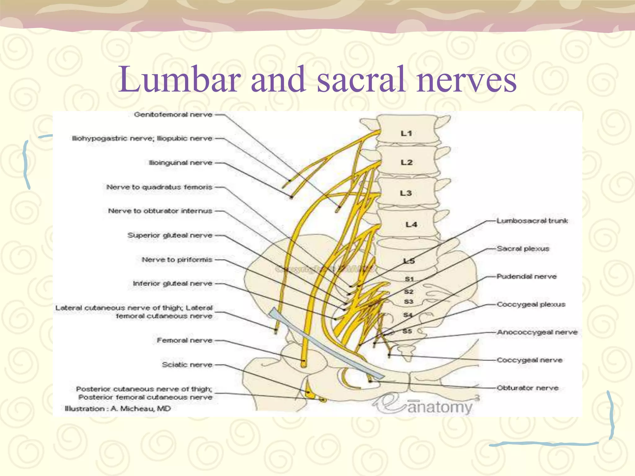
Cervical (neck/arms), Thoracic (chest), Lumbosacral (legs/pelvis) regions outlined.
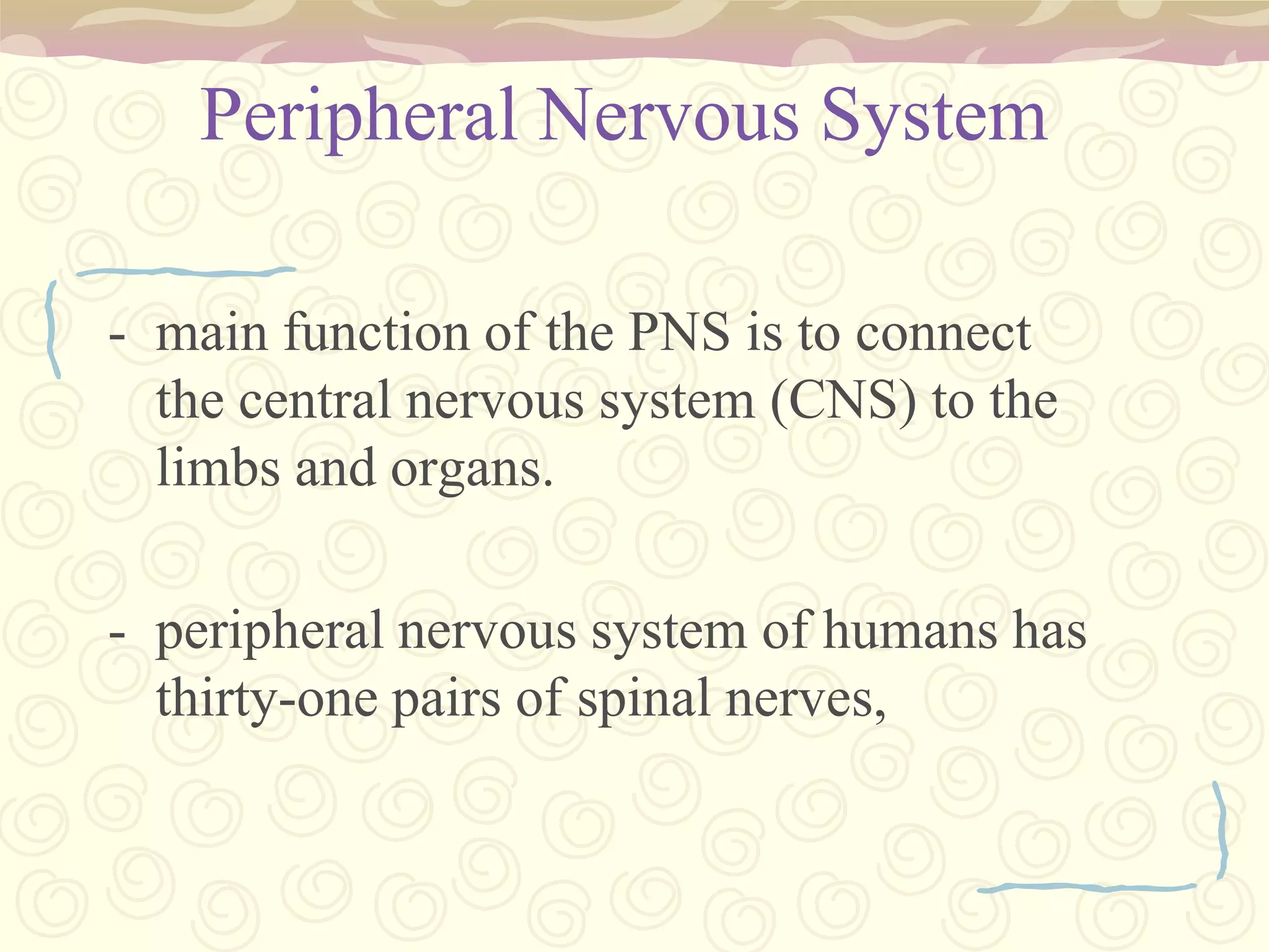
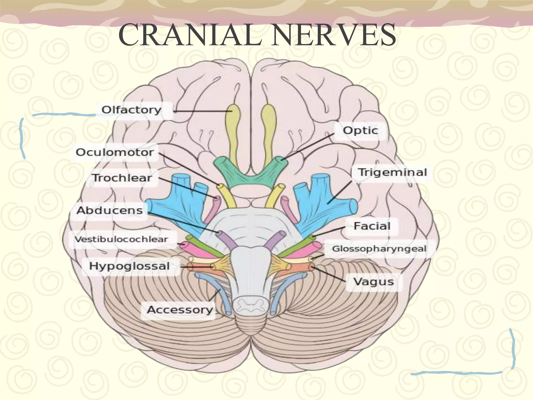
Connects CNS to limbs/organs, includes cranial nerves and 31 spinal nerve pairs.
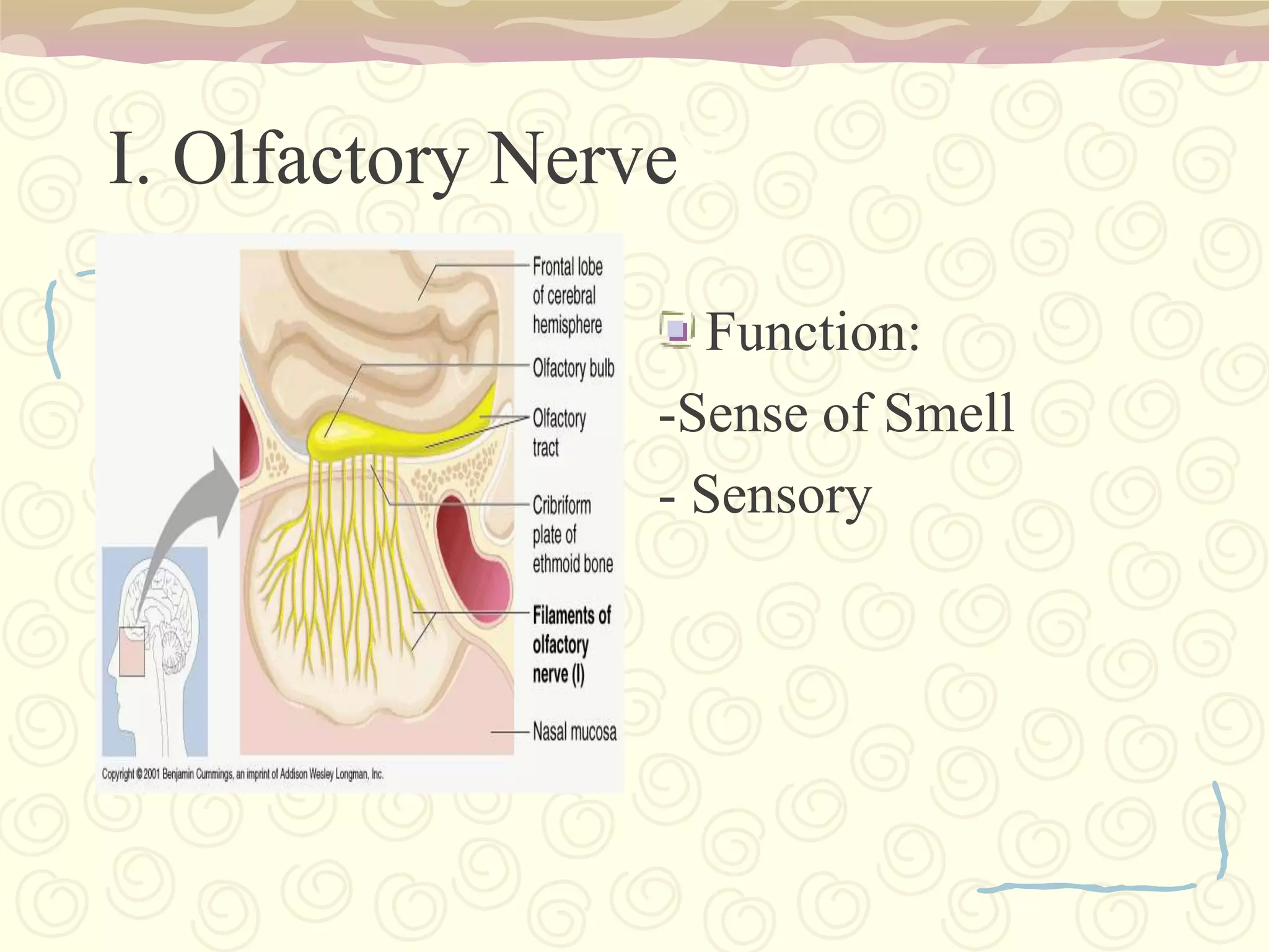
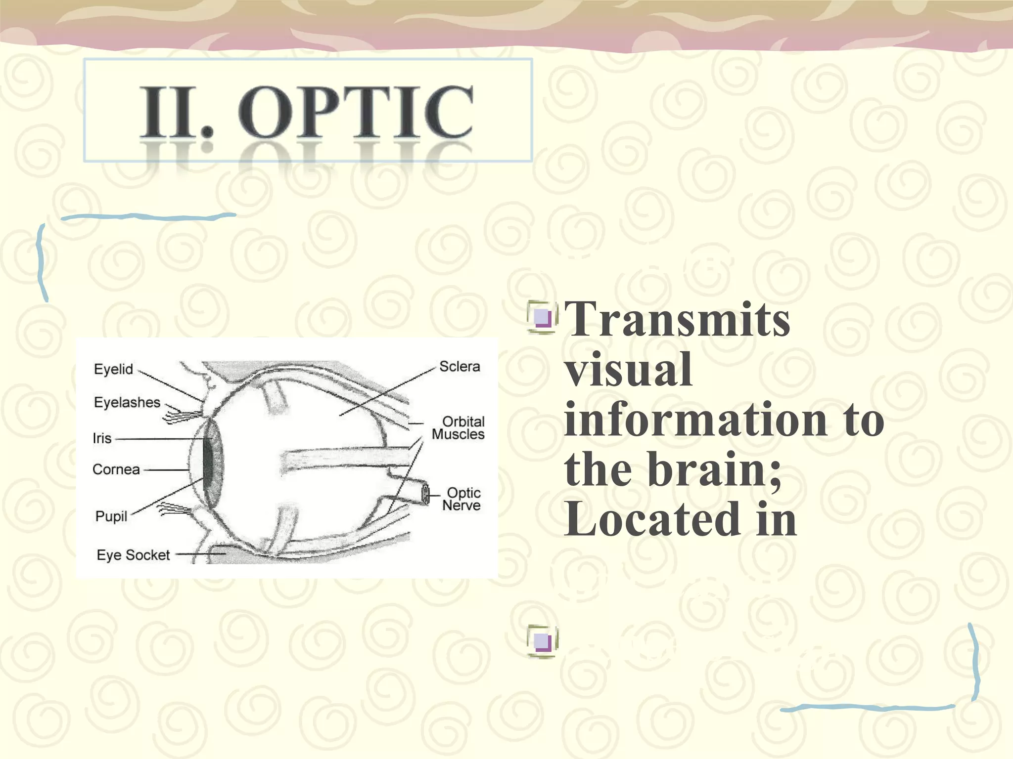
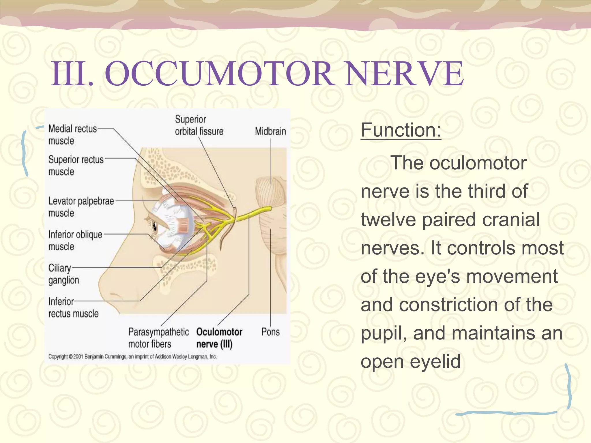
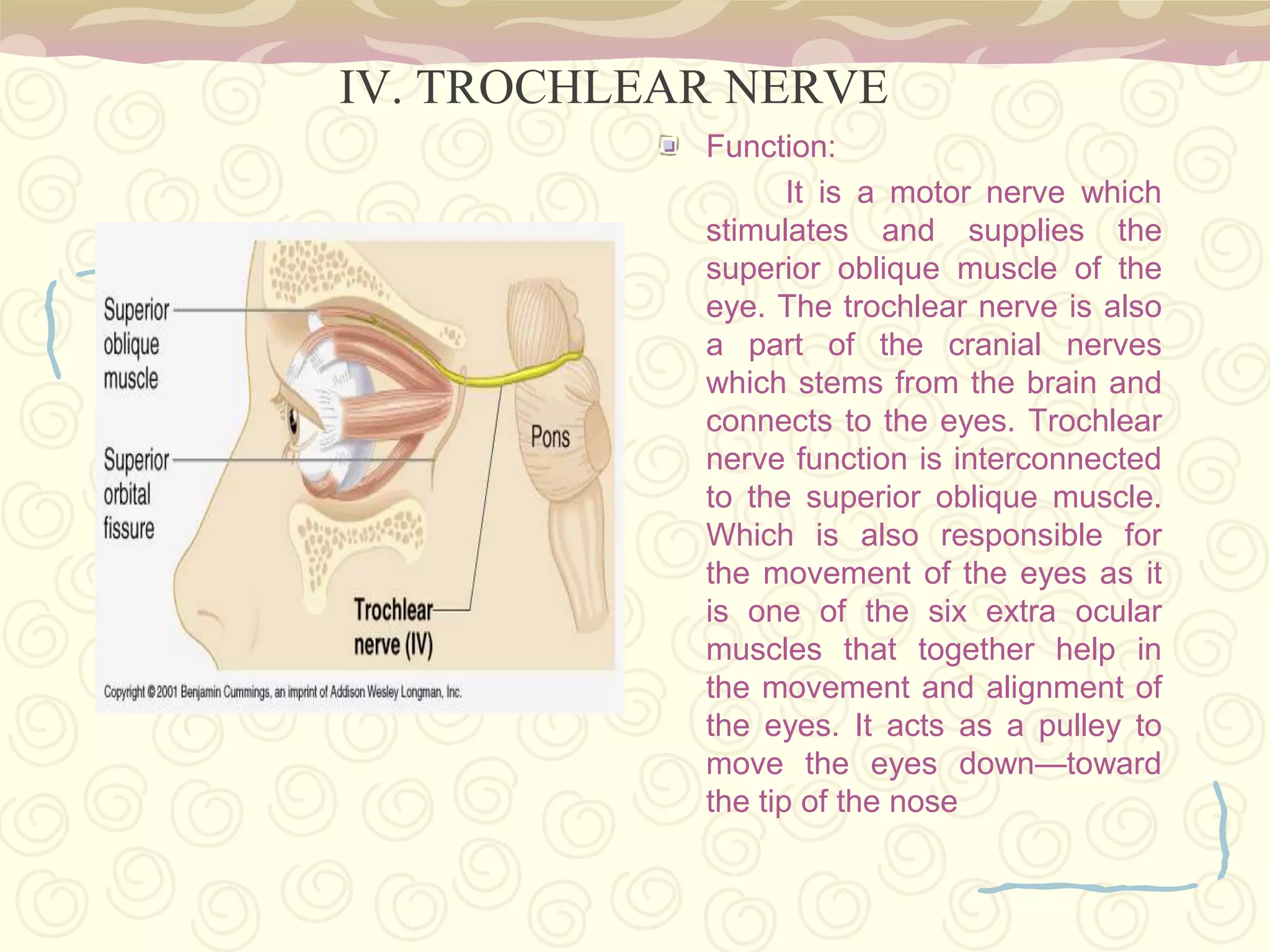
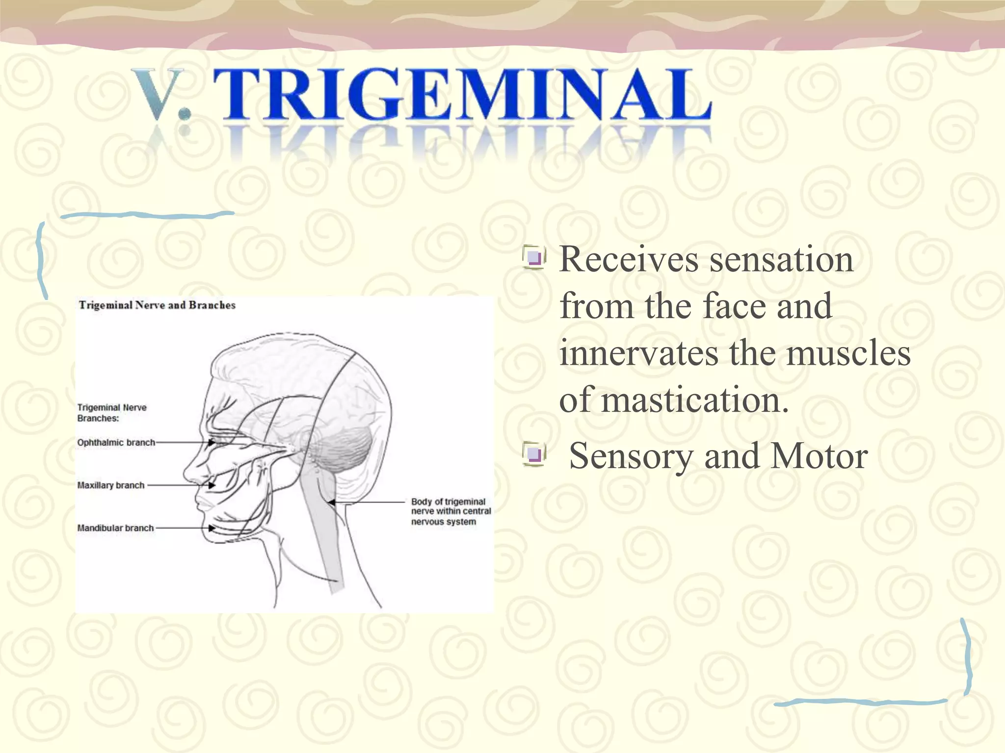
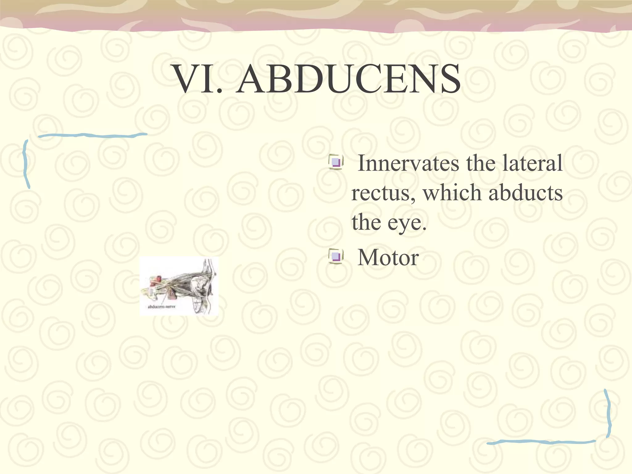
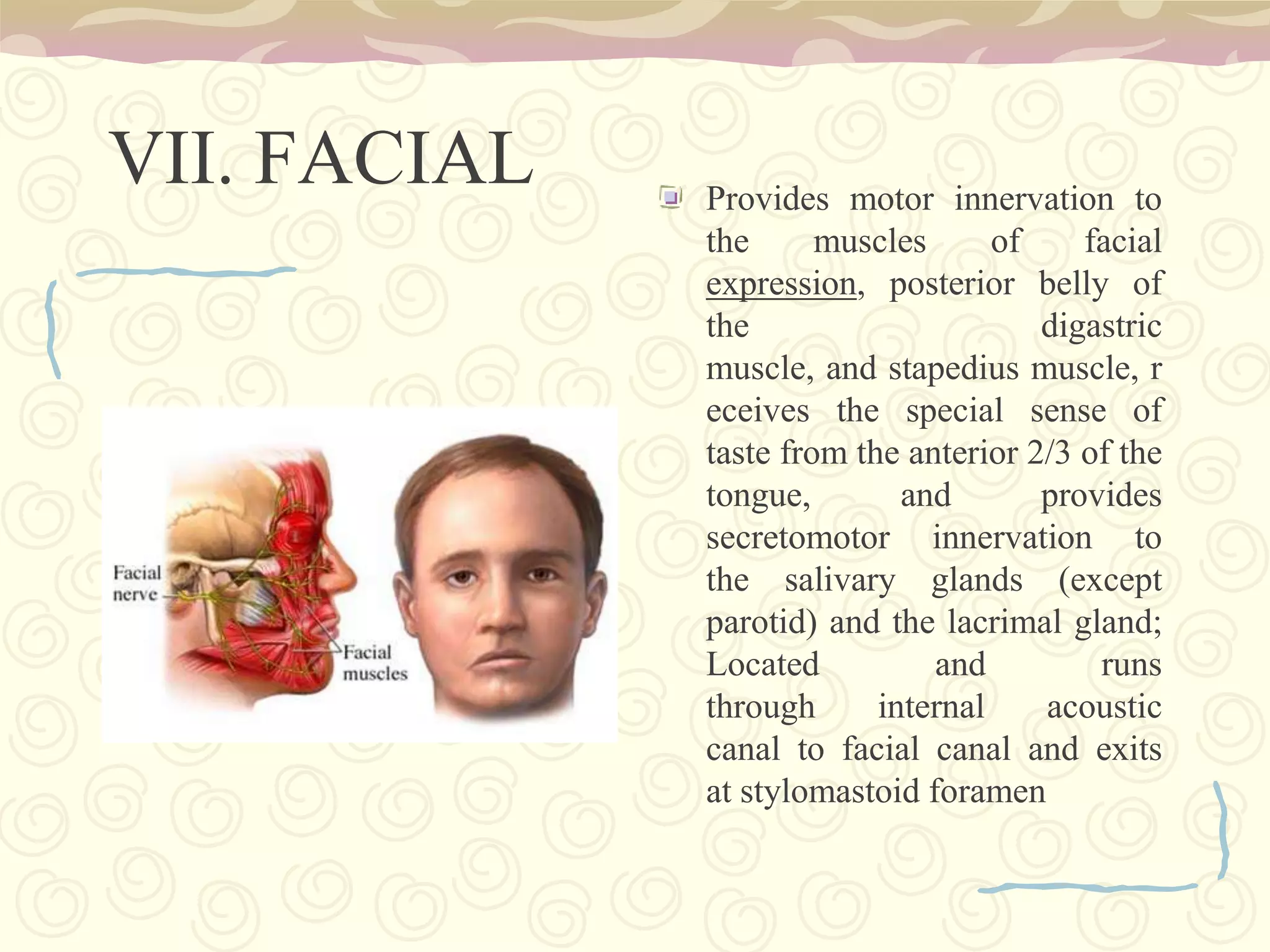
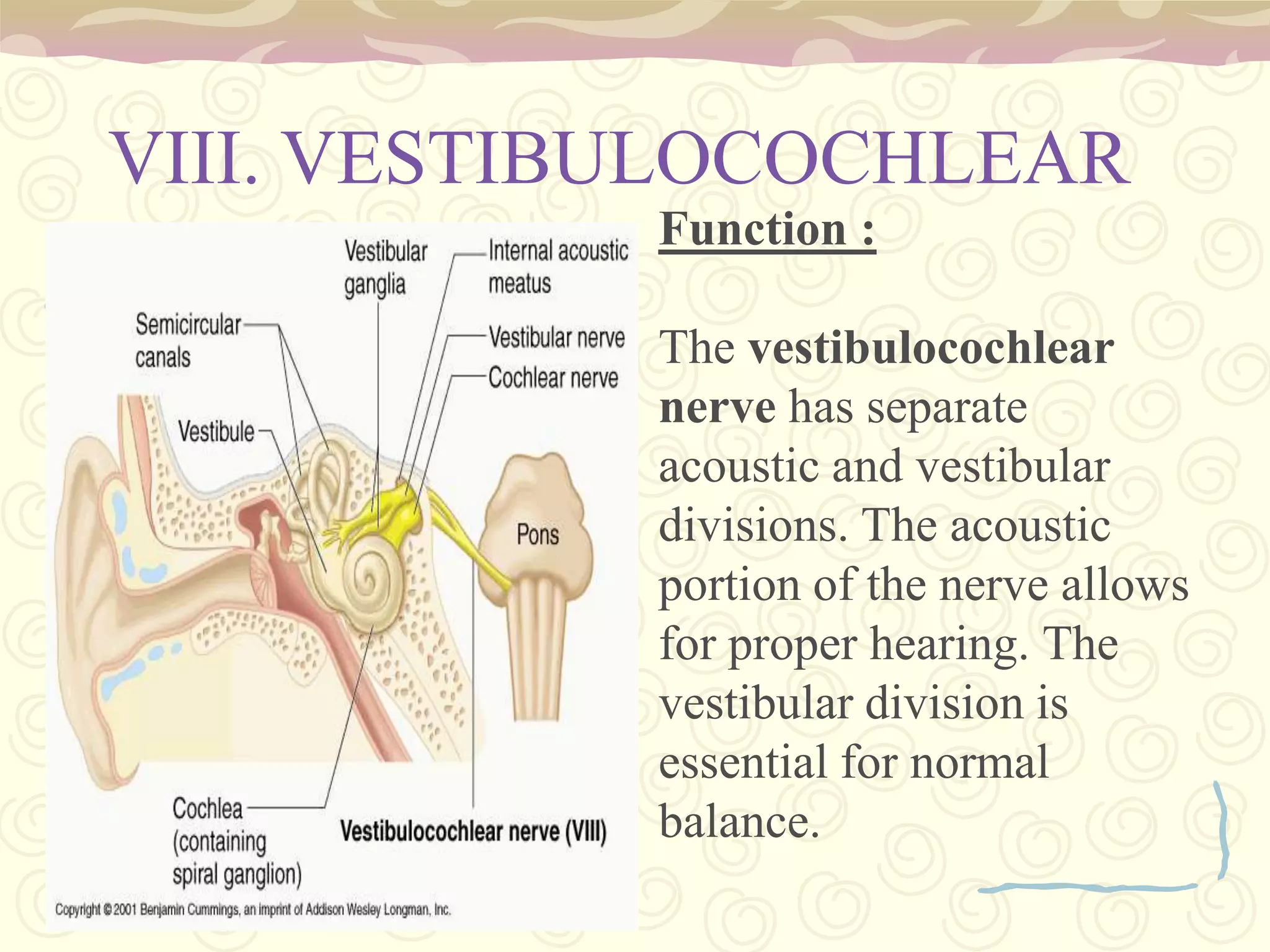
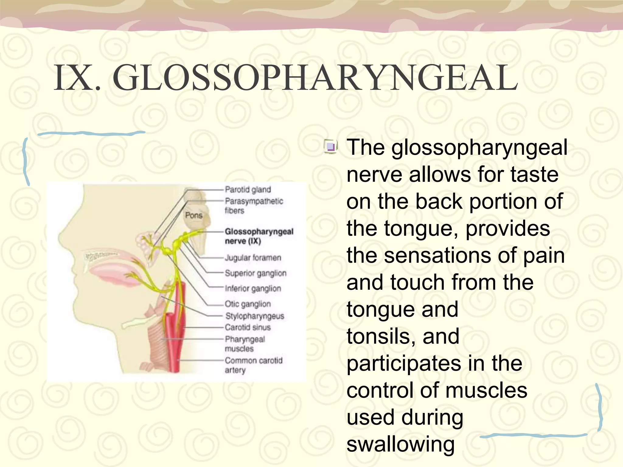
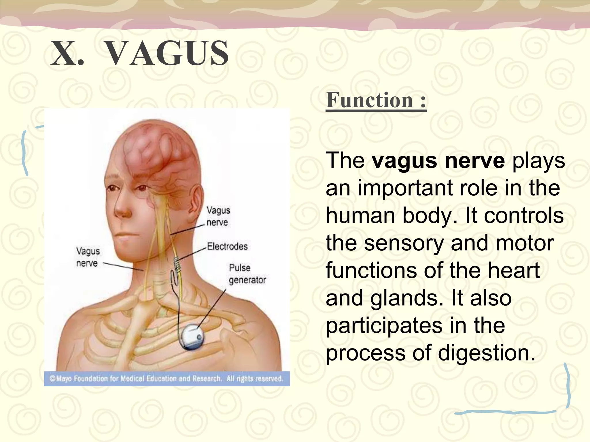
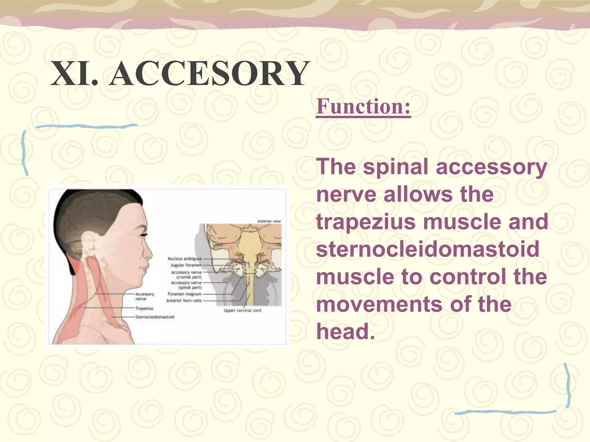
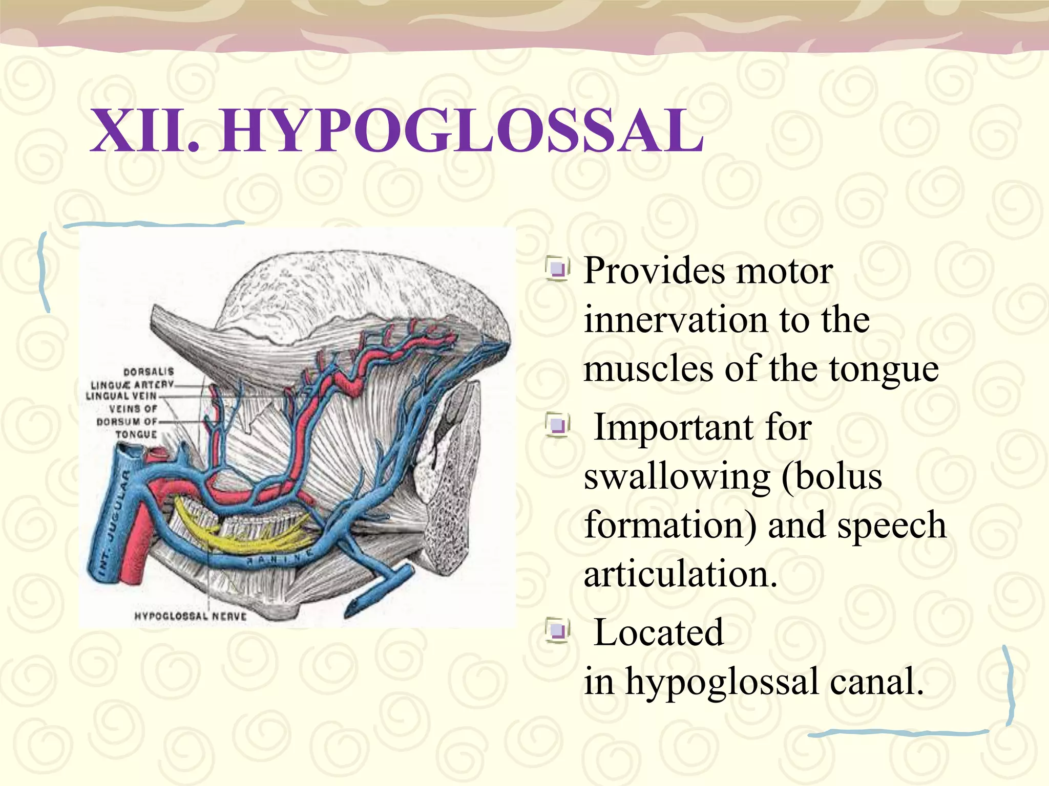
Describes functions of 12 cranial nerves including sensory and motor functions.
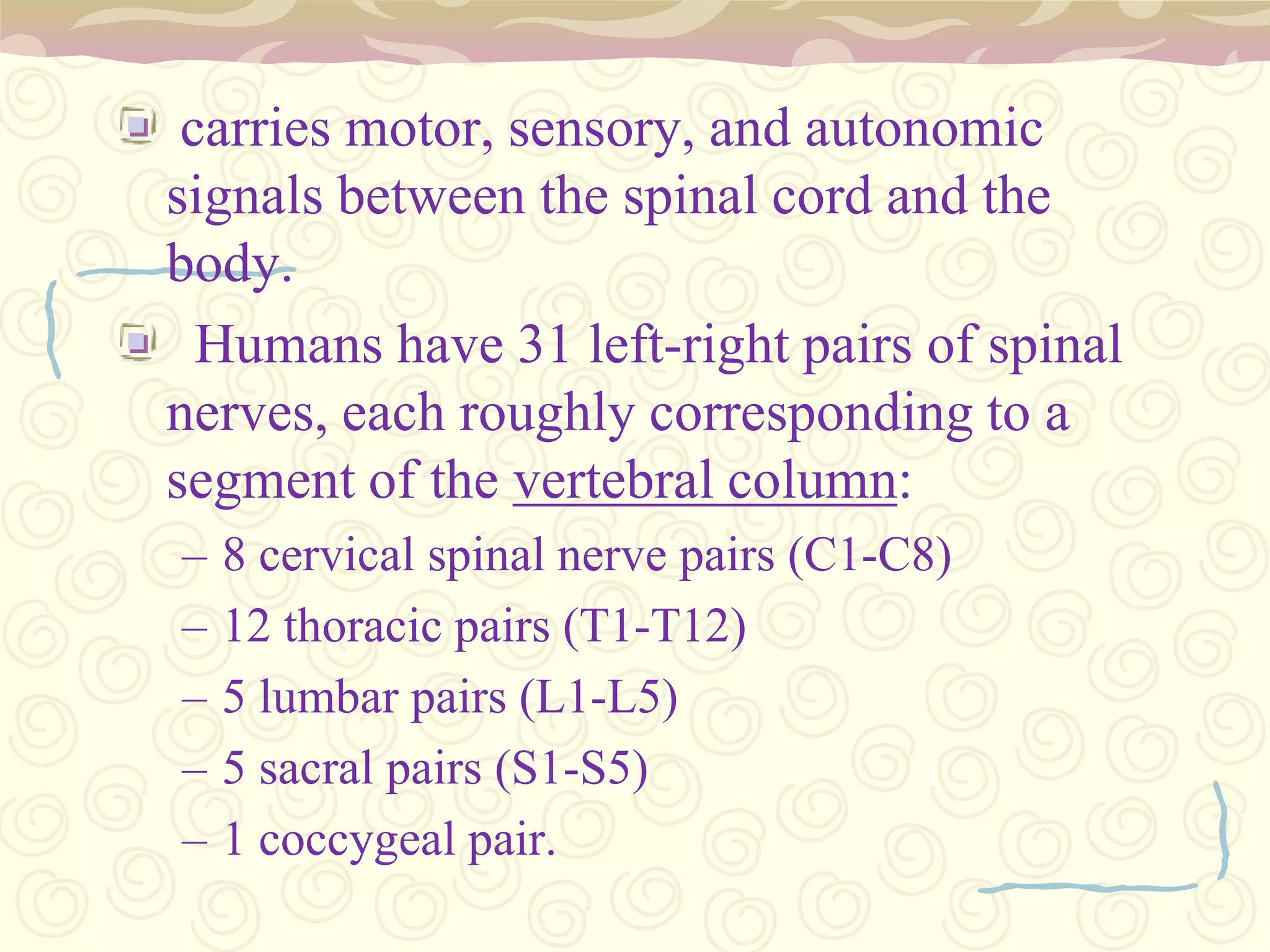
Motor and sensory signals; includes cervical, thoracic, lumbar, and sacral nerves.
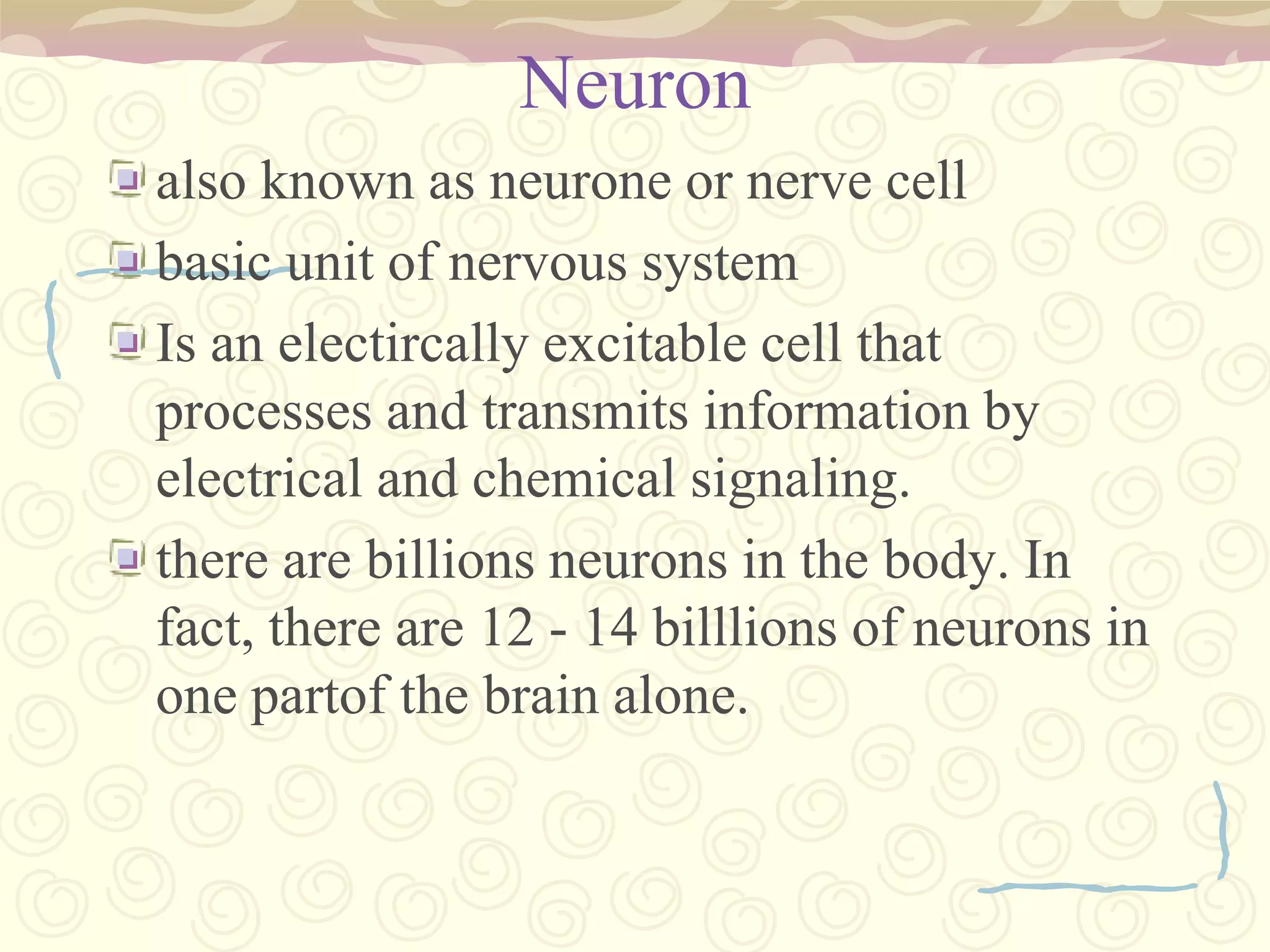
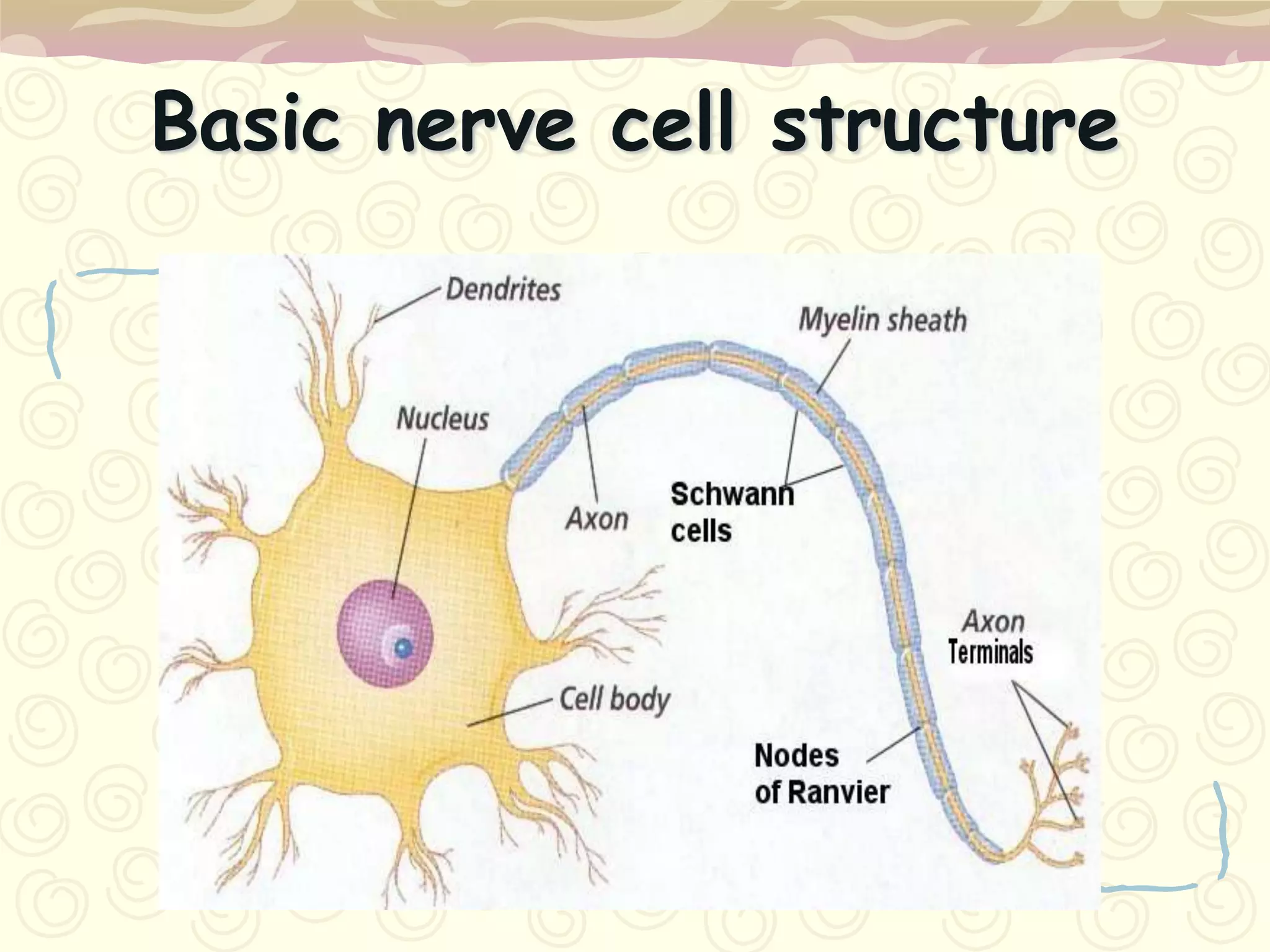
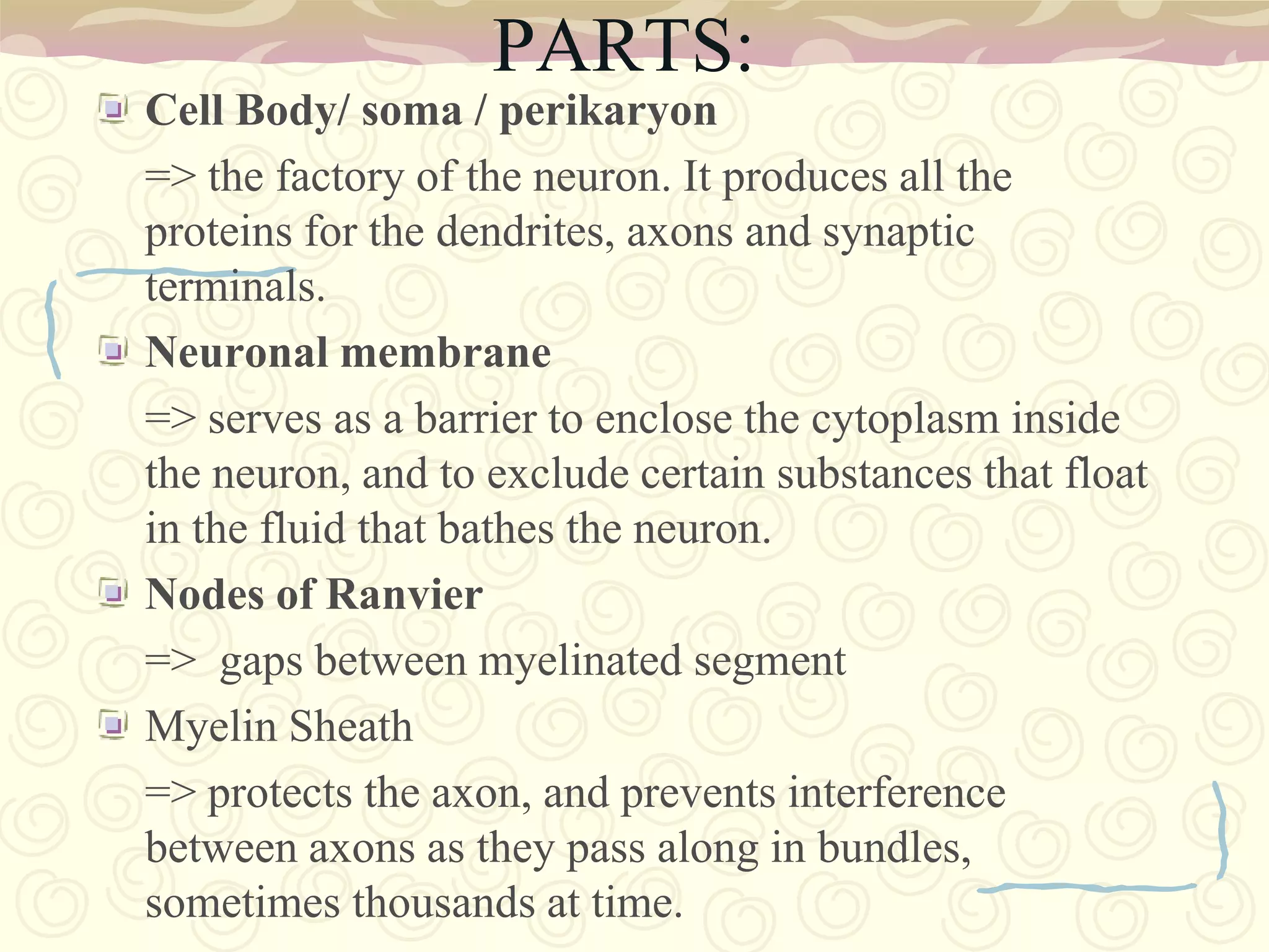
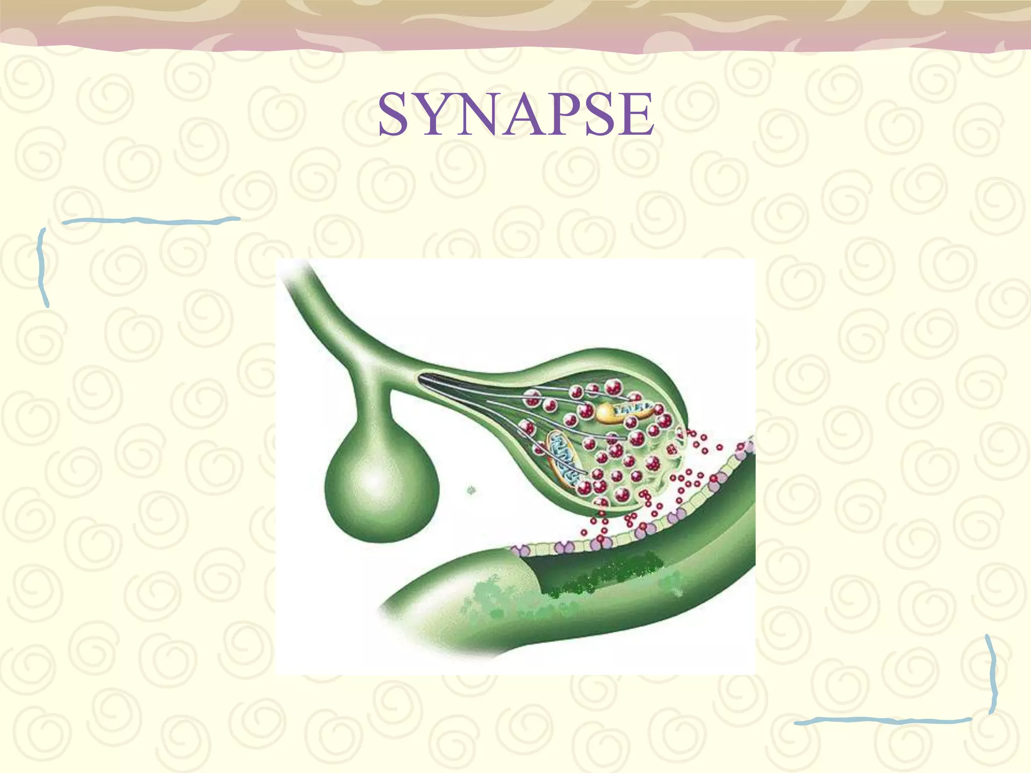
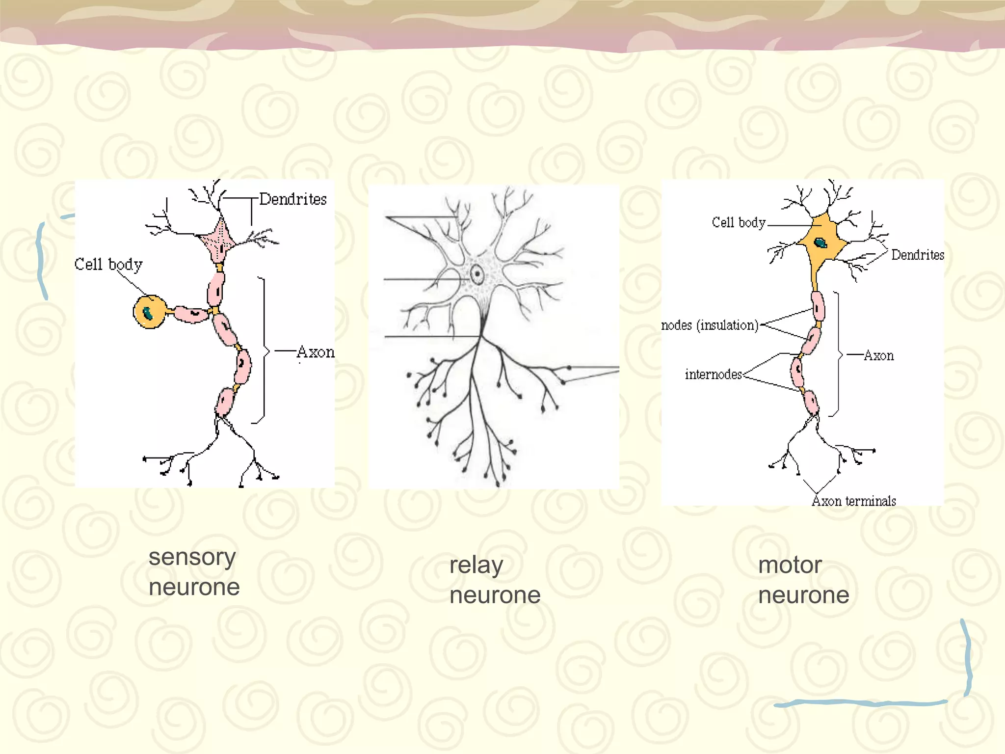
Neurons: basic unit, dendrites, axon, synapses where transmission occurs.
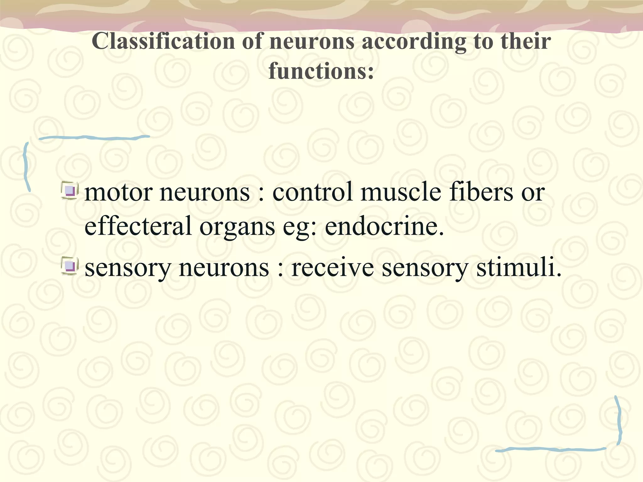
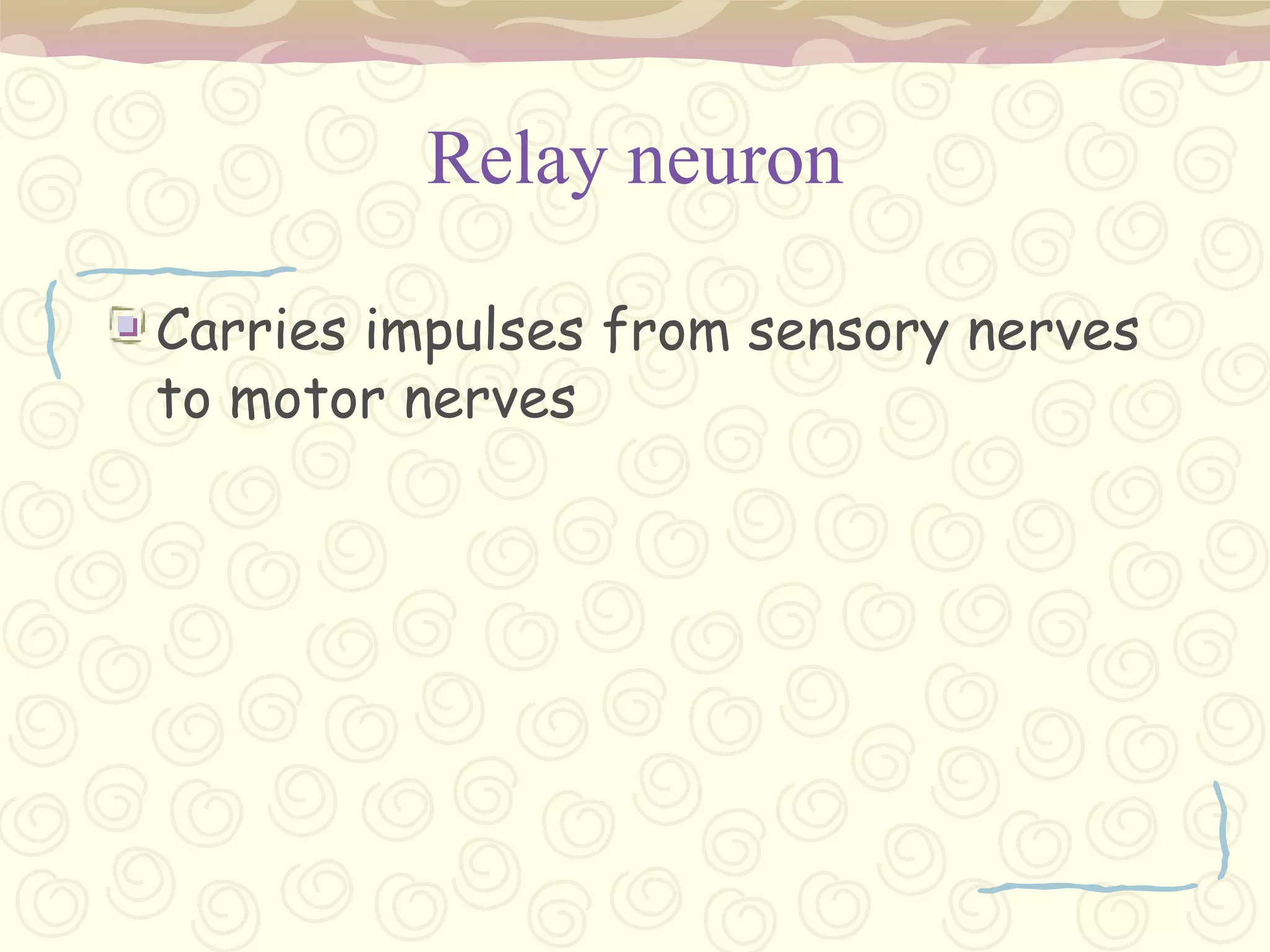
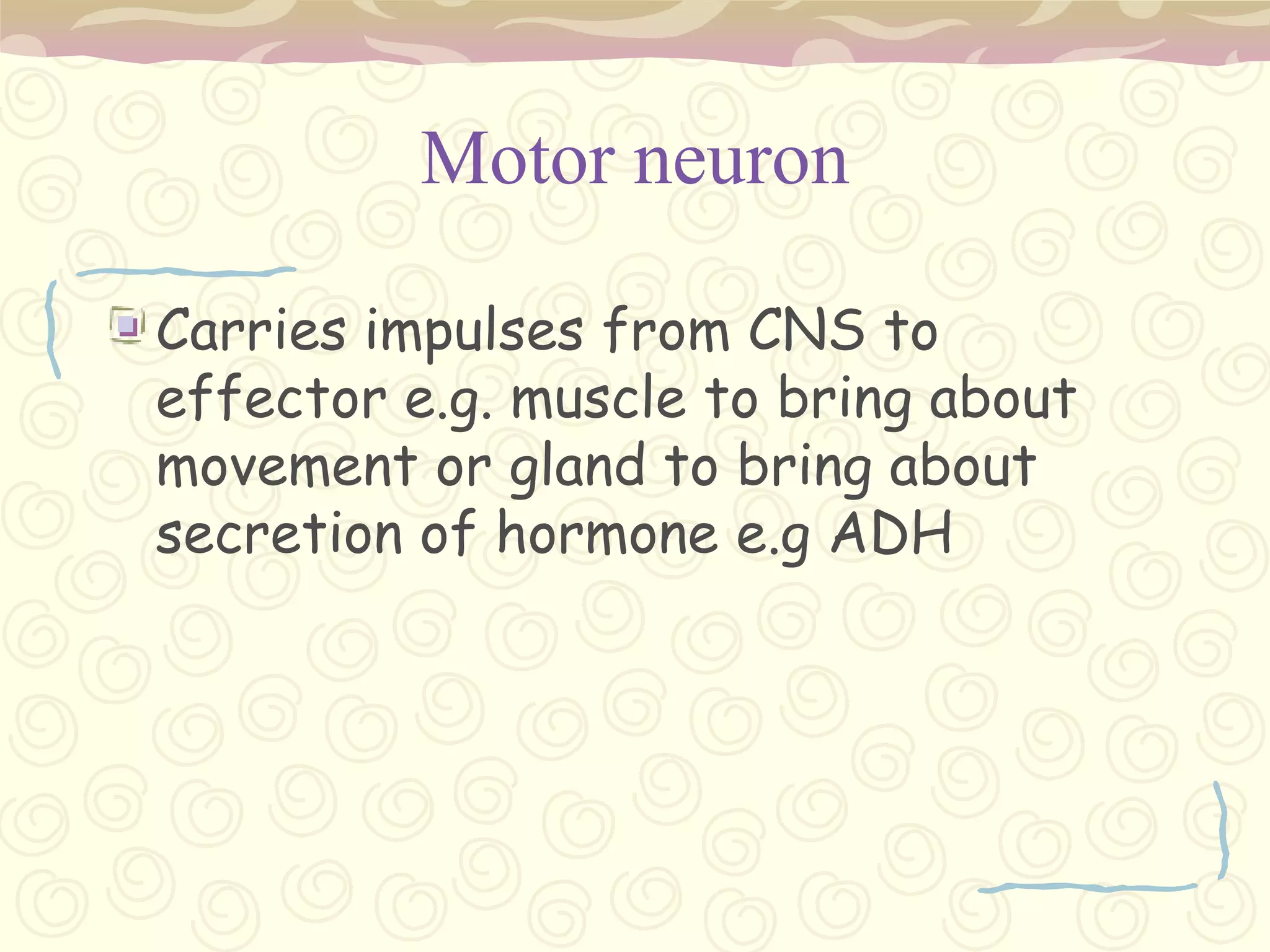
Afferent, efferent, and interneurons distinguished by functions in signal transmission.
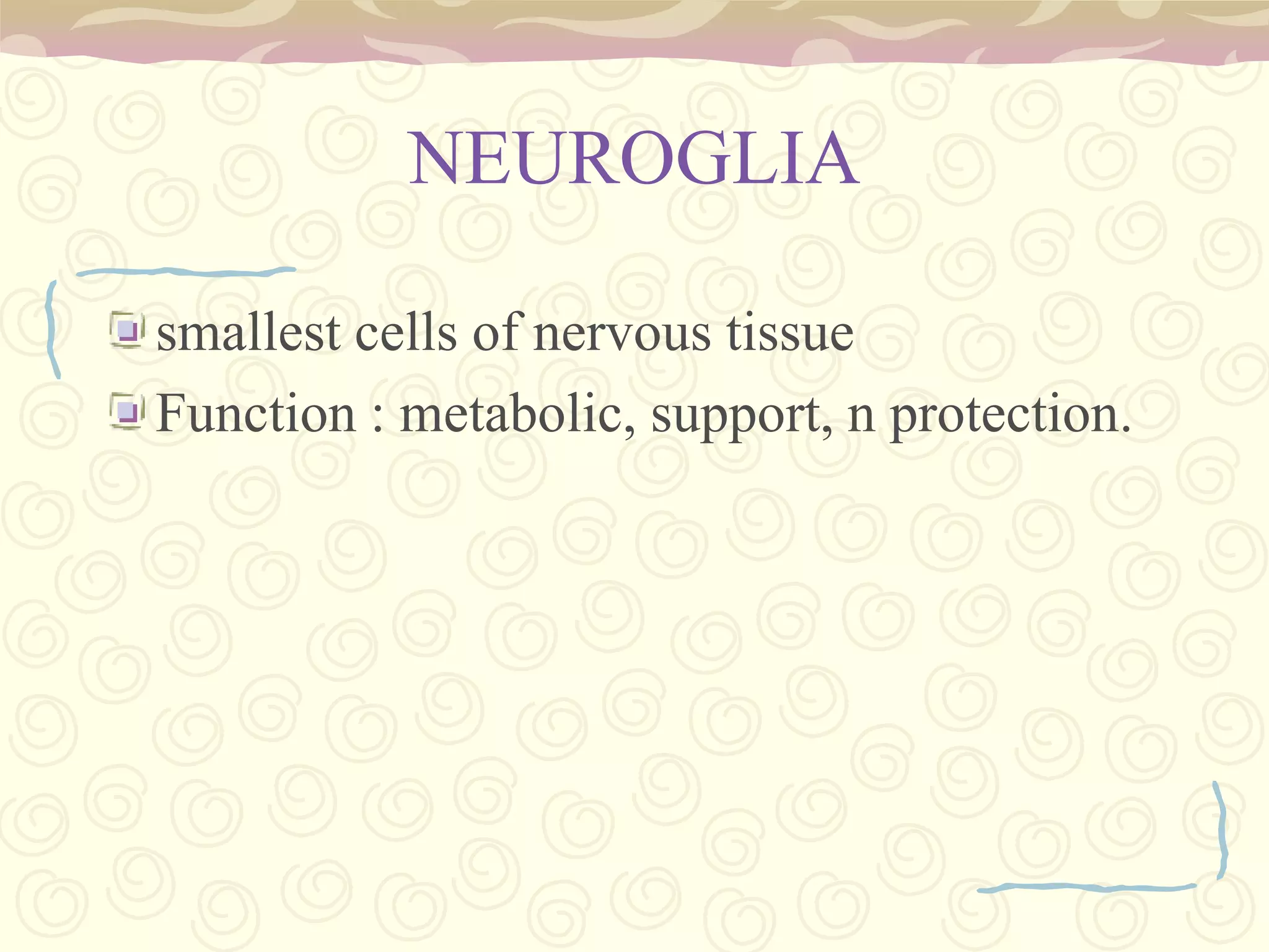
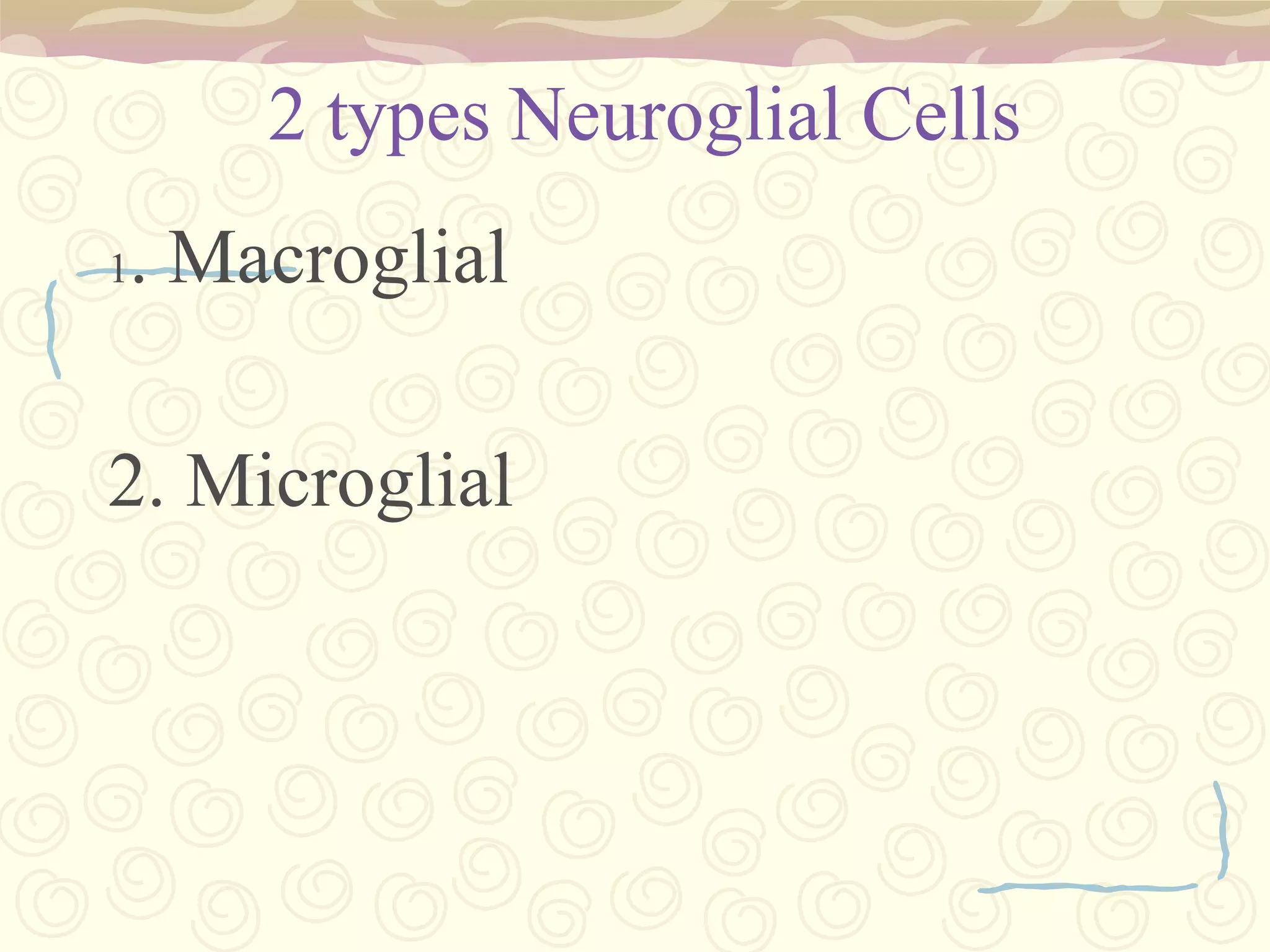
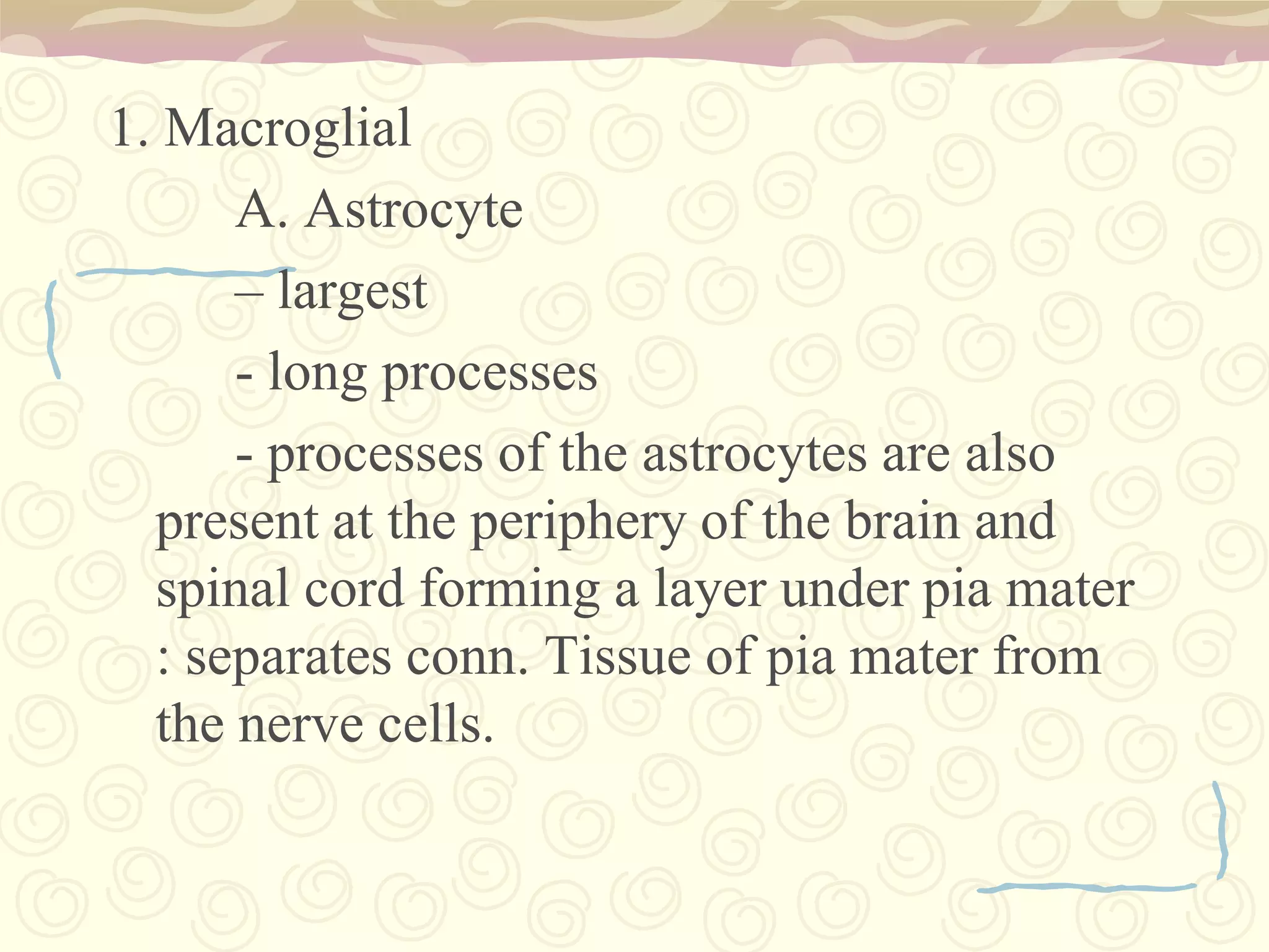
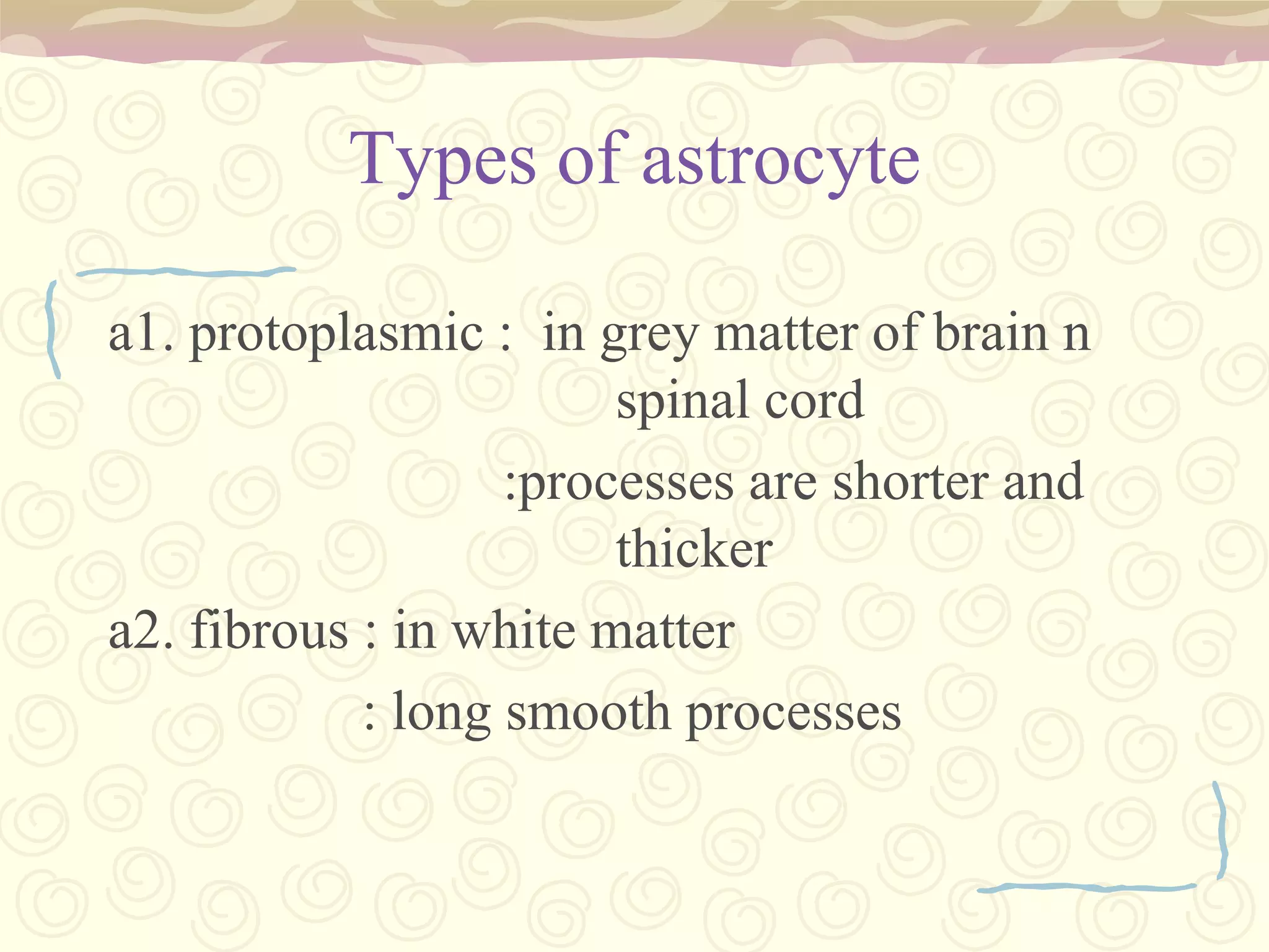
Two glial cell types: Macroglial (astrocytes, oligodendrocytes) and Microglia.
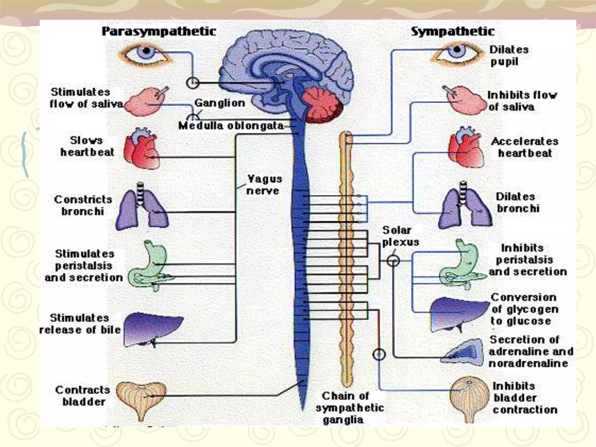
Somatic (voluntary) and Autonomic (involuntary control, e.g., heart rate, digestion).