
![A N AT O M Y A N D P H Y S I O L O G Y O F
N E RV O U S S Y S T E M
S U B M IT T E D TO S U B M I T T E D B Y
D R . PA L L AV I PAT H A N I A M S . K A N C H A N
H . O . D [ M . S . N . ] M . S C . ( N ) 1 S T Y E A R](https://image.slidesharecdn.com/nervoussystem-210215074426/75/Nervous-system-2-2048.jpg)
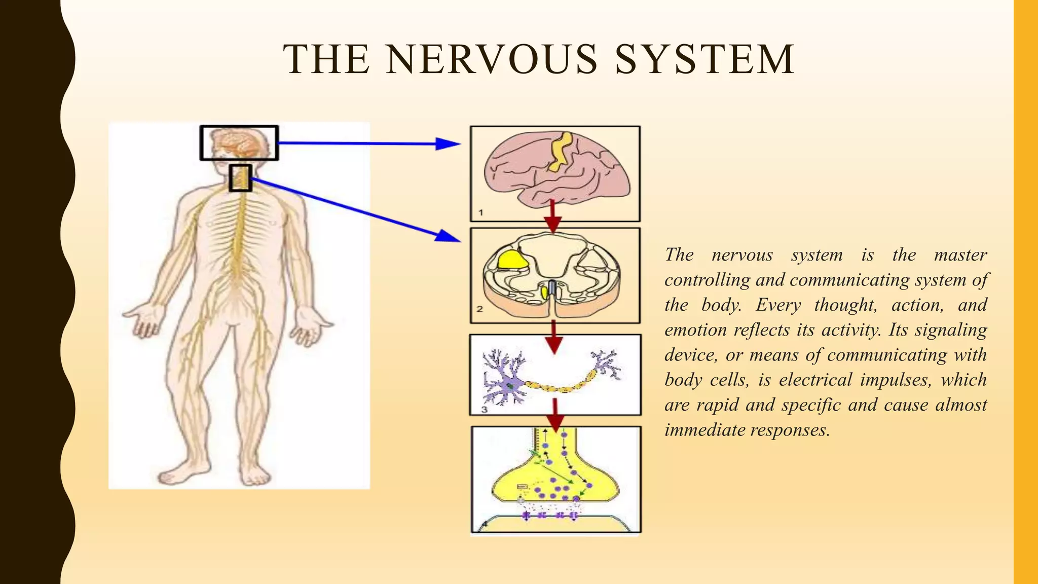
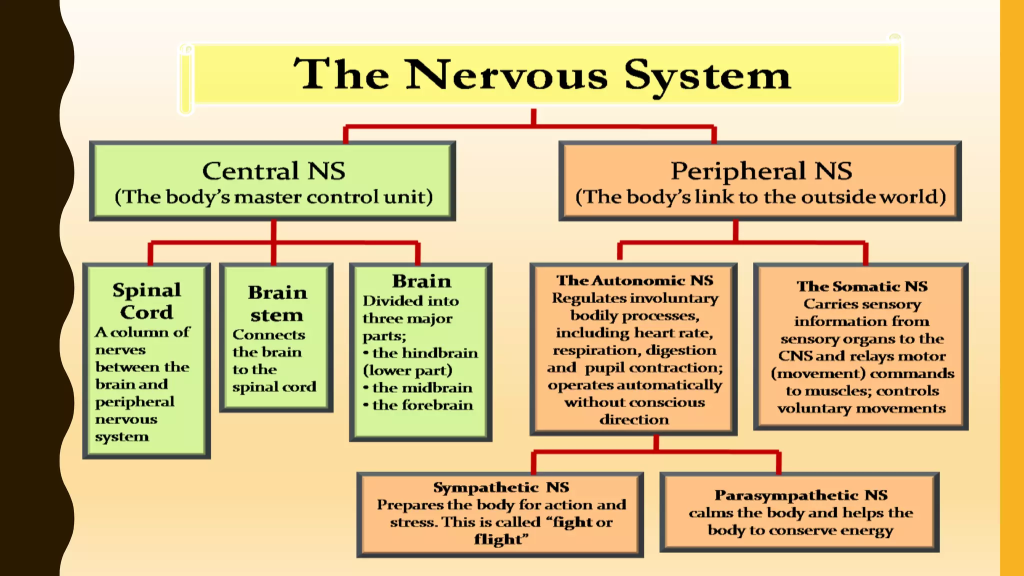


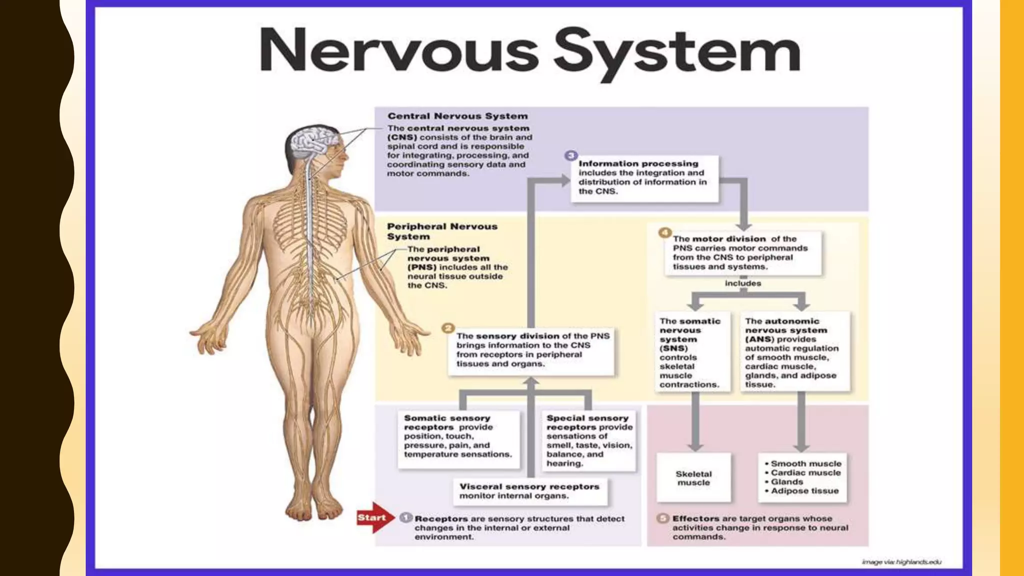


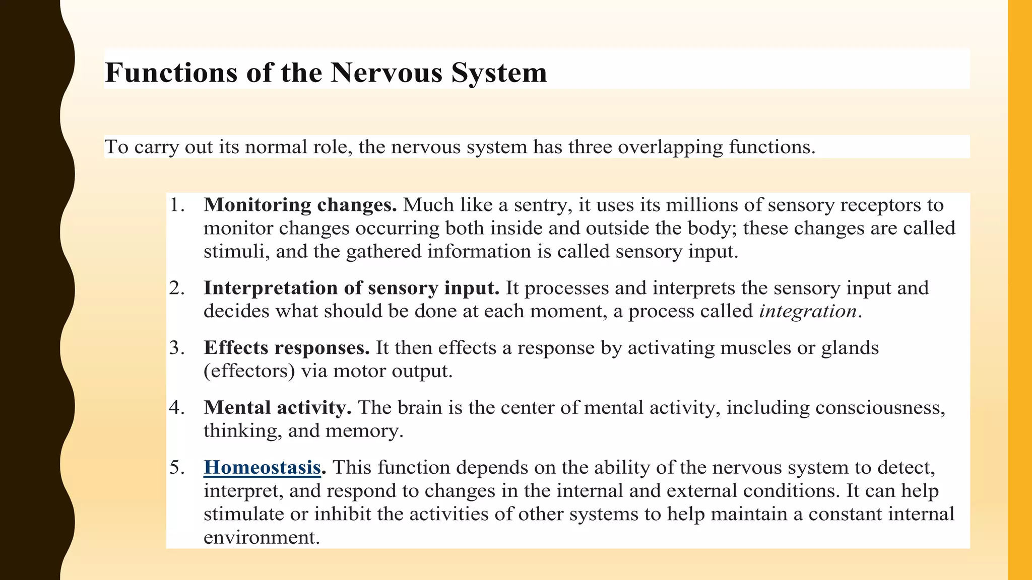


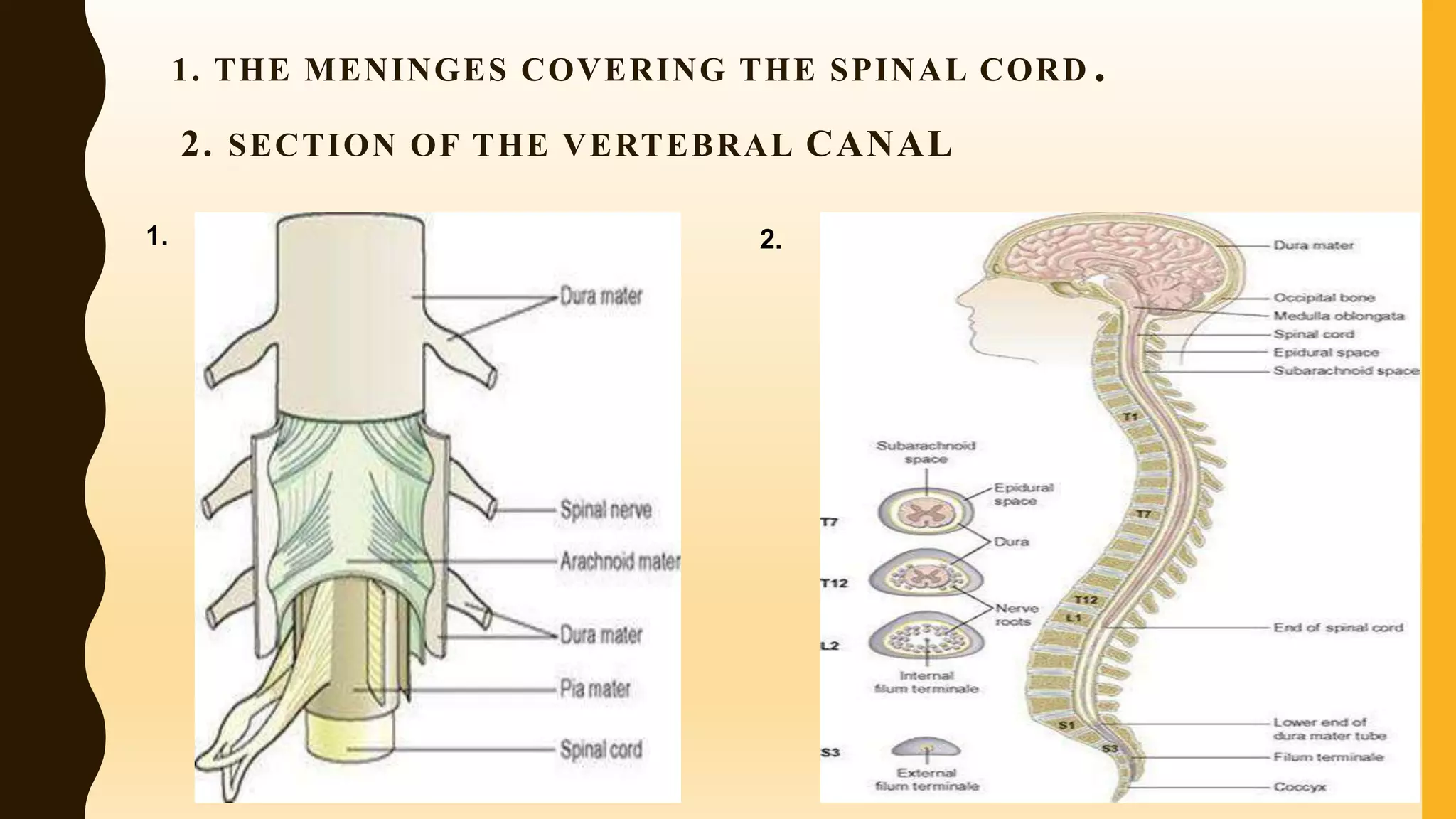

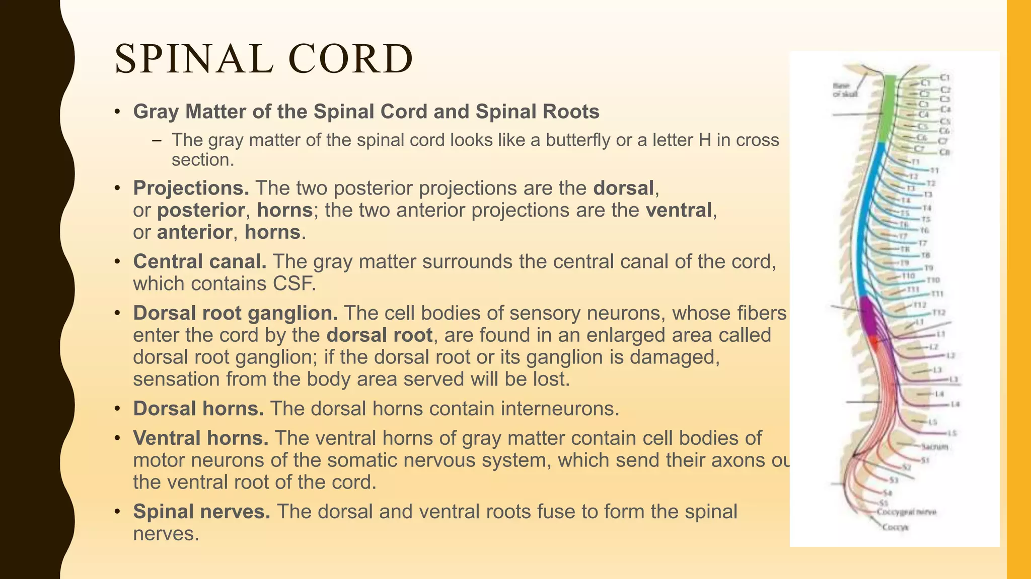


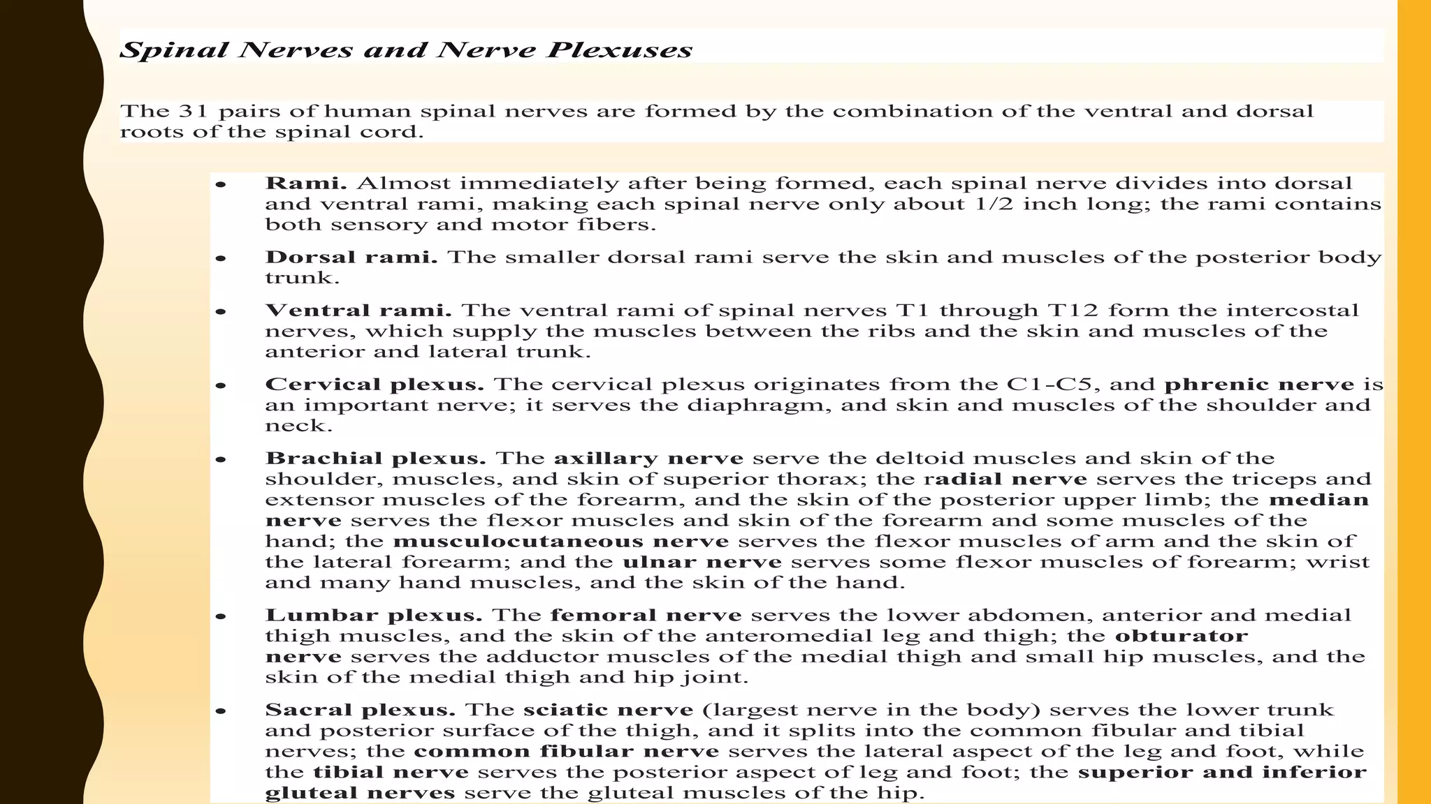

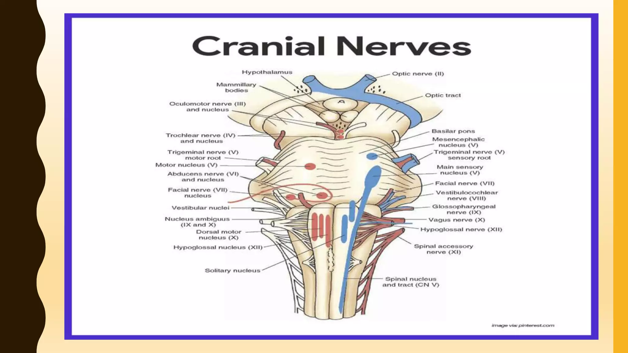


The document provides an overview of the anatomy and physiology of the nervous system, detailing its structure, classification of neurons, and major functions, including homeostasis and mental activity. It discusses the central nervous system (CNS), which comprises the brain and spinal cord, and the peripheral nervous system (PNS), which includes nerves outside the CNS. Key components such as neurons, synapses, and cerebrospinal fluid are explained, alongside the roles of different brain regions and their functions.


