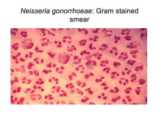
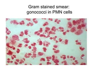
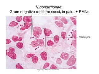
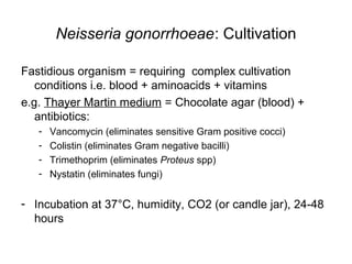
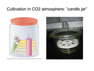
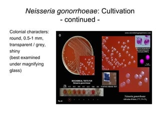
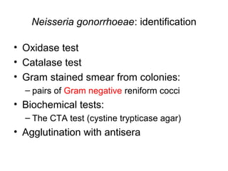
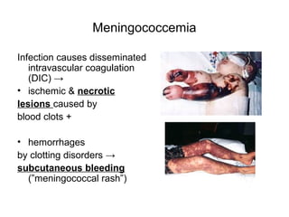
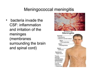
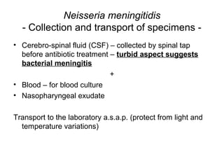
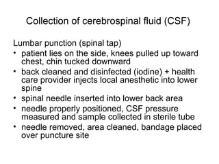
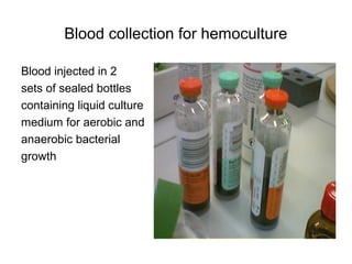
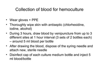
Laboratory diagnosis of infections caused
by the genus Neisseria
http://www.slideshare.net/DanaSinzianaBreharCi/
neisseriaceae-45658928
Family Neisseriaceae
• Comensal; exception: Neisseria gonorrhoeae (always
pathogenic)
• Colonize mucous membranes
• Common characters:
– Gram negative,
– shape: reniform cocci
– arrangement: pairs (diplo) + tetrades/larger groups
– cultivation: optimal growth 35-37°C
– Catalase +
– Oxydase +
Family Neisseriaceae
• Genera:
– Neisseria
• Neisseria gonorrhoeae
• Neisseria meningitidis
– Moraxella
• Branhamella catarrhalis
– Kingella
– Acinetobacter
Neiseria gonorrhoeae
- Clinical Significance -
• Pathogenic germ: gonorrhea = sexually transmited
infection (STI); incubation 2-7 days
• Men:
– acute urethritis (abundant urethral secretion + dysuria);
– Complications (due to lack of / delayed treatment):
• urethral strictures,
• prostatitis,
• epididymitis
Neiseria gonorrhoeae
- Clinical Significance - continued
• Women:
– endocervicitis (purulent vaginal secretion + dysuria)
– OR asymptomatic !!
↓
– further transmission to sexual partner;
– Complications (due to lack of / delayed treatment):
• Salpingitis (inflammation of the falopian tubes) + infertility
• pelvic peritonitis,
• PID (pelvic inflammatory disease)
• Gonococcal arthritis + skin rash (rare complication by
hematogenous dissemination)
Neiseria gonorrhoeae
- Clinical Significance - continued
• Newborns to infected mothers:
gonococcal ophtalmia
neonatorum (may lead to
blindness)
• Prophylactic treatment:
erythromycin ointment / silver
nitrate
Neisseria gonorrhoeae
- Collection of specimens -
• Women – endocervical secretion (during gynecologic
exam):
– insert valves
– use 2 consecutive swabs (1 for microscopy + 1 for inoculation of
culture media);
– insert each swab into cervix and rotate
• Men – urethral secretion (in the morning/at least 1 hour
after last miction):
– Acute urethritis: direct swab/loop collection (abundant secretion)
– Chronic infection: loop / special swab inserted 2 cm into urethra
and rotated
Neisseria gonorrhoeae
- Transport of specimens -
• Ideally: direct inoculation on culture media (gonococci
are very sensitive to external environment; very low
microbial load in collected specimens)
• Alternatively - transport media (semisolid, non-nutritive):
– Stuart‘s medium: agar + sodium thioglycolate (delays oxydation),
Ca, Mg salts (osmolar protection of bacterial cells), methylene
blue (redox indicator; blue colour = oxydation)
– Amies‘ medium: similar + charcoal (neutralize toxic materials)
• Transportation to lab – a.s.a.p.
– tube cap firmly screwed;
– keep tube cool but do not freeze!
Neisseria gonorrhoeae
- Microscopic examination -
Gram stained smears:
- over 95% sensitivity in acute male urethritis;
- less sensitive in:
- chronic infections (increased associated microbial flora)
- endocervicitis (50-70% sensitivity)
- High no of PMN cells with Gram negative reniform
(kidney-like) cocci, arranged in pairs (”coffee beans”;
concavities facing each other)
Neisseria gonorrhoeae: Gram stained
smear
Gram stained smear:
gonococci in PMN cells
N.gonorrhoeae:
Gram negative reniform cocci, in pairs + PMNs
Neisseria gonorrhoeae: Cultivation
Fastidious organism = requiring complex cultivation
conditions i.e. blood + aminoacids + vitamins
e.g. Thayer Martin medium = Chocolate agar (blood) +
antibiotics:
- Vancomycin (eliminates sensitive Gram positive cocci)
- Colistin (eliminates Gram negative bacilli)
- Trimethoprim (eliminates Proteus spp)
- Nystatin (eliminates fungi)
- Incubation at 37°C, humidity, CO2 (or candle jar), 24-48
hours
Cultivation in CO2 atmosphere: ”candle jar”
Cultivation in CO2 atmosphere:
CO2 incubators; gas pack systems
Neisseria gonorrhoeae: Cultivation
- continued -
Colonial characters:
round, 0.5-1 mm,
transparent / grey,
shiny
(best examined
under magnifying
glass)
Neisseria gonorrhoeae: identification
• Oxidase test
• Catalase test
• Gram stained smear from colonies:
– pairs of Gram negative reniform cocci
• Biochemical tests:
– The CTA test (cystine trypticase agar)
• Agglutination with antisera
The Oxidase test
Principle: the enzyme cytochrome c oxidase oxidizes the
reagent TMPD (TetraMethylPhenylenDiamine) to
indophenol – purple/dark blue end product; if enzyme is
not present TMPD remains colourless.
• used to identify bacteria that produce cytochrome c
oxidase e.g. Neisseria
Procedure: filter paper soaked with TMPD is moistened
with sterile distilled water; pick bacterial colony with loop
and smear on filter paper; colour change to dark
blue/purple within 10-30 sec = POSITIVE test
The Oxidase test:
purple colour = cytochrome c oxidase present
The Catalase test
• Principle: the enzyme catalase decomposes
hydrogen peroxide (H2O2) into water and
oxygen:
2H2O2 →2H2O + O2 (gas bubbles)
• 2-3 drops of hydrogen peroxide placed directly
on a colony
• POSITIVE TEST: rapid effervescence
• Neisseria (+)
The CTA (cystine trypticase agar) test
- Neisseria spp. produce acid from carbohydrates
by oxidation → yellow colour
- 4 tubes with CTA base + phenol red indicator
- Add 4 carbohydrates (one in each tube):
- glucose (dextrose) – POSITIVE test (N.gonorrhoeae)
- Maltose - Negative
- Lactose - Negative
- Sucrose - Negative
- Inoculate bacterial culture (3-5 colonies) in each
tube with different disposable loops
Neisseria gonorrhoeae: oxidation of sugars
CTA test:
• Only glucose (dextrose) tube
shows production of acid
(yellow turbidity in upper part
of tube)
• The maltose, sucrose and
lactose tubes show no
acidification (red colour
persists in the whole tube)
API NH gallery
• Only glucose +
Agglutination-based Identification
N.gonorrhoeae:
antibiotic susceptibility testing
• recquired due to strains resistant to:
– Penicillins (production of penicillinase or other mechanisms)
– Spectinomycin (narrow spectrum antibiotic, selectively active
against gonococci)
• therapeutic agents for penicillin-resistant strains:
cephalosporins, fluoroquinolones
Family Neisseriaceae
• Genera:
– Neisseria
• Neisseria gonorrhoeae
• Neisseria meningitidis
– Moraxella
• Branhamella catarrhalis
– Kingella
– Acinetobacter
Neisseria meningitidis
- Clinical significance -
• Comensal germ; may colonize human oro- and
nasopharinx
• Inter-human transmission via contaminated respiratory
droplets
• May cause very severe infections: ”meningococcal
disease”
Meningococcal disease
• Nasopharingeal colonization (from infected person;
transmission via respiratory droplets: coughing,
sneezing)
• Bloodstream invasion:
– Meningitis (~55%)
– Meningitis + meningococcemia (~30%)
– Fulminant meningococcemia (~15%)
Meningococcemia
Infection causes disseminated
intravascular coagulation
(DIC) →
• ischemic & necrotic
lesions caused by
blood clots +
• hemorrhages
by clotting disorders →
subcutaneous bleeding
(”meningococcal rash”)
Meningococcal meningitis
• bacteria invade the
CSF: inflammation
and irritation of the
meninges
(membranes
surrounding the brain
and spinal cord)
Neisseria meningitidis
- Collection and transport of specimens -
• Cerebro-spinal fluid (CSF) – collected by spinal tap
before antibiotic treatment – turbid aspect suggests
bacterial meningitis
+
• Blood – for blood culture
• Nasopharyngeal exudate
Transport to the laboratory a.s.a.p. (protect from light and
temperature variations)
Collection of cerebrospinal fluid (CSF)
Lumbar punction (spinal tap)
• patient lies on the side, knees pulled up toward
chest, chin tucked downward
• back cleaned and disinfected (iodine) + health
care provider injects local anesthetic into lower
spine
• spinal needle inserted into lower back area
• needle properly positioned, CSF pressure
measured and sample collected in sterile tube
• needle removed, area cleaned, bandage placed
over puncture site
Blood collection for hemoculture
Blood injected in 2
sets of sealed bottles
containing liquid culture
medium for aerobic and
anaerobic bacterial
growth
Collection of blood for hemoculture
• Wear gloves + PPE
• Thoroughly wipe skin with antiseptic (chlorhexidine,
iodine, alcohol)
• During 3 hours, draw blood by venipuncture from up to 3
different sites at 1 hour interval (3 sets of 2 bottles each)
– around 5 ml blood per bottle
• After drawing the blood, dispose of the syring needle and
attach new, sterile needle
• Disinfect cap of each culture medium bottle and inject 5
ml blood/bottle
Collection of blood for hemoculture
Automated systems for detection of bacteria in blood and
other normally sterile body fluids
Neisseria meningitidis
- Microscopic examination -
CSF – after centrifugation:
• Supernatant – testing for meningococcal antigens
(antisera for group identication – rapid tests)
• sediment smeared on slides - Gram staining:
↓
• High no of PMNs + pairs of Gram negative, reniform
cocci, concavities facing each other, both intra- and
extracellular
Neisseria meningitidis: Gram stained smear
• Smears should be
examined for at least 10
minutes
• Large no of PMNs –
usually indicative of good
prognosis
Neisseria meningitidis: cultivation
• CSF sediment inoculated on:
– Blood agar/chocolate agar – nonhemolytic, grey, transparent,
smooth colonies, 1 mm
• Blood inoculated on liquid media (see above):
examination and reinoculation during 5-7 days
• Nasopharyngeal exudate
• Muller-Hinton agar; Selective media (antibiotic content:
e.g. vancomycin)
Neisseria meningitidis: identification
• Colonial characters (see above)
• Gram stained smear from suspected colonies
• Tests:
– Oxidase
– Utilisation of sugars (acid production): Glucose (Dextrose),
Maltose, Lactose, Sucrose (CTA sugar test; API id systems)
– Serogroup identification (agglutination with antisera)
Neisseria meningitidis: nonhemolytic
colonies, OX+, Vancomycin resistant
N. meningitidis: CTA sugar test
• POSITIVE tests for
maltose and dextrose
(turbidity + yellow
colour in upper part of
2 tubes on the left)
• Negative tests for
lactose and succrose
(bacterial growth, no
colour change in 2
tubes on the right)
Neisseria meningitidis:
antimicrobial susceptibility
• Not routinely performed in diagnostic laboratories (most
strains are still sensitive to penicillins)
Family Neisseriaceae
• Genera:
– Neisseria
• Neisseria gonorrhoeae
• Neisseria meningitidis
– Moraxella
• Branhamella catarrhalis
– Kingella
– Acinetobacter
Branhamella catarrhalis
- Clinical significance -
• Comensal of the upper respiratory tract
• May cause angina, otitis, synusitis, pulmonary infections,
endocarditis, meningitis
• Often involved in co-infections with Streptococcus
pyogenes, Streptococcus pneumoniae
bronchopneumonia complicating viral infections
(influenza, measles, whooping cough)
Branhamella catarrhalis:
collection of specimens
Sputum (in respiratory infections):
• Challenging! – must avoid contamination of sputum with
saliva and secretions from upper air ways
Optimal moment: in the morning (higher amount of sputum
secreted during the night and stagnant in lower
respiratory ways)
Indirect method:
• Patient energically rinses mouth with saline solution
• Coughs and expectorates in sterile container (Petri dish)
Direct method:
• Bronchoscopy / tracheal punctioning
Branhamella catarrhalis:
- Microscopic examination -
PMNs,
multiple mucus
filaments,
Gram negative cocci,
in diplo
Branhamella catarrhalis:
cultivation and identification
• Blood agar, 35°C, CO2 atmosphere: grey, nonhemolytic,
3-5 mm, colonies
• Identification:
– Gram smear from suspected colonies
– Tests:
• Oxidase +
• Catalase +
• Utilization of sugars (acid production): CTA sugar test
– Negative for all 4 sugars
Branhamella catarrhalis:
nonhemolytic colonies, OX+, no acid from sugars
Neisseria and Moraxella:
Utilization of sugars (acid production)
Germ Glucose Maltose Lactose Sucrose
Neisseria
gonorrhoeae
+
(very weak in
some strains)
- - -
Neisseria
meningitidis
+ + - -
Branhamella
catarrhalis
- - - -

Neisseriaceae

  • 1.
    Laboratory diagnosis ofinfections caused by the genus Neisseria http://www.slideshare.net/DanaSinzianaBreharCi/ neisseriaceae-45658928
  • 2.
    Family Neisseriaceae • Comensal;exception: Neisseria gonorrhoeae (always pathogenic) • Colonize mucous membranes • Common characters: – Gram negative, – shape: reniform cocci – arrangement: pairs (diplo) + tetrades/larger groups – cultivation: optimal growth 35-37°C – Catalase + – Oxydase +
  • 3.
    Family Neisseriaceae • Genera: –Neisseria • Neisseria gonorrhoeae • Neisseria meningitidis – Moraxella • Branhamella catarrhalis – Kingella – Acinetobacter
  • 4.
    Neiseria gonorrhoeae - ClinicalSignificance - • Pathogenic germ: gonorrhea = sexually transmited infection (STI); incubation 2-7 days • Men: – acute urethritis (abundant urethral secretion + dysuria); – Complications (due to lack of / delayed treatment): • urethral strictures, • prostatitis, • epididymitis
  • 5.
    Neiseria gonorrhoeae - ClinicalSignificance - continued • Women: – endocervicitis (purulent vaginal secretion + dysuria) – OR asymptomatic !! ↓ – further transmission to sexual partner; – Complications (due to lack of / delayed treatment): • Salpingitis (inflammation of the falopian tubes) + infertility • pelvic peritonitis, • PID (pelvic inflammatory disease) • Gonococcal arthritis + skin rash (rare complication by hematogenous dissemination)
  • 6.
    Neiseria gonorrhoeae - ClinicalSignificance - continued • Newborns to infected mothers: gonococcal ophtalmia neonatorum (may lead to blindness) • Prophylactic treatment: erythromycin ointment / silver nitrate
  • 7.
    Neisseria gonorrhoeae - Collectionof specimens - • Women – endocervical secretion (during gynecologic exam): – insert valves – use 2 consecutive swabs (1 for microscopy + 1 for inoculation of culture media); – insert each swab into cervix and rotate • Men – urethral secretion (in the morning/at least 1 hour after last miction): – Acute urethritis: direct swab/loop collection (abundant secretion) – Chronic infection: loop / special swab inserted 2 cm into urethra and rotated
  • 8.
    Neisseria gonorrhoeae - Transportof specimens - • Ideally: direct inoculation on culture media (gonococci are very sensitive to external environment; very low microbial load in collected specimens) • Alternatively - transport media (semisolid, non-nutritive): – Stuart‘s medium: agar + sodium thioglycolate (delays oxydation), Ca, Mg salts (osmolar protection of bacterial cells), methylene blue (redox indicator; blue colour = oxydation) – Amies‘ medium: similar + charcoal (neutralize toxic materials) • Transportation to lab – a.s.a.p. – tube cap firmly screwed; – keep tube cool but do not freeze!
  • 9.
    Neisseria gonorrhoeae - Microscopicexamination - Gram stained smears: - over 95% sensitivity in acute male urethritis; - less sensitive in: - chronic infections (increased associated microbial flora) - endocervicitis (50-70% sensitivity) - High no of PMN cells with Gram negative reniform (kidney-like) cocci, arranged in pairs (”coffee beans”; concavities facing each other)
  • 10.
  • 11.
  • 12.
  • 13.
    Neisseria gonorrhoeae: Cultivation Fastidiousorganism = requiring complex cultivation conditions i.e. blood + aminoacids + vitamins e.g. Thayer Martin medium = Chocolate agar (blood) + antibiotics: - Vancomycin (eliminates sensitive Gram positive cocci) - Colistin (eliminates Gram negative bacilli) - Trimethoprim (eliminates Proteus spp) - Nystatin (eliminates fungi) - Incubation at 37°C, humidity, CO2 (or candle jar), 24-48 hours
  • 14.
    Cultivation in CO2atmosphere: ”candle jar”
  • 15.
    Cultivation in CO2atmosphere: CO2 incubators; gas pack systems
  • 16.
    Neisseria gonorrhoeae: Cultivation -continued - Colonial characters: round, 0.5-1 mm, transparent / grey, shiny (best examined under magnifying glass)
  • 17.
    Neisseria gonorrhoeae: identification •Oxidase test • Catalase test • Gram stained smear from colonies: – pairs of Gram negative reniform cocci • Biochemical tests: – The CTA test (cystine trypticase agar) • Agglutination with antisera
  • 18.
    The Oxidase test Principle:the enzyme cytochrome c oxidase oxidizes the reagent TMPD (TetraMethylPhenylenDiamine) to indophenol – purple/dark blue end product; if enzyme is not present TMPD remains colourless. • used to identify bacteria that produce cytochrome c oxidase e.g. Neisseria Procedure: filter paper soaked with TMPD is moistened with sterile distilled water; pick bacterial colony with loop and smear on filter paper; colour change to dark blue/purple within 10-30 sec = POSITIVE test
  • 19.
    The Oxidase test: purplecolour = cytochrome c oxidase present
  • 20.
    The Catalase test •Principle: the enzyme catalase decomposes hydrogen peroxide (H2O2) into water and oxygen: 2H2O2 →2H2O + O2 (gas bubbles) • 2-3 drops of hydrogen peroxide placed directly on a colony • POSITIVE TEST: rapid effervescence • Neisseria (+)
  • 21.
    The CTA (cystinetrypticase agar) test - Neisseria spp. produce acid from carbohydrates by oxidation → yellow colour - 4 tubes with CTA base + phenol red indicator - Add 4 carbohydrates (one in each tube): - glucose (dextrose) – POSITIVE test (N.gonorrhoeae) - Maltose - Negative - Lactose - Negative - Sucrose - Negative - Inoculate bacterial culture (3-5 colonies) in each tube with different disposable loops
  • 22.
    Neisseria gonorrhoeae: oxidationof sugars CTA test: • Only glucose (dextrose) tube shows production of acid (yellow turbidity in upper part of tube) • The maltose, sucrose and lactose tubes show no acidification (red colour persists in the whole tube) API NH gallery • Only glucose +
  • 23.
  • 24.
    N.gonorrhoeae: antibiotic susceptibility testing •recquired due to strains resistant to: – Penicillins (production of penicillinase or other mechanisms) – Spectinomycin (narrow spectrum antibiotic, selectively active against gonococci) • therapeutic agents for penicillin-resistant strains: cephalosporins, fluoroquinolones
  • 25.
    Family Neisseriaceae • Genera: –Neisseria • Neisseria gonorrhoeae • Neisseria meningitidis – Moraxella • Branhamella catarrhalis – Kingella – Acinetobacter
  • 26.
    Neisseria meningitidis - Clinicalsignificance - • Comensal germ; may colonize human oro- and nasopharinx • Inter-human transmission via contaminated respiratory droplets • May cause very severe infections: ”meningococcal disease”
  • 27.
    Meningococcal disease • Nasopharingealcolonization (from infected person; transmission via respiratory droplets: coughing, sneezing) • Bloodstream invasion: – Meningitis (~55%) – Meningitis + meningococcemia (~30%) – Fulminant meningococcemia (~15%)
  • 28.
    Meningococcemia Infection causes disseminated intravascularcoagulation (DIC) → • ischemic & necrotic lesions caused by blood clots + • hemorrhages by clotting disorders → subcutaneous bleeding (”meningococcal rash”)
  • 29.
    Meningococcal meningitis • bacteriainvade the CSF: inflammation and irritation of the meninges (membranes surrounding the brain and spinal cord)
  • 30.
    Neisseria meningitidis - Collectionand transport of specimens - • Cerebro-spinal fluid (CSF) – collected by spinal tap before antibiotic treatment – turbid aspect suggests bacterial meningitis + • Blood – for blood culture • Nasopharyngeal exudate Transport to the laboratory a.s.a.p. (protect from light and temperature variations)
  • 31.
    Collection of cerebrospinalfluid (CSF) Lumbar punction (spinal tap) • patient lies on the side, knees pulled up toward chest, chin tucked downward • back cleaned and disinfected (iodine) + health care provider injects local anesthetic into lower spine • spinal needle inserted into lower back area • needle properly positioned, CSF pressure measured and sample collected in sterile tube • needle removed, area cleaned, bandage placed over puncture site
  • 32.
    Blood collection forhemoculture Blood injected in 2 sets of sealed bottles containing liquid culture medium for aerobic and anaerobic bacterial growth
  • 33.
    Collection of bloodfor hemoculture • Wear gloves + PPE • Thoroughly wipe skin with antiseptic (chlorhexidine, iodine, alcohol) • During 3 hours, draw blood by venipuncture from up to 3 different sites at 1 hour interval (3 sets of 2 bottles each) – around 5 ml blood per bottle • After drawing the blood, dispose of the syring needle and attach new, sterile needle • Disinfect cap of each culture medium bottle and inject 5 ml blood/bottle
  • 34.
    Collection of bloodfor hemoculture
  • 35.
    Automated systems fordetection of bacteria in blood and other normally sterile body fluids
  • 36.
    Neisseria meningitidis - Microscopicexamination - CSF – after centrifugation: • Supernatant – testing for meningococcal antigens (antisera for group identication – rapid tests) • sediment smeared on slides - Gram staining: ↓ • High no of PMNs + pairs of Gram negative, reniform cocci, concavities facing each other, both intra- and extracellular
  • 37.
    Neisseria meningitidis: Gramstained smear • Smears should be examined for at least 10 minutes • Large no of PMNs – usually indicative of good prognosis
  • 38.
    Neisseria meningitidis: cultivation •CSF sediment inoculated on: – Blood agar/chocolate agar – nonhemolytic, grey, transparent, smooth colonies, 1 mm • Blood inoculated on liquid media (see above): examination and reinoculation during 5-7 days • Nasopharyngeal exudate • Muller-Hinton agar; Selective media (antibiotic content: e.g. vancomycin)
  • 39.
    Neisseria meningitidis: identification •Colonial characters (see above) • Gram stained smear from suspected colonies • Tests: – Oxidase – Utilisation of sugars (acid production): Glucose (Dextrose), Maltose, Lactose, Sucrose (CTA sugar test; API id systems) – Serogroup identification (agglutination with antisera)
  • 40.
  • 41.
    N. meningitidis: CTAsugar test • POSITIVE tests for maltose and dextrose (turbidity + yellow colour in upper part of 2 tubes on the left) • Negative tests for lactose and succrose (bacterial growth, no colour change in 2 tubes on the right)
  • 42.
    Neisseria meningitidis: antimicrobial susceptibility •Not routinely performed in diagnostic laboratories (most strains are still sensitive to penicillins)
  • 43.
    Family Neisseriaceae • Genera: –Neisseria • Neisseria gonorrhoeae • Neisseria meningitidis – Moraxella • Branhamella catarrhalis – Kingella – Acinetobacter
  • 44.
    Branhamella catarrhalis - Clinicalsignificance - • Comensal of the upper respiratory tract • May cause angina, otitis, synusitis, pulmonary infections, endocarditis, meningitis • Often involved in co-infections with Streptococcus pyogenes, Streptococcus pneumoniae bronchopneumonia complicating viral infections (influenza, measles, whooping cough)
  • 45.
    Branhamella catarrhalis: collection ofspecimens Sputum (in respiratory infections): • Challenging! – must avoid contamination of sputum with saliva and secretions from upper air ways Optimal moment: in the morning (higher amount of sputum secreted during the night and stagnant in lower respiratory ways) Indirect method: • Patient energically rinses mouth with saline solution • Coughs and expectorates in sterile container (Petri dish) Direct method: • Bronchoscopy / tracheal punctioning
  • 46.
    Branhamella catarrhalis: - Microscopicexamination - PMNs, multiple mucus filaments, Gram negative cocci, in diplo
  • 47.
    Branhamella catarrhalis: cultivation andidentification • Blood agar, 35°C, CO2 atmosphere: grey, nonhemolytic, 3-5 mm, colonies • Identification: – Gram smear from suspected colonies – Tests: • Oxidase + • Catalase + • Utilization of sugars (acid production): CTA sugar test – Negative for all 4 sugars
  • 48.
  • 49.
    Neisseria and Moraxella: Utilizationof sugars (acid production) Germ Glucose Maltose Lactose Sucrose Neisseria gonorrhoeae + (very weak in some strains) - - - Neisseria meningitidis + + - - Branhamella catarrhalis - - - -