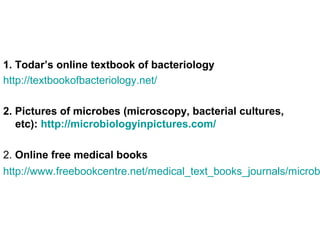
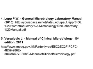
This CV summarizes the educational and professional experience of Malik Alian Mohamed Alian. He holds a Ph.D. in Entomology and Integrated Pest Management from the University of Khartoum. His current position is Associate Professor of Biostatistics at White Nile College of Science and Technology. He has over 30 years of experience in teaching, research, and management roles related to agriculture and health sciences. He has published several papers on topics like biological pest control and the management of insect pests affecting crops.
This document compares monocotyledons and dicotyledons. Monocotyledons have parallel veins in their leaves, fibrous root systems, and flower parts in multiples of three. Dicotyledons have netted veins in their leaves, taproots with lateral roots, and flower parts in multiples of four or five. Both plant groups play important roles in ecosystems and human life.
The document discusses bioinformatics, defining it as the application of information technology to the field of molecular biology. It describes how bioinformatics uses biology, mathematics, and computer science to analyze and manage biological data. Some key applications of bioinformatics mentioned are sequence analysis, prediction of protein structure, genome annotation, comparative genomics, and health/drug discovery. Several important bioinformatics resources are also outlined, including NCBI, PubMed, EMBL, and OMIM.
BRIF: Bioresource Research Impact Factor - Anne Cambon-Thomsen - INSERMLisette Giepmans
This document discusses the Bioresource Research Impact Factor (BRIF) initiative. The BRIF aims to create standardized tools for citing bioresources in scientific literature in order to increase their visibility, assess their usage, and recognize the effort involved in establishing and maintaining them. It outlines barriers to sharing bioresources and incentives needed. It also describes the development of a guideline called CoBRA for standardized bioresource citation and an open access journal for describing bioresources. The presentation concludes by discussing next steps to implement standardized citation practices.
BactFinder Application is an interactive mobile application designed to facilitate bacteria identification based on biochemical tests and bacteria morphology.
Bioinformatics is defined as the field that studies biology using computers and information technology. It involves the collection, storage, and analysis of molecular biological data using techniques from computer science and statistics. Some key events in bioinformatics include Watson and Crick proposing the DNA double helix structure in 1953, and the development of sequence alignment and structure prediction algorithms in the 1970s. Bioinformatics aims to better understand living cells at the molecular level by analyzing raw molecular sequence and structure data. It provides globally accessible databases and analysis tools to enable sharing and study of biological data.
This document discusses the laboratory diagnosis of infections caused by the genus Mycobacterium. It focuses on Mycobacterium tuberculosis and Mycobacterium leprae. Key points include:
- Mycobacteria are acid-fast bacilli that require special staining techniques like Ziehl-Neelsen staining.
- Specimen collection and microscopy are used for diagnosis. Isolation requires slow growth on media like Lowenstein-Jensen. Identification uses tests for niacin, catalase, and tuberculostatic susceptibility.
- Leprosy is caused by M. leprae and presents as lepromatous or tuberculoid forms. Diagnosis involves biopsy staining for acid-fast
presentation is intended for 2nd year medical school students and contains definitions of terms in order to facilitate the understanding of the infectious process, as well as basic elements of biosafety in microgiology laboratories
This document provides an overview of viruses, including their general characteristics, morphology, structure, classification criteria, and methods for laboratory diagnosis. Key points include:
- Viruses are small infectious agents that require a host cell to replicate and are made up of nucleic acids surrounded by a protein capsid.
- Morphology varies between spherical, tubular, and complex shapes depending on the virus. Viruses also have either DNA or RNA genomes.
- Laboratory diagnosis methods examine viral particles, detect viral proteins/genetic material, and measure antibody response. Techniques include electron microscopy, cell culture, serology like ELISA, and molecular methods like PCR.
This document discusses the laboratory diagnosis of infections caused by Treponema, Borrelia, and Leptospira bacteria. It describes the characteristics and pathogenic species of each genus, how they cause diseases like syphilis, Lyme disease, and leptospirosis, and the methods used to diagnose infections through direct examination of clinical samples, culture techniques, and serological tests. Key diagnostic tests include darkfield microscopy, immunofluorescence, culture media, microscopic agglutination, ELISA and Western blot. Penicillin and doxycycline are commonly used for treatment.
This document summarizes the laboratory diagnosis of infections caused by Legionella, Haemophilus, and Bordetella genera. It describes the characteristics, habitats, and modes of transmission. Diagnosis involves microscopy, culture, antigen detection, and molecular methods. Legionella is diagnosed via sputum culture on BCYE agar and urine antigen tests. Haemophilus grows on chocolate or blood agar showing satellitism near Staphylococcus. Bordetella is cultured on Bordet-Gengou medium. Public health control involves identifying and decontaminating water sources for Legionella and vaccination for Haemophilus and Bordetella infections.
Streptococcus and Staphylococcus are common bacterial genera that can cause infections. The document outlines the steps for laboratory diagnosis of infections caused by these bacteria, including specimen collection, culture, staining, and biochemical and antimicrobial testing. Gram staining reveals Gram-positive cocci arranged in clusters for Staphylococcus or chains for Streptococcus. Culture on blood agar shows hemolytic patterns. Biochemical tests help identify pathogenic species like S. aureus and S. pyogenes. Antibiotic susceptibility testing is also important for treatment.
This document discusses bacterial culture techniques for isolating and identifying bacteria from clinical samples. Pure bacterial colonies are necessary to determine their characteristics and properties. Bacteria are streaked in patterns on agar plates to separate them into isolated colonies. The colonies are then analyzed based on morphology, including size, shape, color, and other traits. Hemolysis patterns and reactions in biochemical tests of the colonies provide information to identify the bacterial species. Proper culture methods allow clear observation of colony traits for bacterial identification.
This document discusses laboratory diagnosis of infections caused by obligate anaerobic bacteria. It defines various types of bacteria based on their ability to grow with or without oxygen. Obligate anaerobes cannot grow in the presence of oxygen because they lack enzymes like superoxide dismutase and catalase to break down harmful oxygen byproducts. Specimen collection and transport methods aim to maintain an oxygen-free environment. Identification techniques for certain pathogenic anaerobes that cause infections like gas gangrene, tetanus, and botulism are also outlined.
teaching support for 2nd year medical school students: steps of the laboratory diagnosis of infections caused by bacteria of the genera Staphylococcus and Streptococcus
This document discusses the laboratory diagnosis of infections caused by Vibrio, Campylobacter, Helicobacter, and Pseudomonas genera. Key points include:
Vibrio cholerae causes cholera and is identified through microscopy showing motile comma-shaped bacilli, growth on selective media like TCBS showing yellow colonies, and serological testing. Campylobacter species cause diarrhea and are microaerophilic and spiral-shaped. Helicobacter pylori causes gastric ulcers and is identified through microscopy of gastric biopsies and microaerophilic culture. Pseudomonas aeruginosa is an opportunistic pathogen identified through microscopy, growth on non-selective media, and biochemical testing.
This document discusses the laboratory diagnosis of infections caused by various Gram-positive bacilli, including Corynebacterium, Listeria, Erysipelothrix, and Bacillus. It provides details on specimen collection, microscopic examination, culture methods and media, and biochemical testing for identifying these bacteria, with a focus on Corynebacterium diphtheriae, Listeria monocytogenes, and Bacillus anthracis. Vaccination is emphasized as an important prevention method for diphtheria.
This document provides information on laboratory diagnosis of infections caused by the family Enterobacteriaceae. It discusses the collection and testing of various specimen types, including stool, urine, and blood. A variety of culture media and biochemical tests are described to isolate and identify bacterial pathogens like E. coli, Salmonella, and Shigella from clinical samples. Identification methods include antigenic structure identification using agglutination tests and phage typing for certain Salmonella serotypes. The document provides details on laboratory diagnosis of common infections like urinary tract infections and enteric diseases including typhoid fever.
an introduction to PCR principles and applications in microbiological diagnosis; to serve as a support for students in the second year of medical school
Immunologic methods are used in the laboratory diagnosis of infections by detecting the interaction between antigens and antibodies. Common techniques include agglutination, immunofluorescence, ELISA, and immunoblotting. Agglutination involves clumping of antigens by antibodies that can be seen visually. Immunofluorescence uses antibodies coupled to fluorescent dyes to identify antigens under UV light. ELISA detects antigens or antibodies through an enzymatic reaction, while immunoblotting confirms antibody presence by blotting proteins and detecting binding. These methods exploit the high specificity of the antigen-antibody reaction for diagnostic purposes.
This document summarizes several Gram positive and Gram negative bacilli of medical importance, including Corynebacterium diphtheriae, Bacillus anthracis, Salmonella, Shigella, E. coli, Klebsiella, Proteus, and Yersinia pestis. It describes their morphology, pathogenicity, diseases caused, specimen collection and testing methods, and appearance on common culture media such as blood agar, MacConkey agar, and Hektoen agar. Key identification characteristics include diphtheria's "Chinese letters" morphology, anthrax's spore formation, Salmonella's black-centered colonies on IS media, and Proteus's swarming growth pattern.
The document discusses Mycobacterium, the genus of bacteria that includes Mycobacterium tuberculosis which causes tuberculosis. It provides details on the epidemiology of tuberculosis, noting it is one of the top infectious disease burdens globally and in Tanzania specifically. It describes the pathogenesis and clinical presentation of tuberculosis as well as methods for diagnosis and treatment.
presentation on the most frequently encountered dental healthcare associated infections, including control measures; intended to be used as a support for students in dental medicine (second year of study)
Steps of the bacteriological diagnosis in infections caused by bacteria of the genus Neisseria - for the use of medical school students in the second year of study
This document outlines the topics to be covered in a practical exam in general medicine for the first semester. The topics include sterilization and disinfection methods, antiseptics and disinfectants, culture media, the main steps in bacteriological diagnosis, collection and transport of biological specimens, microscopic and macroscopic examination in bacteriology, identification of bacteria by morphology and biochemical characteristics, antimicrobial susceptibility testing, immunological reactions used in diagnosis, and polymerase chain reaction.
Macroscopic and microscopic examination of clinical specimens is important for orienting the diagnosis of infectious diseases. Macroscopic examination assesses specimen quality and identifies abnormalities that may indicate a pathogen, such as turbid CSF suggesting bacterial meningitis. Microscopic examination uses various staining techniques and microscopy types to visualize pathogens like bacteria, parasites, and fungi directly in specimens. Stains such as Gram and Ziehl-Neelsen are used to classify bacteria and identify acid-fast organisms like Mycobacterium tuberculosis respectively. Together, macroscopic and microscopic examination provide clues for targeting appropriate microbiological tests and presumptive diagnosis.
definitions and description of the main procedures used for antimicrobial susceptibility testing; to support teaching general medicine students in their second year of study
This document discusses antimicrobial susceptibility testing, which determines how well a microbe will respond to different antibiotic drugs. There are two main methods: disk diffusion testing, which uses disks containing antibiotics placed on an agar plate, and minimum inhibitory concentration (MIC) testing, which finds the lowest concentration of antibiotics that prevents microbial growth. The results of these tests guide the selection of effective antibiotic treatment based on whether a microbe is susceptible, resistant, or intermediately resistant to various drugs.
general overview on the main steps of bacteriological diagnosis, including pre-analytical (presumptive clinical diagnosis, collection and transport of specimens), analytical and post-analytical phases (communication of test results).
This document discusses various methods used in clinical microbiology for the diagnosis of bacterial infections, including specimen collection, microscopy, and bacterial culture. It emphasizes the importance of proper specimen collection for accurate diagnosis, describing rules for collecting samples from the correct site and in optimal quantities while avoiding contamination. Microscopy techniques described include staining methods like Gram stain, Ziehl-Neelsen stain, and Giemsa stain to identify bacteria, mycobacteria, and parasites under an optical microscope. Bacterial culture methods involve using various liquid and solid growth media suited to different microbial needs to isolate and identify infectious agents.
1. Antiseptics are used on living tissues to prevent or reduce microbial growth, while disinfectants are used on non-living surfaces.
2. Semmelweis and Lister were pioneers in antisepsis, with Semmelweis reducing childbed fever by having doctors wash hands and Lister advancing aseptic surgery using carbolic acid.
3. Common antiseptics and disinfectants include phenol derivatives, iodine, chlorhexidine, alcohols, acids, metals like silver, and dyes like gentian violet; they act by disrupting microbes or their structures through oxidation, coagulation, or other mechanisms.
presentation for students in medical school: general principles of the most important sterilisation methods applicable in microbiology labs and in healthcare facilities
Executive Directors Chat Leveraging AI for Diversity, Equity, and InclusionTechSoup
Let’s explore the intersection of technology and equity in the final session of our DEI series. Discover how AI tools, like ChatGPT, can be used to support and enhance your nonprofit's DEI initiatives. Participants will gain insights into practical AI applications and get tips for leveraging technology to advance their DEI goals.
ISO/IEC 27001, ISO/IEC 42001, and GDPR: Best Practices for Implementation and...PECB
Denis is a dynamic and results-driven Chief Information Officer (CIO) with a distinguished career spanning information systems analysis and technical project management. With a proven track record of spearheading the design and delivery of cutting-edge Information Management solutions, he has consistently elevated business operations, streamlined reporting functions, and maximized process efficiency.
Certified as an ISO/IEC 27001: Information Security Management Systems (ISMS) Lead Implementer, Data Protection Officer, and Cyber Risks Analyst, Denis brings a heightened focus on data security, privacy, and cyber resilience to every endeavor.
His expertise extends across a diverse spectrum of reporting, database, and web development applications, underpinned by an exceptional grasp of data storage and virtualization technologies. His proficiency in application testing, database administration, and data cleansing ensures seamless execution of complex projects.
What sets Denis apart is his comprehensive understanding of Business and Systems Analysis technologies, honed through involvement in all phases of the Software Development Lifecycle (SDLC). From meticulous requirements gathering to precise analysis, innovative design, rigorous development, thorough testing, and successful implementation, he has consistently delivered exceptional results.
Throughout his career, he has taken on multifaceted roles, from leading technical project management teams to owning solutions that drive operational excellence. His conscientious and proactive approach is unwavering, whether he is working independently or collaboratively within a team. His ability to connect with colleagues on a personal level underscores his commitment to fostering a harmonious and productive workplace environment.
Date: May 29, 2024
Tags: Information Security, ISO/IEC 27001, ISO/IEC 42001, Artificial Intelligence, GDPR
-------------------------------------------------------------------------------
Find out more about ISO training and certification services
Training: ISO/IEC 27001 Information Security Management System - EN | PECB
ISO/IEC 42001 Artificial Intelligence Management System - EN | PECB
General Data Protection Regulation (GDPR) - Training Courses - EN | PECB
Webinars: https://pecb.com/webinars
Article: https://pecb.com/article
-------------------------------------------------------------------------------
For more information about PECB:
Website: https://pecb.com/
LinkedIn: https://www.linkedin.com/company/pecb/
Facebook: https://www.facebook.com/PECBInternational/
Slideshare: http://www.slideshare.net/PECBCERTIFICATION
A review of the growth of the Israel Genealogy Research Association Database Collection for the last 12 months. Our collection is now passed the 3 million mark and still growing. See which archives have contributed the most. See the different types of records we have, and which years have had records added. You can also see what we have for the future.
Exploiting Artificial Intelligence for Empowering Researchers and Faculty, In...Dr. Vinod Kumar Kanvaria
Exploiting Artificial Intelligence for Empowering Researchers and Faculty,
International FDP on Fundamentals of Research in Social Sciences
at Integral University, Lucknow, 06.06.2024
By Dr. Vinod Kumar Kanvaria
This presentation includes basic of PCOS their pathology and treatment and also Ayurveda correlation of PCOS and Ayurvedic line of treatment mentioned in classics.
This presentation was provided by Steph Pollock of The American Psychological Association’s Journals Program, and Damita Snow, of The American Society of Civil Engineers (ASCE), for the initial session of NISO's 2024 Training Series "DEIA in the Scholarly Landscape." Session One: 'Setting Expectations: a DEIA Primer,' was held June 6, 2024.
How to Manage Your Lost Opportunities in Odoo 17 CRMCeline George
Odoo 17 CRM allows us to track why we lose sales opportunities with "Lost Reasons." This helps analyze our sales process and identify areas for improvement. Here's how to configure lost reasons in Odoo 17 CRM