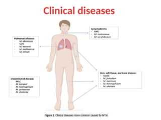
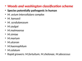
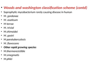
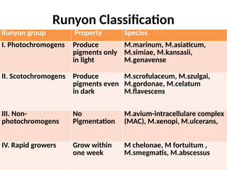
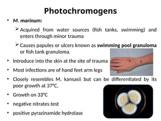
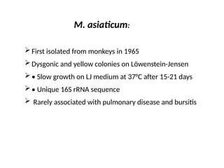
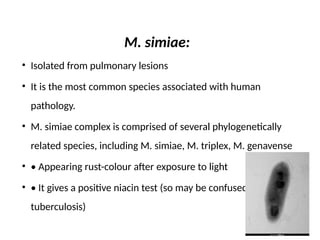
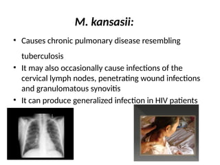
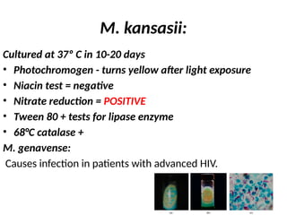
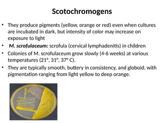
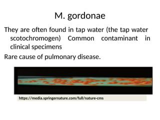
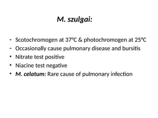
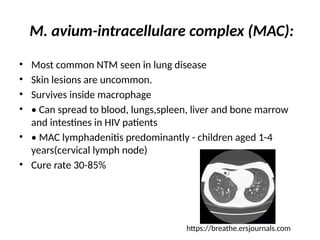
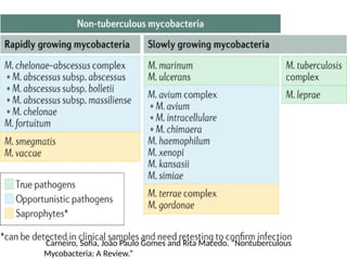
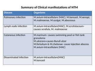
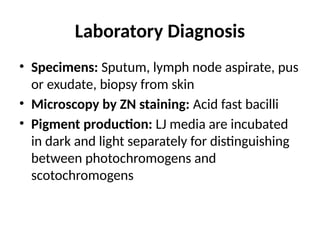
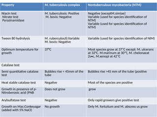
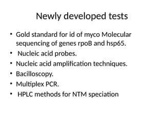
NONTUBERCULOUS MYCOBACTERIA
( NTM)
INTRODUCTION
• Nontuberculous mycobacteria (NTM), also known as
environmental mycobacteria, atypical mycobacteria and
mycobacteria other than tuberculosis (MOTT), are
mycobacteria which do not cause tuberculosis or leprosy
Mycobacterium tuberculosis complex includes
M.tuberculosis
M.bovis
M. africanum
M. caprae
M. microti
M. pinnipedii
Leprosy (M. leprae
NONTUBERCULOUS MYCOBACTERIA
INFECTIONS
• Diverse group of mycobacteria isolated from
birds, animals, environmental sources
• Opportunistic pathogens, occasionally
associated with human infection
• Man-to-man transmission is not known.
• Saprophytic mycobacteria - isolated from soil,
water and other environmental sources. They
do not cause any disease in humans
Common features
• They are not usually transmitted from person to
person
• Source of infection is water, soil, food and animals
• Human infection with NTM is common in some
areas, disease is rare
• Exhibits dysgonic growth on LJ medium
• Niacin and nitrate reduction tests are negative
• Not able to cause progressive disease in guniea pigs
Risk factors
• Immunosuppression (HIV, Medications )
• Aging
• BCG vaccination
• Cystic fibrosis
• Fibronodular bronchiectasis
• Number of inherited and acquired defects in the host
immune response, particularly those that affect the
Th1 cell and macrophage pathway
Risk factors
• Interferon gamma receptor deficiencies
• Signal transducer and activator of
transcription 1 (STAT1) deficiency
• Auto-antibodies to interferon gamma
• CD4 lymphopenia due to HIV or other causes
• Use of TNF-alpha inhibitors
Symptoms
• No typical Manifestations
Fever
Weight loss
Enlarged lymph glands
Diarrhoea
Sweating, excessive -- night sweats
Fatigue
Malaise
Cough
Dyspnoea
Skin lesions
Joint pain, Bone pain
Clinical diseases
• Woods and washington classification scheme
• Species potentially pathogenic in human
• M. avium intercellulare complex
• M. kansasii
• M. scrofulanceum
• M.szulgai
• M.malmoense
• M.simiae
• M.marium
• M.ulceran
• M.haemophilum
• M.celatum
• Rapid growers: M.fortuitum, M.chelonae, M.abscessus
• Woods and washington classification scheme (contd)
• Saprophytic mycobacterium rarely causing disease in human
• M. gordonae
• M. asaticum
• M terrae
• M. trivial
• M.shimoidei
• M. gastri
• M.paratuberculosis
• M. flavescens
• Other rapid growing species
• M.thermoresistible
• M.smegmatis
• M.phlei
Runyon Classification
Runyon group Property Species
I. Photochromogens Produce
pigments only
in light
M.marinum, M.asiaticum,
M.simiae, M.kansasii,
M.genavense
II. Scotochromogens Produce
pigments even
in dark
M.scrofulaceum, M.szulgai,
M.gordonae, M.celatum
M.flavescens
III. Non-
photochromogens
No
Pigmentation
M.avium-intracellulare complex
(MAC), M.xenopi, M.ulcerans,
IV. Rapid growers Grow within
one week
M chelonae, M fortuitum ,
M.smegmatis, M.abscessus
Photochromogens
• M. marinum:
Acquired from water sources (fish tanks, swimming) and
enters through minor trauma
Causes papules or ulcers known as swimming pool granuloma
or fish tank granuloma.
• Introduce into the skin at the site of trauma
• Most infections are of hand feet arm legs
• Closely resembles M. kansasii but can be differentiated by its
poor growth at 37°C,
• Growth on 33°C
• negative nitrates test
• positive pyrazinamide hydrolase
M. asiaticum:
First isolated from monkeys in 1965
Dysgonic and yellow colonies on Löwenstein-Jensen
• Slow growth on LJ medium at 37°C after 15-21 days
• Unique 16S rRNA sequence
 Rarely associated with pulmonary disease and bursitis
M. simiae:
• Isolated from pulmonary lesions
• It is the most common species associated with human
pathology.
• M. simiae complex is comprised of several phylogenetically
related species, including M. simiae, M. triplex, M. genavense
• • Appearing rust-colour after exposure to light
• • It gives a positive niacin test (so may be confused with M.
tuberculosis)
M. kansasii:
• Causes chronic pulmonary disease resembling
tuberculosis
• It may also occasionally cause infections of the
cervical lymph nodes, penetrating wound infections
and granulomatous synovitis
• It can produce generalized infection in HIV patients
M. kansasii:
Cultured at 37º C in 10-20 days
• Photochromogen - turns yellow after light exposure
• Niacin test = negative
• Nitrate reduction = POSITIVE
• Tween 80 + tests for lipase enzyme
• 68°C catalase +
M. genavense:
Causes infection in patients with advanced HIV.
Scotochromogens
• They produce pigments (yellow, orange or red) even when cultures
are incubated in dark, but intensity of color may increase on
exposure to light
• M. scrofulaceum: scrofula (cervical lymphadenitis) in children
• Colonies of M. scrofulaceum grow slowly (4-6 weeks) at various
temperatures (21°, 31°, 37° C).
• They are typically smooth, buttery in consistency, and globoid, with
pigmentation ranging from light yellow to deep orange.
M. gordonae
They are often found in tap water (the tap water
scotochromogen) Common contaminant in
clinical specimens
Rare cause of pulmonary disease.
https://media.springernature.com/full/nature-cms
M. szulgai:
- Scotochromogen at 37°C & photochromogen at 25°C
- Occasionally cause pulmonary disease and bursitis
• Nitrate test positive
• Niacine test negative
• ™M. celatum: Rare cause of pulmonary infection
Nonphotochromogens
M. avium-intracellulare complex (MAC):
• M. avium and M. intracellulare are so similar that that they
have been considered as one group, the M. avium complex
(MAC)
- Opportunistic pathogens – HIV low CD4 T-cell count (<50/μL)
- Lymphadenitis, respiratory infection and disseminated
disease.
M. avium-intracellulare complex (MAC):
• Most common NTM seen in lung disease
• Skin lesions are uncommon.
• Survives inside macrophage
• • Can spread to blood, lungs,spleen, liver and bone marrow
and intestines in HIV patients
• • MAC lymphadenitis predominantly - children aged 1-4
years(cervical lymph node)
• Cure rate 30-85%
https://breathe.ersjournals.com
™M. xenopi
- Hospital water supplies, and associated with
nosocomial outbreaks
- Rarely pulmonary disease in HIV-infected people.
- Though classified as a non-photochromogen, M.
xenopi may form yellow colored colonies similar to
scotochromogens.
- It was originally isolated from toads.
Nonphotochromogens
• M. ulcerans:
- Waterborne skin pathogen
- Buruli ulcer - painless ulcers and nodules  necrotic later
- Osteomyelitis and limb deformities
- Exotoxin: mycolactone toxin
„
- Narrow temperature range (31°–34°C)
• ™M. malmoense:
- Pulmonary disease and rarely lymphadenitis
Rapid Growers
• M. fortuitum and M. chelonae: post-trauma
injection abscess and catheter-related
infections
• ™M. abscessus: pulmonary infection
• Tests are done for rapid growers:
- Arylsulfatase test: Positive for all rapid growers
- Growth on MacConkey agar: M. fortuitum
grows
Carneiro, Sofia, João Paulo Gomes and Rita Macedo. “Nontuberculous
Mycobacteria: A Review.”
Summary of Clinical manifestations of NTM
Disease Organisms
Pulmonary infection M.avium-intracellulare (MAC), M.kansasii, M.xenopi,
M.malmoense, M.szulgai, M.abscessus
Lymph node infection M.avium-intracellulare(MAC), M.scrofulaceum-
causes scrofula, M. malmoense
Cutaneous infection M.marinum- causes swimming pool or fish tank
granuloma
M.ulcerans-causes Buruli ulcer
M.fortuitum & M.chelonae- cause injection abscess
M.avium-intracellulare (MAC)
Disseminated infection M.avium-intracellulare(MAC)
M.kansasii
Laboratory Diagnosis
• ™Specimens: Sputum, lymph node aspirate, pus
or exudate, biopsy from skin
• ™Microscopy by ZN staining: Acid fast bacilli
• ™Pigment production: LJ media are incubated
in dark and light separately for distinguishing
between photochromogens and
scotochromogens
Property M. tuberculosis complex Nontuberculous mycobacteria (NTM)
Niacin test
Nitrate test
Pyrazinamidase
M. tuberculosis: Positive
M. bovis: Negative
Negative (exceptM.simiae}
Variable (used for species identification of
NTM)
Variable (used for species identification of
NTM)
Tween 80 hydrolysis M. ruberculosJS:Variable
M. bovis: Negative
Variable (used for species identification of NlM)
Optimum temperature for
growth
37°C Most species grow at 37"C except: M. ulcerans
at 32°C. M.marinum at 30'°(, M. chelonaeat
2s•c, M.xenopi at 42"C
Catalase test
Semi quantitative catalase
test
Bubbles rise < 45mm of the
tube
Bubbles rise >45 mm of the tube (positive
Heat stable catalase test Negative Most of the species are positive
Growth in presence of p-
Nitrobenzoic acid (PNB
Does not grow grow
Arylsulfatase test Negative Only rapid growers give positive test
Growth on Mac:Conkeyagar
(added with 5% NaCl)
No growth Only M. fortuitum and M. abscess us grow
Newly developed tests
• Gold standard for id of myco Molecular
sequencing of genes rpoB and hsp65.
• Nucleic acid probes.
• Nucleic acid amplification techniques.
• Bacilloscopy.
• Multiplex PCR.
• HPLC methods for NTM speciation
New developments in diagnosis of NTM
disease
• MALDI-TOF mass spectrometry
• Multi-locus sequence typing
• Multiplex PCR assay for detection of MAC
• Serological markers
MALDI-TOF mass spectrometry
• This allows comparison of mass spectral patterns of molecules
specific to an NTM species to an online library of NTM strain
patterns.
• online databases grow its utility is increasing
• advantages in terms of speed
• MALDI-TOF mass spectrometry has been validated as a highly
reproducible method for identification of NTM in a recent
multicentre study.
Multi-locus sequence typing
• multi-locus sequence typing (MLST) allows for comparison of
multiple DNA sequences from an organism to those of an
online dataset.
• Enhancing the accuracy of species and subspecies
identification
• Whole genome sequencing (WGS) methods are also evolving,
both in terms of technology and accessibility, paving the way
for highly specific species identification
• limit the widespread availability of WGS for NTM
• The high costs and expert training, as well as the significant
software and programming capacity required to process the
vast datasets produced.
Multiplex PCR assay for detection of MAC
• PCR assay for simultaneous detection of Mtb and Mycobacterium
avium complex (MAC), using primers for two separate genes
present in MAC species
• Xpert MTB/RIF molecular assay for the rapid detection of Mtb in
clinical samples prior to culture revolutionised diagnosis of
tuberculosis worldwide.
• rapid PCR methods to detect NTM directly from clinical specimens
have not been previously available
• rapid detection of MAC infection is feasible. As MAC is the most
common NTM causing pulmonary disease, this may have a
dramatic impact on clinical practice if their results are replicated in
clinical validation studies.
Serological markers
• Detection of IgA antibody against the 31 serotype-specific
glycopeptidolipid (GPL) core surface antigens of MAC has a
reported 69.9% sensitivity and 90.6% specificity for the
detection of MAC pulmonary disease .
• the utility of this method needs further validation before it is
endorsed in clinical practice guidelines
Identification
• Species of NTM can be differentiated from M.
tuberculosis complex by:
- MPT64 antigen by ICT – Negative
„
- Positive for tests - catalase test
(semiquantitative and heat-stable catalase
tests) and tween 80 hydrolysis test
- Newer methods: MALDI-TOF (matrix-assisted
laser desorption ionization-time of flight), PCR
Treatment Nontuberculous mycobacterial
disease
• Associated with emergence of drug resistance &
relapse
• Multidrug therapy
• ‰M. avium-intracellulare complex (MAC), M.
kansasii & M.marinum - multidrug therapy with
macrolide (clarithromycin or azithromycin),
ethambutol, and a rifamycin (rifampin or rifabutin)
• NTM are
‰ resistant to most of the first and second
line intitubercular drugs
REFERENCES
• Konemans color atlas & textbook of diagnosticsc
microbiology – 7th
edition
• Essential of medical microbiology, Apurva Shankar Sastry, 4rd
edition.
• An Official ATS/IDSA Statement: Diagnosis, Treatment, and
Prevention of Nontuberculous Mycobacterial Disease.
• Thoracic Society Guideline for the management of non-
tuberculous mycobacterial pulmonary disease (NTM-PD)BMJ
Open Respiratory Research .
• João Paulo Gomes and Rita Macedo. “Nontuberculous
Mycobacteria: A Review.” Clin Infect Dis 7 (2023): 206.
THANKYOU

NONTUBERCULOUS MYCOBACTERIA. .pptx

  • 1.
  • 2.
    INTRODUCTION • Nontuberculous mycobacteria(NTM), also known as environmental mycobacteria, atypical mycobacteria and mycobacteria other than tuberculosis (MOTT), are mycobacteria which do not cause tuberculosis or leprosy Mycobacterium tuberculosis complex includes M.tuberculosis M.bovis M. africanum M. caprae M. microti M. pinnipedii Leprosy (M. leprae
  • 3.
    NONTUBERCULOUS MYCOBACTERIA INFECTIONS • Diversegroup of mycobacteria isolated from birds, animals, environmental sources • Opportunistic pathogens, occasionally associated with human infection • Man-to-man transmission is not known. • Saprophytic mycobacteria - isolated from soil, water and other environmental sources. They do not cause any disease in humans
  • 4.
    Common features • Theyare not usually transmitted from person to person • Source of infection is water, soil, food and animals • Human infection with NTM is common in some areas, disease is rare • Exhibits dysgonic growth on LJ medium • Niacin and nitrate reduction tests are negative • Not able to cause progressive disease in guniea pigs
  • 5.
    Risk factors • Immunosuppression(HIV, Medications ) • Aging • BCG vaccination • Cystic fibrosis • Fibronodular bronchiectasis • Number of inherited and acquired defects in the host immune response, particularly those that affect the Th1 cell and macrophage pathway
  • 6.
    Risk factors • Interferongamma receptor deficiencies • Signal transducer and activator of transcription 1 (STAT1) deficiency • Auto-antibodies to interferon gamma • CD4 lymphopenia due to HIV or other causes • Use of TNF-alpha inhibitors
  • 7.
    Symptoms • No typicalManifestations Fever Weight loss Enlarged lymph glands Diarrhoea Sweating, excessive -- night sweats Fatigue Malaise Cough Dyspnoea Skin lesions Joint pain, Bone pain
  • 8.
  • 9.
    • Woods andwashington classification scheme • Species potentially pathogenic in human • M. avium intercellulare complex • M. kansasii • M. scrofulanceum • M.szulgai • M.malmoense • M.simiae • M.marium • M.ulceran • M.haemophilum • M.celatum • Rapid growers: M.fortuitum, M.chelonae, M.abscessus
  • 10.
    • Woods andwashington classification scheme (contd) • Saprophytic mycobacterium rarely causing disease in human • M. gordonae • M. asaticum • M terrae • M. trivial • M.shimoidei • M. gastri • M.paratuberculosis • M. flavescens • Other rapid growing species • M.thermoresistible • M.smegmatis • M.phlei
  • 11.
    Runyon Classification Runyon groupProperty Species I. Photochromogens Produce pigments only in light M.marinum, M.asiaticum, M.simiae, M.kansasii, M.genavense II. Scotochromogens Produce pigments even in dark M.scrofulaceum, M.szulgai, M.gordonae, M.celatum M.flavescens III. Non- photochromogens No Pigmentation M.avium-intracellulare complex (MAC), M.xenopi, M.ulcerans, IV. Rapid growers Grow within one week M chelonae, M fortuitum , M.smegmatis, M.abscessus
  • 12.
    Photochromogens • M. marinum: Acquiredfrom water sources (fish tanks, swimming) and enters through minor trauma Causes papules or ulcers known as swimming pool granuloma or fish tank granuloma. • Introduce into the skin at the site of trauma • Most infections are of hand feet arm legs • Closely resembles M. kansasii but can be differentiated by its poor growth at 37°C, • Growth on 33°C • negative nitrates test • positive pyrazinamide hydrolase
  • 13.
    M. asiaticum: First isolatedfrom monkeys in 1965 Dysgonic and yellow colonies on Löwenstein-Jensen • Slow growth on LJ medium at 37°C after 15-21 days • Unique 16S rRNA sequence  Rarely associated with pulmonary disease and bursitis
  • 14.
    M. simiae: • Isolatedfrom pulmonary lesions • It is the most common species associated with human pathology. • M. simiae complex is comprised of several phylogenetically related species, including M. simiae, M. triplex, M. genavense • • Appearing rust-colour after exposure to light • • It gives a positive niacin test (so may be confused with M. tuberculosis)
  • 15.
    M. kansasii: • Causeschronic pulmonary disease resembling tuberculosis • It may also occasionally cause infections of the cervical lymph nodes, penetrating wound infections and granulomatous synovitis • It can produce generalized infection in HIV patients
  • 16.
    M. kansasii: Cultured at37º C in 10-20 days • Photochromogen - turns yellow after light exposure • Niacin test = negative • Nitrate reduction = POSITIVE • Tween 80 + tests for lipase enzyme • 68°C catalase + M. genavense: Causes infection in patients with advanced HIV.
  • 17.
    Scotochromogens • They producepigments (yellow, orange or red) even when cultures are incubated in dark, but intensity of color may increase on exposure to light • M. scrofulaceum: scrofula (cervical lymphadenitis) in children • Colonies of M. scrofulaceum grow slowly (4-6 weeks) at various temperatures (21°, 31°, 37° C). • They are typically smooth, buttery in consistency, and globoid, with pigmentation ranging from light yellow to deep orange.
  • 18.
    M. gordonae They areoften found in tap water (the tap water scotochromogen) Common contaminant in clinical specimens Rare cause of pulmonary disease. https://media.springernature.com/full/nature-cms
  • 19.
    M. szulgai: - Scotochromogenat 37°C & photochromogen at 25°C - Occasionally cause pulmonary disease and bursitis • Nitrate test positive • Niacine test negative • ™M. celatum: Rare cause of pulmonary infection
  • 20.
    Nonphotochromogens M. avium-intracellulare complex(MAC): • M. avium and M. intracellulare are so similar that that they have been considered as one group, the M. avium complex (MAC) - Opportunistic pathogens – HIV low CD4 T-cell count (<50/μL) - Lymphadenitis, respiratory infection and disseminated disease.
  • 21.
    M. avium-intracellulare complex(MAC): • Most common NTM seen in lung disease • Skin lesions are uncommon. • Survives inside macrophage • • Can spread to blood, lungs,spleen, liver and bone marrow and intestines in HIV patients • • MAC lymphadenitis predominantly - children aged 1-4 years(cervical lymph node) • Cure rate 30-85% https://breathe.ersjournals.com
  • 22.
    ™M. xenopi - Hospitalwater supplies, and associated with nosocomial outbreaks - Rarely pulmonary disease in HIV-infected people. - Though classified as a non-photochromogen, M. xenopi may form yellow colored colonies similar to scotochromogens. - It was originally isolated from toads.
  • 23.
    Nonphotochromogens • M. ulcerans: -Waterborne skin pathogen - Buruli ulcer - painless ulcers and nodules  necrotic later - Osteomyelitis and limb deformities - Exotoxin: mycolactone toxin „ - Narrow temperature range (31°–34°C) • ™M. malmoense: - Pulmonary disease and rarely lymphadenitis
  • 24.
    Rapid Growers • M.fortuitum and M. chelonae: post-trauma injection abscess and catheter-related infections • ™M. abscessus: pulmonary infection • Tests are done for rapid growers: - Arylsulfatase test: Positive for all rapid growers - Growth on MacConkey agar: M. fortuitum grows
  • 25.
    Carneiro, Sofia, JoãoPaulo Gomes and Rita Macedo. “Nontuberculous Mycobacteria: A Review.”
  • 26.
    Summary of Clinicalmanifestations of NTM Disease Organisms Pulmonary infection M.avium-intracellulare (MAC), M.kansasii, M.xenopi, M.malmoense, M.szulgai, M.abscessus Lymph node infection M.avium-intracellulare(MAC), M.scrofulaceum- causes scrofula, M. malmoense Cutaneous infection M.marinum- causes swimming pool or fish tank granuloma M.ulcerans-causes Buruli ulcer M.fortuitum & M.chelonae- cause injection abscess M.avium-intracellulare (MAC) Disseminated infection M.avium-intracellulare(MAC) M.kansasii
  • 27.
    Laboratory Diagnosis • ™Specimens:Sputum, lymph node aspirate, pus or exudate, biopsy from skin • ™Microscopy by ZN staining: Acid fast bacilli • ™Pigment production: LJ media are incubated in dark and light separately for distinguishing between photochromogens and scotochromogens
  • 28.
    Property M. tuberculosiscomplex Nontuberculous mycobacteria (NTM) Niacin test Nitrate test Pyrazinamidase M. tuberculosis: Positive M. bovis: Negative Negative (exceptM.simiae} Variable (used for species identification of NTM) Variable (used for species identification of NTM) Tween 80 hydrolysis M. ruberculosJS:Variable M. bovis: Negative Variable (used for species identification of NlM) Optimum temperature for growth 37°C Most species grow at 37"C except: M. ulcerans at 32°C. M.marinum at 30'°(, M. chelonaeat 2s•c, M.xenopi at 42"C Catalase test Semi quantitative catalase test Bubbles rise < 45mm of the tube Bubbles rise >45 mm of the tube (positive Heat stable catalase test Negative Most of the species are positive Growth in presence of p- Nitrobenzoic acid (PNB Does not grow grow Arylsulfatase test Negative Only rapid growers give positive test Growth on Mac:Conkeyagar (added with 5% NaCl) No growth Only M. fortuitum and M. abscess us grow
  • 29.
    Newly developed tests •Gold standard for id of myco Molecular sequencing of genes rpoB and hsp65. • Nucleic acid probes. • Nucleic acid amplification techniques. • Bacilloscopy. • Multiplex PCR. • HPLC methods for NTM speciation
  • 30.
    New developments indiagnosis of NTM disease • MALDI-TOF mass spectrometry • Multi-locus sequence typing • Multiplex PCR assay for detection of MAC • Serological markers
  • 31.
    MALDI-TOF mass spectrometry •This allows comparison of mass spectral patterns of molecules specific to an NTM species to an online library of NTM strain patterns. • online databases grow its utility is increasing • advantages in terms of speed • MALDI-TOF mass spectrometry has been validated as a highly reproducible method for identification of NTM in a recent multicentre study.
  • 32.
    Multi-locus sequence typing •multi-locus sequence typing (MLST) allows for comparison of multiple DNA sequences from an organism to those of an online dataset. • Enhancing the accuracy of species and subspecies identification • Whole genome sequencing (WGS) methods are also evolving, both in terms of technology and accessibility, paving the way for highly specific species identification • limit the widespread availability of WGS for NTM • The high costs and expert training, as well as the significant software and programming capacity required to process the vast datasets produced.
  • 33.
    Multiplex PCR assayfor detection of MAC • PCR assay for simultaneous detection of Mtb and Mycobacterium avium complex (MAC), using primers for two separate genes present in MAC species • Xpert MTB/RIF molecular assay for the rapid detection of Mtb in clinical samples prior to culture revolutionised diagnosis of tuberculosis worldwide. • rapid PCR methods to detect NTM directly from clinical specimens have not been previously available • rapid detection of MAC infection is feasible. As MAC is the most common NTM causing pulmonary disease, this may have a dramatic impact on clinical practice if their results are replicated in clinical validation studies.
  • 34.
    Serological markers • Detectionof IgA antibody against the 31 serotype-specific glycopeptidolipid (GPL) core surface antigens of MAC has a reported 69.9% sensitivity and 90.6% specificity for the detection of MAC pulmonary disease . • the utility of this method needs further validation before it is endorsed in clinical practice guidelines
  • 35.
    Identification • Species ofNTM can be differentiated from M. tuberculosis complex by: - MPT64 antigen by ICT – Negative „ - Positive for tests - catalase test (semiquantitative and heat-stable catalase tests) and tween 80 hydrolysis test - Newer methods: MALDI-TOF (matrix-assisted laser desorption ionization-time of flight), PCR
  • 36.
    Treatment Nontuberculous mycobacterial disease •Associated with emergence of drug resistance & relapse • Multidrug therapy • ‰M. avium-intracellulare complex (MAC), M. kansasii & M.marinum - multidrug therapy with macrolide (clarithromycin or azithromycin), ethambutol, and a rifamycin (rifampin or rifabutin) • NTM are ‰ resistant to most of the first and second line intitubercular drugs
  • 37.
    REFERENCES • Konemans coloratlas & textbook of diagnosticsc microbiology – 7th edition • Essential of medical microbiology, Apurva Shankar Sastry, 4rd edition. • An Official ATS/IDSA Statement: Diagnosis, Treatment, and Prevention of Nontuberculous Mycobacterial Disease. • Thoracic Society Guideline for the management of non- tuberculous mycobacterial pulmonary disease (NTM-PD)BMJ Open Respiratory Research . • João Paulo Gomes and Rita Macedo. “Nontuberculous Mycobacteria: A Review.” Clin Infect Dis 7 (2023): 206.
  • 38.