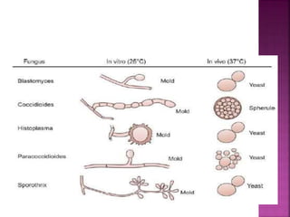
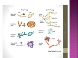
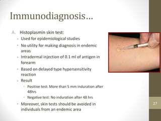
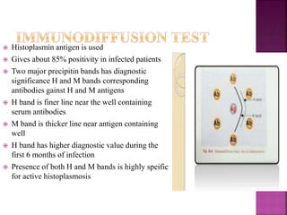
Sundar Khadka,
Microbiologist
HIV Reference Unit, NPHL
 Super kingdom: Eukaryota
 Kingdom: Fungi
 Phylum: Ascomycota
 Class: Eurotiomycetes
 Order: Onygenales
 Family: Onygenaceae
 Genus: Ajellomyces
 Histoplasmosis is a systemic granulomatous disease caused by a dimorphic
fungi Histoplasma capsulatum.
 Caused by three varieties of dimorphic fungus
1) H.c var capsulatum
2) H.c var duboisii
3) H.c. var farciminosum
 H.c var capsulatum - Classic histoplasmosis-
primary pathogenic to human

 H.c var duboisii – african histoplsmosis- primary
pathogenic to human
 H.c. var farciminosum- epizootic histoplasmosis-
pathogenic to animals
 Most important clinical infection is caused by H.C
var capsulatum
 Most commonly it occurs as an asymptomatic or
relatively mild self limiting pulmonary infection
 An intracellular uninucleate organism involving
cells of RE system ( macrophages and giant cells)
 First case reported on December 7 1905
 By samuel Taylor Darling while performing autopsy on a patient
 Also called Darling’s disease
 First believed it as encapsulated plasmodium
 When encountered two more cases despite similarities to
kalaazar it was different organism
 In tissue section cells were seen as hyaline halos
around stained cytoplasm – capsulated organism
 Believed he was observing new organism and
named Histoplasma capsulata
 Literal meaning of nomenclature
 He saw intracellular organism in histio –cytes
resembled plasmo-dium and also seen capsule
 Dimorphic fungus
 Takes 6 weeks to grow in SDA at 25-30 0 c
 On SDA at 250c colonies are initially white
or buff brown
 Mycelial phase has septate hyphae –
produces two types of unicellular asexual
conidia
Characteristics and
Exists as mycelial form(mould) in
environment (25-300C)
Mycelial phase has septate hyphae
producing two types of unicellular
conidia;
Macroconidia: 8-15 µm diameter,
thick walled with distinctive
projections on surface
• Microconidia: 2-4 µm diameter,
smooth structure, infectious form
Exists as yeast in vitro and in tissues
(35-370C)
• Composed of tiny 2-4µm oval budding
yeasts
Non capsulated (although in tissues it appears to
have a clear zone that was misinterpreted by Samuel
Darling.)
Morphology
•
•
•
7
 Macroconidia
 prominent
 Thick walled larger in size 8-16um
 Emerge from short, tubular conidophores
 Microconidia
 smooth walled
 small in size 2-4um
 arise from short stalks from undifferentiated hyphae
 abundant in primary culture
 Worldwide distribution, common in temperate climate
 Fungus lives in the environment, particularly in soil that
contains large amounts of bird or bat droppings.
 Soil is the reservoir where organism exist as multicellular
filamentous form
 Infection occurs by inhalation of microconidia
 Accidental infection may occur to lab workers while handling
culture
 Most common in North and Central America
 Endemic in the Mississippi and Ohio River valleys and also
exists in localized areas in many mideastern states of United states
of America.
 Called classic histoplasmosis
 Highly infectious
 Primarily affects lungs
 Intracellular mycosis of RE system involving spleen, liver,
adrenals, kidneys, skin ,CNS and other body organs
 Yeast cells are found within histiocytes , neutrophils and
lymphocytes
 Molecular genetics have shown that calcium binding protein
CBP1 is required for virulence
 Microscopic morphology of mycelial form of Histoplasma capsulatum showing large,
rounded, single-celled, tuberculate macroconidia formed on short, hyaline, undifferentiated
conidiophores.
Causes and risk factors
Histoplasmosis typically results from exposure during
activities that disturb soil harboring the organism,
generating aerosols of spores
Examples of so-called microfoci of histoplasmosis
include the following: caves, chicken coops, bird
roosts, school yards, prison grounds, decayed wood
piles, dead trees, contaminated chimneys, and old
buildings
Living in an endemic area predisposes to
histoplasmosis
Pre-existing lung disease (eg, COPD) and cigarette
use predispose to chronic pulmonary histoplasmosis
Those who are immunosuppressed,
•
•
•
•
9
•
Pathogenesis
• Infection begins with inhalation of microconidia or hyphal
fragments
Mycelial form transforms into yeast form in alveolar
macrophages
Yeast cells are phagocytized by host immune system
H. capsulatum is able to survive phagocytosis
Calcium-binding protein(CBP1), a cytoplasmic enzyme, a
peroxisomal enzyme
Apoptosis of infected macrophages allow H. capsulatum to
spread
Infection is usually self-limiting in immunocompetent
individuals
•
•
•
•
•
10
Pathogenesis…
11
 Infection remains asymptomatic in about 90-95% of cases
 Asymptomatic form is indicated by presence of positive
histoplasmin skin test without any focus of infection
 Development of asymptomatic or symptomatic disease
depends on intensity of exposure to conidia and status of
cellular response
 Illness may appear after years leaving endemic areas –
reactivation histoplasmosis- occurs in pt with serious
underlying disorders as organ transplantation or AIDS
 flu-like symptoms
 headache
 cough
 lymphadenopathy
 caseating necrosis
 fever
 muscle pain
 chest pain
 Hepatomegaly
 erythema nodosum
(inflammation of the fat cells
under the skin, resulting in
tender red nodules)
 malaise
 anorexia
 dyspnea
 splenomegaly
 coin lesion
 Duration of infection
 Acute
 Subacute
 Chronic
 Site of involvement
 Pulmonary
 Extrapulmonary or disseminated
 Pattern of infection
 Primary
 Reactivation
1. Acute pulmonary histoplasmosis
2. Chronic pulmonary histoplasmosis
3. Cutaneous ,Mucocutaneous histoplasmosis
4. Disseminated histoplasmosis
 Acute type covers entire range of infection in normal host
 More tham 95% of immunocompetent person remain asymptomatic
 In symptomatic cases clinical manifestation of the disease is
nonspecific
 i.p – 10-16 days
 Onset resembles influenza –like disease ie generalized malaise with
fever
 Headache
 Chills
 Profuse sweating
 Sorethroat
 Dry cough
 Chestpain and dyspnoea
 Remains latent for long time and gradully
produces similar symptoms to acute disease but in
more pronounced form
 Disease presents with hemoptysis and apical and
subapical cavities
 Pt lose wt and may have ulcerative lesion over the
lips , mouth ,nose, larynx and intestine
 There is peteche usually in the abdominal wall and
thorax
 Mucous lesions are predominantly found on oral
cavity
 Commonly seen in children under 2years and adolescent
 Manifested as fever, anorexia, weight loss, deterioration
of general condition, anaemia, leukopenia, constant
hepatosplenomegaly and lymphadenopathy
 Pt infected with HIV are greater risk of developing
disseminated histoplasmosis
 Has also been reported in pt with organ transplantation,
infections of thyroid gland, sinuses,prostate , epididymis
 In gastrointestinal histoplasmosis distal ileum most
common site followed by colon and stomach
 Specimen
1) Sputum
2) Bone marrow aspirate/biopsy
3) Lymph node aspirate/biopsy
4) Peripheral blood film- blood smears can be made from buffy coat
or sediment
5) Biopsy of lesions from skin, mucous membrane , lymph nodes
6) Tissues from autopsy
Direct Microscopy
a) KOH Mount
• KOH wet mount prepared from the clinical sample shows tiny
yeast cells.
Staining methods
b)
a)
b)
c)
d)
Calcoflour staining
Giemsa or Wright stains
PAS staining
Gomori’s methanamine silver staining
Fungus appears as small, oval yeast cells, 2-4µm, within
mononuclear or poly-morphonuclear cells and ocassionally in
giant cells.
The flourescent antibody technique has been found useful in
diagnosis of histoplasmosis in tissues.
20
Direct Microscopy…
In Giemsa stain, shows
cell wall as light blue
with clear space
between wall and dark
blue protoplasm
21
Direct Microscopy…
• In PAS(Periodic Acid schiff’s)
stain, wall is stained voilet, red
to pink with pallor protoplasm
filling cell
• In GMS (Gomori
silver) stain, the
black
Methanamine
wall is stained
22
Fungal Culture
Culture medias:
Saboraud Dextrose Agar with antibiotics
Brain Heart infusion Agar with antibiotics
Yeast extract phosphate Agar with ammonium hydroxide
Kelly’s medium
Potato Dextrose Agar
Temperature requirements:
250C : for demonstration of mycelial form
370C : for demonstration of yeast form
•All cultures should be examined daily for one week
and twice a week further three weeks
•Conversion of mycelial –to –yeast phase is useful
for confirmation
23
Fungal culture…
24
Ref. :American Academy of Pediatrics, Red book: 2012. Report of the committee on infectious Diseases. Pickerine LK, ed.29th ed. Elk
Grobe Village, IL
 On SDA at 250C, colonies are initially white (albino) or buff brown with fine arial
3.Immunodiagnosis
• The antigens used in immunological test for histoplasmosis are
derived from two sources
a) Sterile culture filtrate(histoplasmin) of asparagine-glucose
broth in which mycelial form of H. capsulatum has been grown
for a period of 3-6 months
Merthiolated suspension of whole yeast cells in saline.
b)
Histoplasmin contains two antigens
1. H antigen: Antibodies against this antigen are formed during
active histoplasmosis
M antigen: Abundant in mycelial form and antibodies are
produced against this antigen in both active and chronic
histoplasmosis
2.
26
Immunodiagnosis…
A. Histoplasmin skin test:
Used for epidemiological studies
No utility for making diagnosis in endemic
areas
Intradermal injection of 0.1 ml of antigen in
forearm
Based on delayed type hypersensitivity
reaction
Result
• Positive test: More than 5 mm induration after
48hrs
• Negative test: No induration after 48 hrs
Moreover, skin tests should be avoided in
individuals from an endemic area
•
•
•
•
•
• 27
Immunodiagnosis
B. Serological test:
• Immunodiffusion, Latex agglutination and CFT are commonly
used.
Complement fixation test is more sensitive than Immunodiffusion
test.
H-band
Positive control
Sample
Sample
M-band
Positive control
28
Fig. Ochterlony’s Double diffusion in two dimension
 Histoplasmin antigen is used
 Gives about 85% positivity in infected patients
 Two major precipitin bands has diagnostic
significance H and M bands corresponding
antibodies gainst H and M antigens
 H band is finer line near the well containing
serum antibodies
 M band is thicker line near antigen containing
well
 H band has higher diagnostic value during the
first 6 months of infection
 Presence of both H and M bands is highly speific
for active histoplasmosis
 A number of animals are susceptible to infection with both
phases of organism
 Dogs, guine pigs, hamsters and rabbits are used as models
 Mouse is an ideal laboratory animal for isolation of organism
from pathological material as well as from air, soil or bat and
bird guano
 Inoculation of yeast forms suspended in saline by intravenous
route is reproducible method in establishing systemic
infection
 Amphotericin B is comonly used for treatment of disseminated
and other severe forms of histoplasmosis
 Oral itraconazole is an effective alternative
Supportive care :
 Oxygen for respiratory care
 Glucorticoids to support adrenal function
 Antipyretics to support treatment with antifungals
 Surgery may be needed to treat fibrosis
 Prevention includes regular cleaning of farm buildings and
chicken houses
CLS Histoplasmosis.pptx

CLS Histoplasmosis.pptx

  • 1.
  • 2.
     Super kingdom:Eukaryota  Kingdom: Fungi  Phylum: Ascomycota  Class: Eurotiomycetes  Order: Onygenales  Family: Onygenaceae  Genus: Ajellomyces
  • 3.
     Histoplasmosis isa systemic granulomatous disease caused by a dimorphic fungi Histoplasma capsulatum.  Caused by three varieties of dimorphic fungus 1) H.c var capsulatum 2) H.c var duboisii 3) H.c. var farciminosum
  • 6.
     H.c varcapsulatum - Classic histoplasmosis- primary pathogenic to human   H.c var duboisii – african histoplsmosis- primary pathogenic to human  H.c. var farciminosum- epizootic histoplasmosis- pathogenic to animals
  • 7.
     Most importantclinical infection is caused by H.C var capsulatum  Most commonly it occurs as an asymptomatic or relatively mild self limiting pulmonary infection  An intracellular uninucleate organism involving cells of RE system ( macrophages and giant cells)
  • 8.
     First casereported on December 7 1905  By samuel Taylor Darling while performing autopsy on a patient  Also called Darling’s disease  First believed it as encapsulated plasmodium  When encountered two more cases despite similarities to kalaazar it was different organism
  • 9.
     In tissuesection cells were seen as hyaline halos around stained cytoplasm – capsulated organism  Believed he was observing new organism and named Histoplasma capsulata  Literal meaning of nomenclature  He saw intracellular organism in histio –cytes resembled plasmo-dium and also seen capsule
  • 10.
     Dimorphic fungus Takes 6 weeks to grow in SDA at 25-30 0 c
  • 11.
     On SDAat 250c colonies are initially white or buff brown  Mycelial phase has septate hyphae – produces two types of unicellular asexual conidia
  • 12.
    Characteristics and Exists asmycelial form(mould) in environment (25-300C) Mycelial phase has septate hyphae producing two types of unicellular conidia; Macroconidia: 8-15 µm diameter, thick walled with distinctive projections on surface • Microconidia: 2-4 µm diameter, smooth structure, infectious form Exists as yeast in vitro and in tissues (35-370C) • Composed of tiny 2-4µm oval budding yeasts Non capsulated (although in tissues it appears to have a clear zone that was misinterpreted by Samuel Darling.) Morphology • • • 7
  • 13.
     Macroconidia  prominent Thick walled larger in size 8-16um  Emerge from short, tubular conidophores  Microconidia  smooth walled  small in size 2-4um  arise from short stalks from undifferentiated hyphae  abundant in primary culture
  • 14.
     Worldwide distribution,common in temperate climate  Fungus lives in the environment, particularly in soil that contains large amounts of bird or bat droppings.  Soil is the reservoir where organism exist as multicellular filamentous form  Infection occurs by inhalation of microconidia  Accidental infection may occur to lab workers while handling culture  Most common in North and Central America  Endemic in the Mississippi and Ohio River valleys and also exists in localized areas in many mideastern states of United states of America.
  • 15.
     Called classichistoplasmosis  Highly infectious  Primarily affects lungs  Intracellular mycosis of RE system involving spleen, liver, adrenals, kidneys, skin ,CNS and other body organs  Yeast cells are found within histiocytes , neutrophils and lymphocytes  Molecular genetics have shown that calcium binding protein CBP1 is required for virulence
  • 16.
     Microscopic morphologyof mycelial form of Histoplasma capsulatum showing large, rounded, single-celled, tuberculate macroconidia formed on short, hyaline, undifferentiated conidiophores.
  • 17.
    Causes and riskfactors Histoplasmosis typically results from exposure during activities that disturb soil harboring the organism, generating aerosols of spores Examples of so-called microfoci of histoplasmosis include the following: caves, chicken coops, bird roosts, school yards, prison grounds, decayed wood piles, dead trees, contaminated chimneys, and old buildings Living in an endemic area predisposes to histoplasmosis Pre-existing lung disease (eg, COPD) and cigarette use predispose to chronic pulmonary histoplasmosis Those who are immunosuppressed, • • • • 9 •
  • 18.
    Pathogenesis • Infection beginswith inhalation of microconidia or hyphal fragments Mycelial form transforms into yeast form in alveolar macrophages Yeast cells are phagocytized by host immune system H. capsulatum is able to survive phagocytosis Calcium-binding protein(CBP1), a cytoplasmic enzyme, a peroxisomal enzyme Apoptosis of infected macrophages allow H. capsulatum to spread Infection is usually self-limiting in immunocompetent individuals • • • • • 10
  • 20.
  • 21.
     Infection remainsasymptomatic in about 90-95% of cases  Asymptomatic form is indicated by presence of positive histoplasmin skin test without any focus of infection  Development of asymptomatic or symptomatic disease depends on intensity of exposure to conidia and status of cellular response  Illness may appear after years leaving endemic areas – reactivation histoplasmosis- occurs in pt with serious underlying disorders as organ transplantation or AIDS
  • 22.
     flu-like symptoms headache  cough  lymphadenopathy  caseating necrosis  fever  muscle pain  chest pain  Hepatomegaly  erythema nodosum (inflammation of the fat cells under the skin, resulting in tender red nodules)  malaise  anorexia  dyspnea  splenomegaly  coin lesion
  • 23.
     Duration ofinfection  Acute  Subacute  Chronic  Site of involvement  Pulmonary  Extrapulmonary or disseminated  Pattern of infection  Primary  Reactivation
  • 24.
    1. Acute pulmonaryhistoplasmosis 2. Chronic pulmonary histoplasmosis 3. Cutaneous ,Mucocutaneous histoplasmosis 4. Disseminated histoplasmosis
  • 25.
     Acute typecovers entire range of infection in normal host  More tham 95% of immunocompetent person remain asymptomatic  In symptomatic cases clinical manifestation of the disease is nonspecific  i.p – 10-16 days  Onset resembles influenza –like disease ie generalized malaise with fever  Headache  Chills  Profuse sweating  Sorethroat  Dry cough  Chestpain and dyspnoea
  • 26.
     Remains latentfor long time and gradully produces similar symptoms to acute disease but in more pronounced form  Disease presents with hemoptysis and apical and subapical cavities  Pt lose wt and may have ulcerative lesion over the lips , mouth ,nose, larynx and intestine
  • 27.
     There ispeteche usually in the abdominal wall and thorax  Mucous lesions are predominantly found on oral cavity
  • 28.
     Commonly seenin children under 2years and adolescent  Manifested as fever, anorexia, weight loss, deterioration of general condition, anaemia, leukopenia, constant hepatosplenomegaly and lymphadenopathy  Pt infected with HIV are greater risk of developing disseminated histoplasmosis  Has also been reported in pt with organ transplantation, infections of thyroid gland, sinuses,prostate , epididymis  In gastrointestinal histoplasmosis distal ileum most common site followed by colon and stomach
  • 29.
     Specimen 1) Sputum 2)Bone marrow aspirate/biopsy 3) Lymph node aspirate/biopsy 4) Peripheral blood film- blood smears can be made from buffy coat or sediment 5) Biopsy of lesions from skin, mucous membrane , lymph nodes 6) Tissues from autopsy
  • 30.
    Direct Microscopy a) KOHMount • KOH wet mount prepared from the clinical sample shows tiny yeast cells. Staining methods b) a) b) c) d) Calcoflour staining Giemsa or Wright stains PAS staining Gomori’s methanamine silver staining Fungus appears as small, oval yeast cells, 2-4µm, within mononuclear or poly-morphonuclear cells and ocassionally in giant cells. The flourescent antibody technique has been found useful in diagnosis of histoplasmosis in tissues. 20
  • 31.
    Direct Microscopy… In Giemsastain, shows cell wall as light blue with clear space between wall and dark blue protoplasm 21
  • 32.
    Direct Microscopy… • InPAS(Periodic Acid schiff’s) stain, wall is stained voilet, red to pink with pallor protoplasm filling cell • In GMS (Gomori silver) stain, the black Methanamine wall is stained 22
  • 33.
    Fungal Culture Culture medias: SaboraudDextrose Agar with antibiotics Brain Heart infusion Agar with antibiotics Yeast extract phosphate Agar with ammonium hydroxide Kelly’s medium Potato Dextrose Agar Temperature requirements: 250C : for demonstration of mycelial form 370C : for demonstration of yeast form •All cultures should be examined daily for one week and twice a week further three weeks •Conversion of mycelial –to –yeast phase is useful for confirmation 23
  • 34.
    Fungal culture… 24 Ref. :AmericanAcademy of Pediatrics, Red book: 2012. Report of the committee on infectious Diseases. Pickerine LK, ed.29th ed. Elk Grobe Village, IL  On SDA at 250C, colonies are initially white (albino) or buff brown with fine arial
  • 35.
    3.Immunodiagnosis • The antigensused in immunological test for histoplasmosis are derived from two sources a) Sterile culture filtrate(histoplasmin) of asparagine-glucose broth in which mycelial form of H. capsulatum has been grown for a period of 3-6 months Merthiolated suspension of whole yeast cells in saline. b) Histoplasmin contains two antigens 1. H antigen: Antibodies against this antigen are formed during active histoplasmosis M antigen: Abundant in mycelial form and antibodies are produced against this antigen in both active and chronic histoplasmosis 2. 26
  • 36.
    Immunodiagnosis… A. Histoplasmin skintest: Used for epidemiological studies No utility for making diagnosis in endemic areas Intradermal injection of 0.1 ml of antigen in forearm Based on delayed type hypersensitivity reaction Result • Positive test: More than 5 mm induration after 48hrs • Negative test: No induration after 48 hrs Moreover, skin tests should be avoided in individuals from an endemic area • • • • • • 27
  • 37.
    Immunodiagnosis B. Serological test: •Immunodiffusion, Latex agglutination and CFT are commonly used. Complement fixation test is more sensitive than Immunodiffusion test. H-band Positive control Sample Sample M-band Positive control 28 Fig. Ochterlony’s Double diffusion in two dimension
  • 38.
     Histoplasmin antigenis used  Gives about 85% positivity in infected patients  Two major precipitin bands has diagnostic significance H and M bands corresponding antibodies gainst H and M antigens  H band is finer line near the well containing serum antibodies  M band is thicker line near antigen containing well  H band has higher diagnostic value during the first 6 months of infection  Presence of both H and M bands is highly speific for active histoplasmosis
  • 39.
     A numberof animals are susceptible to infection with both phases of organism  Dogs, guine pigs, hamsters and rabbits are used as models  Mouse is an ideal laboratory animal for isolation of organism from pathological material as well as from air, soil or bat and bird guano  Inoculation of yeast forms suspended in saline by intravenous route is reproducible method in establishing systemic infection
  • 40.
     Amphotericin Bis comonly used for treatment of disseminated and other severe forms of histoplasmosis  Oral itraconazole is an effective alternative Supportive care :  Oxygen for respiratory care  Glucorticoids to support adrenal function  Antipyretics to support treatment with antifungals  Surgery may be needed to treat fibrosis  Prevention includes regular cleaning of farm buildings and chicken houses