
More Related Content
What's hot
What's hot (20)
Similar to Mycobacteria.pptx
Similar to Mycobacteria.pptx (20)
More from Yadav Raj
More from Yadav Raj (20)
Recently uploaded
Recently uploaded (20)
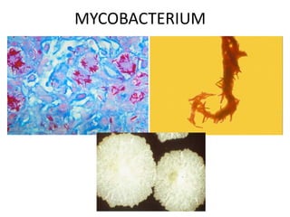
Mycobacteria.pptx
- 2. • Scientific classification • Kingdom: Bacteria • Phylum: Actinobacteria • Class: Actinomycetes • Order: Actinomycetales • Family: Mycobacteriaceae • Genus: Mycobacterium • Species: M. tuberculosis
- 3. • In Greek Mykes means mushroom/fungus, • Fungus like bacteria forming pellicle when grown in liquid medium. General properties: • Non- motile, non-sporing (except M.marinum), • Slender slightly curved or straight, 0.2 to 0.6 × 1 to 10 μm, some species may display branching morphology. • Strictly aerobic (some may grow in reduced oxygen concentration). • Have unusual cell wall as they have high content of lipid (Mycolic acid), N- glycolyl muramic acid in lieu of N-acetyl muramic acid. Because of this, do not stain well with other ordinary stains and are weakly gram positive and Acid Fast. • Relatively slow growth with colonies being visible in 2 to 20 days in optimal temperature. This is because of the hydrophobicity, organisms tend to clump, so that nutrients are not easily allowed into the cell. Generation time may range from approximately 20 hours to 36 hours • Robert Koch isolated M. tuberculosis (Koch’s bacillus) in 1882. • Hansen discovered M.leprae (Hansen’s bacillus) in 1868. • Johne (in 1985 ) discovered M.paratuberculosis (Johne’s bacillus) as a causative agent of Johne’s disease in cattle. M. paratuberculosis has also been suspected as a cause of Crohn’s disease, an illness in human similar to Johne’s disease in cattle
- 4. • The genus Mycobacterium contains over 100 well defined species which includes causative agents of tuberculosis, leprosy, chronic hypertrophic enteritis of cattle (Johne’s disease) and saprophytes.
- 5. Classification 1. Strict pathogens a. Mycobacterium tuberculosis complex (all species cause tuberculosis): Bacteria of M. tuberculosis complex are always pathogenic to human. M. tuberculosis (human type) , M. bovis (bovine type , also infectious to human, including the vaccination strain bacillus Calmette Guerin), M. africanum (human type), M.microti (murine type), M. pinnipedii (primarily infects seals) M.caprie (cattle and human)and M.canetti (an emerging disease in the Horn of Africa, natural reservoir, host range, and mode of transmission of the organism are still unknown). All of these are slow growers and non-pigmented. b. Lepra bacilli: M. leprae (human type), M. microti (murine type) c. Other animal pathogens: M.microti (murine type), M. paratuberculosis (Cattle type)
- 6. 2. Atypical mycobacteria/ MOTT (free living) Group I: Photochromogens Group II: Scotochromogens Group III: Non-chromogens Group IV: Rapid growers 3. Saprophytic mycobacteria (non-pathogenic) M. Smegmatis: present in smegma, thermophiles (grow at 52 degree C) M. phlei: Present in grass M. stercoris: Present in dung M. thermoresistible: tropical evironment
- 7. M. Tuberculosis complex • Initially in 1886, Koch’s bacillus referred to both human and bovine bacillus. But later in 1970, the name M.bovis was specified for bovine bacilli. • Both cause human lesions like pulmonary tuberculosis. • M.bovis is zoonotic and is excreted in milk of cows. • M. tuberculosis and M. bovis are typical tubercle bacilli while M.microti, M. canetti, M.africanum, M.caprae, and M.pinnipedii also belong to this group because all of these species are so closely related as shown by both antigenic and DNA studies, that they all are sometimes regarded as variants of a single species.
- 8. • Species of M. tuberculosis complex can be distinguished on some basis like Niacin accumulation, Oxygen preference, Nitrate reduction, drug sensitivity like Pyrazinamide, Cycloserine etc. Species identification is required for epidemiologic and public health reasons only. 1. Morphology • 0.5 to 10 μ long, slender, straight or slightly curved, non-motile, non- sporing, non-capsulated, arranged singly or in groups , acid fast due to mycolic acid (a long chain fatty acid) in cell wall, weakly gram positive due to presence of high amount of lipids (upto 60% of cell wall). • M. bovis are straighter, stouter and show more uniform staining. 2. Culture • Organisms belonging to this group are considered slow growers (generation time12-24 hours) and colonies are non-pigmented. Visible colony usually appear in 2-3 weeks, optimum temperature 37 degree C, pH 6.4- 7.0. They are fastidious and require enriched media like egg based media, serine based media etc. for growth. • Growth is enhanced by 5- 10%CO2. • Both solid (for routine isolation) and liquid media (for drug sensitivity, biochemical tests, Ag and vaccine preparation) are available.
- 9. A. Liquid media: Bacteria grow faster, Eg. Middlebrook 7H9 ,7H12, 7H13 broth , where growth is enhanced by inclusion of biotin and catalase. B. Solid media: • Egg- based media, eg. Lowenstein-Jensen medium (L J medium), Ogawa media, Petragnani, American Thoracic Society Media, etc. • The basic ingredient is inspissated egg. • Each contains malachite green to suppress the growth of Gram Positive bacteria.
- 10. • Rate of growth and pigmentation are the basis of categorizing mycobacteria. • Rapid growers grow in less than 7 days • Slow growers require more than 7 days • Specimen is inoculated on Lowenstein – Jensen’s medium and incubated at 370C for 2 – 8 weeks • Colonies of Mycobacterium tuberculosis appear as buff coloured, dry, irregular colonies with wrinkled surface and not easily emulsifiable (Buff, rough and tough colonies) • Colonies of Mycobacterium bovis appear creamy white to yellow colour with smooth surface and are easily emulsifiable M.Tuberculosis M.bovis
- 11. Components LJ, modified Ogawaa Ogawa, modified Kudohb Monopotassium dihydrophosphate (KH2PO4), anhydrous (buffer) 2.4 g 6 g 12 g Magnesium sulfate (MgSO4 ·7H2O) (buffer) 0.24 g – Magnesium citrate (selective agent of Mycobacteria) 0.6 g – 0.6 g L-Asparagine (nitrogen source) 3.6 g – Sodium glutamate (nitrogen source) – 6 g 3 g Distilled water up to 600 ml 600 ml 600 ml Glycerol (ml) or pyruvate (carbon source) 12 ml or 7.2 g 36 ml 24 ml Egg homogenate (accelerating factor, protein and fatty acid) 1000 ml 1200 ml 1200 ml Malachite green (2%) (inhibitory to contaminanats pH indicator) 20 ml 36 ml 24 ml pH (about) 6.8 6.8 6.4 a This medium is cheaper than Löwenstein–Jensen because it is made without asparagine. b Recommended media for direct inoculation of alkaline-decontaminated specimens. Various modifications of these media with addition of antimicrobials, antifungal, RNA etc are available to make the medium more selective.
- 12. • Agar-based media: Serum albumin agar media such as Middlebrook 7H10 and 7H11 medium, are prepared from basal medium of defined salts, vitamins, cofactors, glycerol, malachite green and agar combined with enrichment consisting oleic acid, bovine albumin, glucose, etc.
- 13. Difference between M.tuberculosis and M. bovis Characteristics M.Tuberculosis M.bovis Morphology Long, slender, usually curved Short, stout, straight Staining Barred or beaded Uniform Growth on LJ medium Eugonic Dysgonic Presence of glycerol in medium Enhances Inhibits Presence of pyruvate in medium Inhibits Enhances Colony characters Dry, rough, tough, raised and wrinkled, creamy white or buff, difficult to emulsify Moist, smooth, flat, white and friable Biochemical reactions Niacin test + - Nitrate reduction + - Animal pathogenicity In guinea pig + (progressive and fatal disease + (similar to that of M tuberculosis In rabbit - or mild lesion + (generalized lesion)
- 14. 3. Staining Acid fast in ZN stain due to presence of mycolic acid (N- glycolyl muramic acid) and other cell wall superficial lipids which comprise almost 60% of dry weight.
- 15. 4. Cell wall structure and components Mycobacteria have a complex outer envelope having following distinct layers. I. The innermost layer is the plasma membrane, which is a lipid bilayer structure. Two proteins namely phosphatidylinositol mannosides (PIM, a precursor of lipoarabinomannan), and lipoarabinomannan (LAM) are inserted into it. II. Next is the peptidoglycan layer, which determines the shape of the cell and is similar to peptidoglycans of other gram-positive bacteria. It contains repeating units of N-acetylglucosamine-(β1–4)-N- glycolylmuramic acid cross-linked by tetra-peptides bridges. III. About 10 percent of the N-glycolylmuramic acid residues are covalently attached to a branched-chain polysaccharide, arabinogalactan, via phosphodiester bonds. IV. Distinct arabinose residues of the arabinogalactan molecules are esterified to high-molecular-weight mycolic acids. V. Finally, the outer surface of the mycobacterium is formed by the interlink of medium-chain and short-chain lipids (mycosides) , glycolipids, and peptidoglycolipids into the uneven hydrophobic layer of mycolic acids. VI. Proteins (e.g. porins, transport proteins) are found throughout the various layers.
- 16. Fig. diagrammatic representation of mycobacterial cell wall
- 17. 5. Antigens i. Several components of the mycobacterial envelope are strongly immunologically active. Tuberculins (old tuberculin): a crude preparation of 6-8 week culture filtrate of M tuberculosis grown in 5% glycerol media concentrated by evaporation and are highly antigenic. Purified protein derivative, a purified preparation of the active tuberculoprotein by precipitation with 50%ammonium sulphate. ii. LAM: structurally similar to lipopolysachharide layer in Gram negative bacteria iii. Mycosides: heterogeneous group of biologically and immunologically active medium- and short-chain lipids, present on the outer surface. iv. Soluble antigens: These are the antigens found in the supernatant following high speed centrifugation of a lysate of Mycobacteria. These are: cytoplasmic and secreted proteins , and soluble carbohydrates. The sharing of antigens by the various species accounts for the lack of specificity of serological tests for tuberculosis.
- 18. 6. Virulent factors: • No spore, no flagellum, no exotoxin, no endotoxin, no invasive enzyme a. Cell surface glycolipids: Permit intracellular survival of the bacteria. Lipoarabinomannan (LAM) is a lipoglycan and a major virulent factor with following functions. i. It serves as a modulin with immunoregulatory and anti- inflammatory effects. ii. Inhibits T-cell proliferation and macrophage microbiocidal activity. iii. Neutralizes cytotoxic oxygen free radicals produced by macrophages.
- 19. b. Cording factor (glycolipid and peptidoglycolipid derived mycolic acid, trehalose dimycolate present on outer surface): I. Causes bacilli to grow in culture in serpentine cords (parallel arrangements of bacilli) II. Is anti-phagocytic and antigenic in nature. III. Also responsible for inhibition of migration of polymorphological leucocytes and elicits granuloma formation. c. Mutation is common (antibacterial resistance can be easily achieved).
- 20. 4. Biochemical tests i. Niacin accumulation test • Niacin (nicotinic acid) is a precursor in biosynthesis of NAD and NADP. • All species of Mycobacteria produce nicotinic acid. However, most mycobacteria possess the enzyme (transphosphoribolysase) that converts free niacin to niacin ribonucleotide. But 95% of M. tuberculosis lack this enzyme . Due to this, large amount of Niacin accumulates in the culture medium. Niacin is detected by addition of equal volumes of 10% cyanogen bromide and 4% ethanolic aniline in aqueous extract of culture. • Reagent impregnated strips have eliminated the need to handle and dispose of cyanogen bromide which is both caustic and toxic. • Positive reaction – canary yellow • M. tuberculosis – Positive • M. bovis – Negative • Positive Niacin test is also seen with M.chelonei and M.simiae. Note: niacin test may be negative when performed on young culures with few colonies. So it is recommended that the test be done in cultures 3 to 4 weeks old.
- 21. ii. Nitrate reduction test • M. tuberculosis produces an enzyme nitro reductase which reduces nitrate to nitrite • This is detected by colorimetric reaction. •The reaction is detected by inoculating a nitrate broth with a loopful of bacterial colony and alpha-naphthalamine and sulfanilic acid that will react with the released NO2 to produce colour. •Positive reaction – pink or red colour •M. tuberculosis – Positive •M. bovis – Negative •M. kansasii, M. szulgai and M. fortuitum are also positive.
- 22. iii. Resistance to 10µg/ml TCH (Thiophene - 2 - carboxylic acid hydrazide) M. tuberculosis : resistant M. bovis : susceptible M. bovis M. tuberculosis Growth in presence of TCH
- 23. Iv. Arylsulfatase test: This test is positive in culture of atypical bacteria as they form the enzyme arylsulphatase. Arylsulfatase breaks down phenolphthalein disulfate into phenolphthalein (which forms a red color in the presence of sodium bicarbonate) . 3 day arylsulfatase test is used to identify potentially pathogenic rapid growers such as M. fortuitum and M. chelonae. Slow growing M. marinum and M. szulgai are positive in the 14 day arylsulfatase test. M. Tuberculosis complex: Negative v. Catalase test: Most of the atypical mycobacteria are strongly catalase positive and peroxidase negative. M. tuberculosis and M.bovis, however, are peroxidase positive and weakly catalase positive. The reagent is prepared by mixing equal volume of 30% H2O2 and 0.2% catechol in d/w. The reagent is added to 5ml test culture in phosphate buffer (pH7) at 68 degree C in water bath and left for a few minutes. Catalase production is indicated by bubble formation and peroxidase activity is denoted by browning of colonies. vi. Tellurite reduction test: The ability of Mycobacterial species to reduce tellurite in 3 to 4 days is used to distinguish members of M. avium complex from most other non-chromogenic species.
- 24. vii. Tween 80 hydrolysis test: Tween 80 is the trade name of the detergent polyethylene derivative of sorbitan mono-oleate. Some Mycobacterium species posses an enzyme-lipase, that splits the compound and releases oleic acid and polyoxyethylated sorbitol. Positive reactions occur in 1 to 4 days of incubation, and 10 days of incubation are required for confirmation of negative reactions. The color change from orange yellow to red is the positive test. Color change is not due to shift is pH but is due to hydrolysis of Tween 80, which modifies the optical rotation of light passing through the substrate. Result: M. tuberculosis complex : negative M. kansasii : Positive Also useful to differentiate two similar appearing scotochromogens M.gordonae (positive) and M.scrofulaceum(negative). viii. Iron uptake: This is mainly used to identify M.fortuitum which when incubated with 20% ferric ammonium citrate for upto 3 weeks on LJ media will turn into a dark, rusty brown colour. M.tuberculosis complex: negative for iron uptake.
- 25. • Others – Pyrazinamidase: M. tuberculosis : Positive, M. bovis: negative – Urease: M.tuberculosis complex: positive – 5% NaCl tolerance: M.tuberculosis complex: no growth
- 26. 7. Tuberculosis (The disease)
- 27. History: One of the oldest infectious diseases (known at the time of Hippocrates in 400 BC and even in the Hindu Vedas termed rajayakshma), known to have caused more suffering and death than any other infection.. The transmissible nature of TB was established by Villemin in 1868 by inoculating rabbits with tuberculous material from humans and cattle. He also established that scrofula (tuberculous lymphadenitis) and pulmonary TB were manifestations of the same disease process. In 1882, Robert Koch cultivated the bacillus on inspissated serum and transmitted the disease to many animals of different species by inoculation with pure cultures of the bacillus. In addition to cultivating the causative organism, Koch succeeded in staining it by treatment with an alkaline solution of methylene blue for 24 h. Subsequently, Ehrlich improved the technique by using a solution of aniline basic fuchsin followed by decolorization with a mineral acid, and it is this technique, slightly modified by Ziehl and Neelsen whose names it bears, that is still widely used today.
- 28. Epidemiology of disease According to WHO (2015), 10.4 million people got infected and 1.8 million died from TB (including 0.4 million among HIV infected people). Every year, more than eight million new cases occur. Ninety-five percent of cases and 98 percent of deaths occur in low income countries. It is a major cause of death of children, killing at least 500 000 annually. In1993, the WHO took the unprecedented step of declaring TB a ‘global emergency’. Impact of HIV coexistence HIV is the most important factor fueling a TB epidemic. The co- existence of HIV and TB is known to be the most serious threat to the human health and leads to a rapid development of AIDS as explained by the following evidences: • TNF-α and other immunological mediators released in TB lead to transactivation of the HIV provirus and its subsequent replication. • TB causes a CD4+ T-cell lymphopenia, which may synergize with that induced by the HIV.
- 29. Mode of infection • Direct inhalation of aerosolised bacilli contained in the droplet usually of less than 5µm diameter of expectorated sputum. • Patients with sputum that is positive on direct microscopic examination, and thus contains at least 5 000 bacilli/ ml, are the principal sources of infection. • Only 10 % of non-immunocompromised infected people eventually develop active/overt TB, 5 % within the first 2 years following infection and remaining 5% during lifetime. • Infection also occurs infrequently by ingestion for example, through infected milk or by inoculation in an open wound.
- 30. Transmission of M. tuberculosis Millions of tubercle bacilli in lungs (mainly in cavities) Coughing projects droplet nuclei into the air that contain tubercle bacilli. One cough can release 3,000 droplet nuclei. One sneeze can release tens of thousands of droplet nuclei
- 32. Primary tuberculosis • Disease occurring in a person never previously exposed to a tubercle bacillus. • In endemic countries, it usually occurs in young children. • The site of the initial infection is usually the lungs, but sometimes it can be any organ such as tonsil, intestine or skin. The bacilli engulfed by alveolar macrophages, multiply and give rise to a sub-pleural focus of tuberculous inflammation which is commonly located in the lower lobe or lower part of the upper lobe to form the initial lesion or Ghon focus, named after Anton Ghon, an Austrian pathologist. • Some bacilli are carried to the hilar lymphnodes through macrophages, where additional foci of infection develops. The Ghon focus, together with the enlarged hilar lymph-nodes, form the primary complex.
- 33. This occurs for about 3-8 weeks from the time of infection and is associated with the development of tuberculin hypersensitivity. In the majority of the non- immunocompromised cases, most of the cases are asymptomatic and the lesion heals in about 4 months time leaving a calcified nodule. Careful search of lung tissue of adults may show healed primary TB lesion. The bacilli in the lesion slowly die, however, a few bacilli may survive even in the healed lesion and remain latent for decades. • In some people, particularly children under 3 years of age, these foci progress to serious, even fatal, disease principally involving the meninges, kidney, bones and pleurae. Foci developing in the endothelium of major blood vessels may rupture and give rise to widespread small granulomata, a disease termed ‘miliary TB‘ (Latin: milium, a millet seed).
- 35. Post-primary tuberculosis: Post-primary TB develops in previously infected people either as a result of progression of primary TB, endogenous reactivation of latent disease or of exogenous re-infection. Reactivation usually occurs within 5 years after primary infection. The characteristic feature of such disease is extensive tissue necrosis. Very large tumor-like lesions termed tuberculomas develop and, in common with primary lesions, the conditions within them do not favor mycobacterial growth. The necrotic tissue is softened and eventually liquefied by macrophage-derived proteases and, if the lesion erodes , the liquefied contents are discharged and a cavity is formed. In distinct contrast to closed lesions, the well-oxygenated cavities are ideal environments for bacillary replication, and their walls are lined by numerous bacilli. Bacilli escaping from cavities may enter the sputum and be expectorated, thereby infecting other people. They may also spread through the respiratory tract and cause secondary lesions. On the other hand, spread of disease to lymph nodes and more distant organs is uncommon in post-primary disease, probably due to the obliteration of draining lymphatics and capillaries by the tissue necrosis and subsequent deposition of scar tissue. The formation of cavities and localization of the disease process characteristic of post-primary TB are dependent on immune reactivity. Cavity formation is often limited or absent in immunocompromised people, and dissemination of disease to many organs frequently occurs in them.
- 36. Thus, TB in HIV-positive people with relatively limited immunosuppression resembles that in HIV-negative people but, in the more profoundly immunosuppressed, atypical forms of the disease are commonly seen
- 37. Pathogenesis and immunology • Protective immune reactions and tissue necrotising hypersensitivity (Delayed Type) are two aspects of immunlogy in TB. • Both of these reactions are principally cell-mediated, relying on macrophage activation and granuloma formation. • Tubercle bacilli entering the tissues are taken up by macrophages. Entry into macrophages is receptor mediated. Recent results showed that heparin- or fibronectin-binding proteins, present on the bacterial surface, play a role to facilitate their binding to epithelial cells or macrophages. • Once inside the macrophage, the intracellular mycobacteria employ a variety of survival strategies, which include: (1) prevention of an oxidative burst in phagocytosing cells and inhibition of phagosome-lysosome fusion. (2) resistance to lysosomal enzymes and reactive oxygen intermediates (by means of cell-wall lipids, including peptidoglycolipids (mycosides) and LAM and secretion of superoxide dismutase; and (3) escape from the phagosome into the cytoplasm.
- 38. • If the bacilli within macrophages are not destroyed, they replicate and kill the cell. A local area of inflammation is thus established and more phagocytes are attracted to the site by inflammatory mediators and cytokines. • Some bacilli are transported, probably within phagocytes, to the regional lymph nodes, where they are engulfed by antigen-presenting cells (APC). Epitopes from mycobacteria lying within phagosomes within the APC are presented on the cell surface by the major histocompatibility complex (MHC) class II molecules to CD4+ helper T-cells. • Helper T cells now undergo maturation and clonal proliferation into two distinct cells, Th1 and Th2 which secrete cytokines like IFN-gamma, interleukins 1 and 2, TNF-alpha, and others exerting different biological effects. • Th1 dependant cytokines activate macrophages resulting in protective immunity and killing the organisms. • Th2 mediated cytokines induce delayed type hypersensitivity, tissue destruction, called tissue necrotizing hypersensitivity. If the tubercle bacilli proliferate within the APC and escape from the phagosomes, their epitopes are presented to CD8+ T-cells by MHC class I molecules. The CD8+ lymphocyte population contains cytotoxic T-cells that are able to lyse any cell presenting antigen in this manner.
- 39. Macrophage activation and granuloma formation • A single human macrophage, though fully activated, is not capable of killing tubercle bacilli. • Hence aggregation of activated cells called granuloma, which is a characteristic lesion of TB (and other chronic infections too) is formed. All granulomas, regardless of cause, may contain additional cells and matrix which include lymphocytes, neutrophils, eosinophils, multinucleated giant cells (Langhan’s cells), fibroblasts and collagen (fibrosis). • Microscopically, these activated macrophages resemble epithelial cells and are thus termed epithelioid cells. • Fusion of epitheloid cells is called multinucleate giant cell (Langhan’s cell) • Entire granuloma kills bacteria more effectively than single macrophage. The metabolically active macrophages in granuloma consume oxygen and create hypoxia and anoxia leading to tissue necrosis producing cheesy material (caseous material) and the formation of such substance is called caseation. • The anoxic and caseous centre creates an unfavorable environment and many of the bacilli die out.
- 40. IMMUNOPATHOLOGY OF TB M. tuberculosis Macrophage Class II MHC Activated Macrophage (Phagocytosis) Bactericidal activity T–Cell Receptor CD4+ T- Cell CD8+ T- Cell Delayed Hypersensitivity Class I MHC Macrophage Caseous Necrosis
- 41. Tubercle or granuloma formation in tuberculosis ROI: Reactive Oxygen Intermedias RNI: Reative Nitrogen Intermediaes LT α : lymphotoxin α, also known as TNF beta
- 43. When fully developed, tubercle/granuloma consists of 3 zones i. A central area of large, multinucleated giant cells containing tubercle bacilli. ii. A mid zone of pale epitheloid cells, often arranged radially iii. A peripheral zone of fibroblasts, eosinophils, neutrophils, lymphocytes and monocytes Later, peripheral fibrous tissue develops, and the central area undergoes caseation necrosis. It may subsequently heal by fibrosis or calcification
- 44. Characteristics Primary Postprimary Local lesion Small Large Cavity formation Rare Frequent Lymphatic involvement Yes Minimal Infectivity* Uncommon Usual Local spread Uncommon Frequent *Pulmonary cases Differences between primary and post- primary tuberculosis
- 45. Koch’s phenomenon It explains the contrast between primary and secondary tuberculosis. When a normal guinea pig is injected with virulent tubercle bacilli, the injection site heals quickly and there is no immediate visible reaction. But after 10 – 14 days, a nodule appears at the site of injection which ulcerates soon and persists till the animal dies due to pulmonary tuberculosis after a few months. This is an example of progressive tuberculosis. In this period, there is also presence of regional lymphnode enlargement and caseous necrosis. After 4 to 6 weeks of first injection, when the same animal is injected with tubercle bacilli in different site, a dark indurated area of about 1cm diameter appears at the site of injection in 1-2 days which soon undergoes rapid necrosis to form shallow ulcer. This ulcer heals up rapidly without involvement of regional lymph nodes or tissue.
- 46. LAB DIAGNOSIS
- 47. The highest priority for TB control is the identification and cure of infectious cases i.e. patients with sputum smears positive PTB. Therefore, all patients with clinical features suggestive of PTB must submit sputum for diagnostic sputum smear microscopy. Failure to diagnose TB may not only delay appropriate therapy, but may lead to the spread of TB in the community or the health care setting. And the diagnosis of TB is based on the detection of AFB in clinical specimens.
- 48. Specimen: • Must be handled in a safe manner. M. tuberculosis has a low infective dose for humans (infection rate of approx.50% with exposure to <10 bacilli). • Specimens are collected in sterile, leak-proof, disposable, and appropriately labeled containers and placed into bags to contain leakage should it occur. • CDC of US recommends BSL-2 practices for AFB smears and BSL-3 for culture and other tests requiring bacteria propagation. • Specimens should be sent promptly to the laboratory to avoid being overgrown with organisms other than mycobacteria. • If delay is unavoidable, specimens should be stored in the refrigerator. Specimens sent through the post in warm weather should be packed in dry ice or with an ice pack.
- 49. Specimen collection and transport: 1. Pulmonary specimens: May be collected by following methods: Spontaneously produced or induced Sputum, Gastric Lavage, Broncho Alveolar Lavage, Transtracheal Aspiration, Laryngeal Swab. • Sputum: To raise sputum, patients must be instructed to take a deep breath, hold it momentarily, and then cough deeply and vigorously. Ten milliliters of sputum should be collected. Saline-induced sputum specimens is collected from children as young as 5 years of age. • Gastric lavage: Used to collect sputum from patients who may have swallowed sputum during the night. The procedure is limited to senile, non-ambulatory patients, children younger than three years of age, and patients who fail to produce sputum by saline induction.
- 50. 2. Urine specimen: • About 2% to 3% of patients with pulmonary tuberculosis show urinary tract involvement/ renal tuberculosis. • Using clean-catch collection technique, early morning voided urine specimens (40 mL minimum) in sterile containers should be submitted daily for at least 3 days. • The 24-hour urine specimen is undesirable because of excessive dilution, higher contamination, and difficulty in concentrating. 3. Faecal specimen: • In patients with AIDS. • Clinical utility of this practice, remains controversial 4. Tissue and body fluid: CSF for tuberculous meningitis, 5. Blood specimen: Immunocompromised patients particularly HIV can have disseminated tuberculosis. Conventional methods are unacceptable, specialized automated systems are available for growth of Mycobacterium spp., including the Bactec MGIT 960 system (Becton-Dickinson, Franklin Lakes, N.J.), and the BacT/ALERT 3D (Biomerieux,Durham, N.C.).
- 51. Specimen processing • While processing the samples especially for cultures, specimen from the sterile sites can be inoculated directly or after concentration into the media. • But when the sample like sputum that has organic debris, such as mucin, tissue, serum,and other proteinaceous material contaminated with other organisms. has to be cultured for tubercle bacilli, then lab must process such specimen to kill or reduce contaminating bacteria, by a method called decontamination, and also should dissolve the mucin that trap tubercle bacilli by mucolysis/digestion and finally should lead to concentration of the sample. • The digestion-decontamination procedures must be as gentle as possible in order to avoid killing of tubercle bacillus. Various methods are available for this purpose.
- 52. a. Petroff’s method: • Simple and widely used • Sputum mixed with equal volume of 4% NaOH and incubated at 37 C for 30 min. Mixture is then frequently shaken till it gets completely liquefied. NaOH acts as liquefying and also kills contaminating bacteria. The mixture is then centrifuged for 30 min at 3,000rpm, neutralised with 8% HCl in presence of a drop of phenol red indicator. The deposit is used for culture and animal inoculation if needed. b. Homogenization method: • Specimen treated with equal volume of dilute acids such as 6%H2SO4 and then clearing the acids by repeated washing with sterile normal saline.
- 53. c. N-Acetyl-L-Cysteine-Sodium hydroxide method (NALC-NaOH)
- 54. Lab. diagnosis of pulmonary tuberculosis A PTB suspect should submit three sputum samples (1st spot, 2nd early morning, 3rd spot) for microscopy. Nowadays , a two sample concept is being used. 1st early morning sample for microscopy, 2nd spot sample for microscopy/PCR (gene xpert). Secretions build up in the airways overnight. So an early morning sputum sample is more likely to contain TB bacilli than one taken later in the day. Sputum specimen must be free of food particles, residues and other extraneous matter. Saliva and nasal secretions are not to be collected nor should the patient use oral antiseptics during the period of collection
- 55. 1. Sputum smears stained by Z-N stain Advantage: - Cheap – rapid - Easy to perform - Sensitivity > 90% - Specificity of 98% Disadvantages: - Sputum ( need to contain 5000-10000 AFB/ ml.) - Young children, elderly & HIV infected persons may not produce cavities & sputum containing AFB.
- 56. Interpretation of sputum stained by Z N Stain (WHO ) More than 10 bacilli / field ------- +++ From 1 – 10 bacilli / field ------- ++ From 10 – 99 bacilli / 100 fields ----- + From 1 -9 bacilli/100 fields ------ write the number No bacilli seen in 300 fields ---------- negative
- 57. 2. Detecting AFB by fluorochrome stain using fluorescence microscopy: The smear may be stained by auramine-O dye. In this method the TB bacilli are stained yellow against dark background & easily visualized using florescent microscope. Advantages: - More sensitive - Rapid Disadvantages: - Hazards of dye toxicity - More expensive - Must be confirmed by Z-N stain
- 58. a. Cultures on L J media Lowenstein –Jensen medium is an egg based media with addition of salts, glycerol, malachite green & various antibiotics Advantages: - Specificity about 99 %. - More sensitive (lower no. of bacilli 10-100 / ml) - Can differentiate between TB complex & NTM using biochemical reactions. - Source of bacteria for sensitivity tests. Disadvantages: - Slowly growing ( up to 8 weeks ) 3. Culture of M. tuberculosis
- 59. b. Culture on Middlebrook Agar Base 7H10 Media • Defined salts, Vitamins and Cofactors, Oleic acid, Albumin, Catalase, Glycerol, Dextrose, Malachite Green (0.0025g/100 mL) c. Culture on Middlebrook Agar Base 7H11 Medium • Same composition as Middlebrook 7H10 except 0.1% casein hydrolysate added for enhanced recovery of fastidious isoniazid-resistant Mycobacterium tuberculosis • Modified 7H11 contains carbenicillin, amphotericin B, polymixin B, and trimethroprim to inhibit oropharyngeal commensals.
- 60. d. Growth rate and growth at 25°C and 42°C • Tubercle bacilli grow slowly compared to other non- pathogenic acid-fast organisms, taking more than 7 days to appear on culture media. • M. tuberculosis complex do not grow at 25°C and 42°C. • The optimum temperature for the growth of these mycobacteria is 37°C.
- 61. e. Growth on medium containing p-nitrobenzoic acid (PNB) • Para-nitrobenzoic acid has been used for the selective screening of M. tuberculosis. • Human and bovine type of tubercle bacilli can be differentiated from all other mycobacteria in their inability to grow in L-J medium containing PNB.
- 62. 4. Biochemical Tests for the Identification of Mycobacteria
- 63. 5. Recent Methods for Diagnosis a. BACTEC 460 ( rapid radiometric culture system) Specimens are cultured in a liquid medium (Middle brook7H9 broth base )containing C14 – labeled palmitic acid & PANTA antibiotic mixture. Growing mycobacteria utilize the acid, releasing radioactive CO2 which is measured as growth index (GI) in the BACTEC instrument.
- 64. The PANTA antibiotic mixture P ---- Polymyxin B A ---- Amphotericin B N ---- Nalidixic acid T ---- Trimethoprim A ---- Azlocillin The antibiotic mixture inhibits the growth of contaminating bacteria.
- 65. Advantages - Rapid (mycobacteria can be detected within 12 days.) - To determine drug susceptibility . - To differentiate between TB complex & NTM by NAP test. - This method is based on the selective growth inhibition of the TB complex in the presence of p-nitro-α-acetylamino-β- hydroxypropiophenone (NAP). - Specificity is very high Disadvantages: - Expensive - Hazards of using radioactive material.
- 66. b. Mycobacteria Growth Indicator Tube (MGIT) • Tube contains modified Middlebrook 7H9 broth base with OADC enrichment & PANTA antibiotic mixture. • All types of clinical specimens, pulmonary as well as extra- pulmonary can be cultured on this type of media.
- 67. The OADC supplement O: Oleic acid ( Metabolic stimulant) A: Albumin ( to bind toxic free fatty acid ) D: Dextrose (Energy source ) C: Catalase (Destroys toxic peroxides that may be present in the medium).
- 68. Principle: • A fluorescent compound (which is sensitive to O2 eg. an oxygen quenched fluorochrome like Tris 4,7-diphenyl-1,10 phenonthroline ruthenium chloride pentahydrate) is embedded in silicone on the bottom of the tube. • The actively respiring microorganisms consume the oxygen & allow the fluorescence to be observed using UV trans- illuminator lamp.
- 69. MGIT 960 System • The MGIT 960 system is an automated system that uses the MGIT media & sensors to detect the fluorescence. Advantages: The system holds 960 plastic tubes which are continuously monitored. Early detection as the machine monitoring & reading the tubes every hour.
- 70. c. Polymerase Chain Reaction (PCR) Both conventional and real time PCR methods have been devised. Nucleic acid probes & nucleic acid amplification tests in which polymerase enzymes are used to amplify ( make many copies of specific DNA or RNA sequences extracted from mycobacterial cells. Advantages: - Rapid procedure ( 3 – 4 hours) - High sensitivity (1-10 bacilli / ml sputum) Disadvantages: Very expensive. - Require specialist training & equipments. - False positive results. - Can not differentiate between living & dead bacilli.
- 71. d. FASTplaque TB Test -Patient’ s sputum is mixed with myco-bacteriophage. - A virucide (30 mM ferrous ammonium sulfate )is added which destroys any phages outside the TB bacilli. - Lysis of cells & release of phages after replication within the tubercle bacilli. - Non-pathogenic mycobacteria are added & the sample incorporated in agar mixture( over night incubation) - Zones of clearing indicate that patient’ s sputum contained viable M. tuberculosis.
- 72. The MGIT 960 System The MGIT 960 system is a non-radiometeric automated system that uses the MGIT media & sensors to detect the fluorescence. Advantages: -The system holds 960 plastic tubes which are continuously monitored. - Early detection as the machine monitoring & reading the tubes every hour.
- 73. e. Interferon –γ Tests An in vitro T-cell-based assays tests for diagnosis of latent TB infection : These whole-blood assays measure IFN-γ production by previously sensitised lymphocytes in response to M.tuberculosis- specific protein antigens (Early Secreted Antigen Target)ESAT-6 and (Culture Filtrate Protein)CFP-10.
- 74. g. MycoDot antibody test • It is dipstick test, manufactured by Mossman associates. Mycodot uses purified lipoarabinomannans (LAMs) as antigen (highly immunogenic lipopolysaccharide found in the cell wall of all mycobacteria). • Mycodot has been designed for diagnostic use, i.e. it detects anti-mycobacterial antibody levels likely to be found in those with active disease, i.e. high levels. • A mycodot test should therefore only be preformed on those with suspected TB (it is not a screening test). • A positive test indicates active TB.
- 75. h. AccuProbe Acridinium ester-labeled DNA probes are utilized that hybridize to Mycobacterium tuberculosis complex-specific 16S rRNA. Target 16S rRNA released by sonication of organisms from culture. Acridinium ester is chemiluminescent, and DNA probe-16S rRNA hybrids emit light when acridinium ester hydrolyzed to ground state by alkaline peroxidation Chemiluminescence measured in a luminometer Amount of light emitted proportional to amount of DNA-RNA hybrids formed Total time for AccuProbe test is 2 hours
- 76. Antimicrobial susceptibility testing (AST) • Standard conventional diffusion techniques not suitable due to growth rate. • CDC recommends in vitro susceptibility testing on all M. tuberculosis isolates. • Poor clinical outcomes are predicted with an agent when more than 1% of bacilli in a test population are resistant. • Susceptibility testing is used to determine the resistance of strains isolated before the commencement of treatment (initial or primary resistance); or to discover whether resistance has arisen during treatment (secondary resistance).
- 77. • Initial isolates of M.tuberculosis are tested against five antimicrobials, which are referred to as primary (essential) drugs. The five primary drugs are Isoniazid (H), Rifampicin (R), Pyrazinamide (Z), Ethambutol (E), Streptomycin (S).
- 78. • Isoniazid kills 90% of the total population of bacilli during the first few days of treatment. It is most effective against the metabolically active, continuously growing bacilli. RIFAMPICIN can kill the semi-dormant bacilli which isoniazid cannot. PYRAZINAMIDE kills bacilli in an acid environment inside cells, e.g. macrophages.
- 79. • Multidrug-resistant M. tuberculosis (MDR-TB) are those bacilli resistant to both isoniazid and rifampicin as these two drugs are two primary components for the treatment . • Second line drugs are used for treatment of MDR-TB. Second line drugs are ethionamide, cycloserine, kanamycin, capreomycin etc. • Extensively drug resistant (XDR-TB) are those that are not only resistant to rifampicin and isoniazid, but also to some second line drugs.
- 80. Direct vs. Indirect susceptibility testing • Susceptibilities may be performed by either the direct method or the indirect method. • The direct method uses as inoculum, a smear positive concentrate containing more than 50 AFB /100 OIF; the indirect method uses a culture as the inoculum source. • Although, the direct testing provides earlier rapid results, the results may be unsatisfactory because of contamination or low numbers of colony-forming units. • So, direct method is less standardized
- 81. Conventional methods • Different conventional methods used for determination of antimicrobial susceptibility pattern are – resistance ratio – proportion – absolute concentration – disc diffusion methods
- 82. a. Resistance ratio method (RR method) • The RR method is performed either on solid media (egg or Middlebrook agar) or in liquid media (Middlebrook or Dubos broth). • This method compares the resistance (MIC) of unknown strains of tubercle bacilli (test organism) with that of a standard laboratory strain H37Rv, or preferably three wild strains, taking their modal resistance as control.
- 83. b. Proportion method (PR method) • It is a quantitative test, in which several dilutions of standardized inoculum are inoculated onto control (drug free) and drug containing agar medium. • The decrease of growth in drug-containing media and drug-free media are compared. • If growth at the critical concentration of a drug is more than 1%, the isolate is considered clinically resistant. • The critical concentration of a drug is the amount of the drug required to prevent growth above the 1% threshold of the test population of tubercle bacilli.
- 84. c. Absolute concentration method • This method is used in the USA and parts of Europe to determine test strain MICs. The tests are usually performed on Middlebrook’s 7H10 medium.
- 85. d. Disc diffusion method • This method is possible if the mycobacterium is a “rapid grower”. • Agar plates are flood-seeded with the test organism, antibiotic discs are applied.
- 86. Modern methods • Faster, more reliable, easier. a. Bactec® • Radiometric method is commonly used • Uses proportion method principle • Growth indicated by the rate and amount of CO2 produced • Resistance: Growth rate at the critical concentration of a drug is > 1% b. Bactec® MGIT system c. Molecular methods: example, hybridization-based probe assay for detection of rifampicin-resistant gene (rpoB) and its mutations leading to rifampicin resistance. Molecular methods have also been developed for identification of resistance to other drugs. d. Luciferase-reporter mycobacteriophage test: only viable bacteria can become infected, it is an assay based on chemiluminescence.
- 87. Other names of MOTT • Nontuberculous Mycobacteria (NTM), Anonymous, Atypical, Unclassified, Unknown, Tuberculoid, Environmental, Opportunistic NTM are present everywhere in the environment and are also present as normal flora in healthy human skin, GI tract, UR tract etc. Opportunistic pathogens generally in immune suppression. Risk factors for NTM infection are: lowered CMI, COPD 120 different species omnipresent in our environment. Human pathogenicity varies for different species. Infections acquired from an environmental reservoir of organisms. Mycobacteria Other Than Tuberculosis (MOTT)
- 88. There are three typical forms of NTM infection : a tuberculosis-like pattern observed mainly among older men with COPD : presentation with nodules seen in slender older ladies known as the ‘Lady Windermere syndrome’. : hypersensitivity associated with exposure to water-containing systems such as hot tubs or baths. Many patients, however, will have more varied or mixed presentations that do not fit into these prototypic categories Ernst Runyon in 1959, classified MOTT in four groups on the basis of pigment production and growth rate.
- 90. Runyon Classification of MOTT Organisms: Pigment Production Photochromogens (Runyon group I) Produce non-pigmented colonies when grown in the dark and pigmented colonies only after exposure to light. Scotochromogens (Runyon group II) Produce deep yellow to orange pigmented colonies when grown in either the light or dark (some strains show increased pigment production on continuous exposure to light). Nonchromogens (Runyon group III) Non-pigmented in both the light and dark or have only a pale yellow, tan, or buff pigment that does not intensify with light exposure .
- 91. Detection of Pigment Production • Three LJ slants are inoculated with organism. • Two slants are completely shielded from light with cardboard tube or aluminum foil. • When growth is detected in unshielded tube, growth is examined in one of the shielded tubes. • If colonies are not pigmented, then the tube is exposed to light (100-W tungsten bulb for 5 hr) with cap loosened (maximal oxygenation required for pigment production). • Tube is reshielded and incubated for 24 hr. • Colonies in the light-exposed tube and non-exposed tube are compared for pigmentation.
- 92. Runyon Classification of MOTT Organisms: Rate of Growth • Rapid Growers (Rounyon group IV) Mycobacteria forming colonies within 7 days are termed rapid growers, those requiring longer periods are termed slow growers (Runyon group I, II and III). Unlike other mycobacteria, they can be grown on routine bacteriologic media. Determination of growth rate • Inoculate well isolated colony of organism to 7H9 broth containing Tween 80 • Incubate broth for several days until medium is faintly turbid. • Dilute broth 1:100, streak inoculate to Middlebrook 7H10 agar plate. Observe cultures at 5 to 7 days and (if no growth) weekly thereafter for visible colonies
- 94. Group I: Photochromogens • Slow grower (2-3 weeks). Grow at 25oC to 41oC. • Pigment is chemically beta-carotene in nature. • Mycobacterium kansasii: Causes pulmonary disease as well as skin and lymphadenitis. Progression is slow. Detection: yellow pigment, rapid hydrolysis of tween 80, Nitrate reduction is strong, rapid catalase reaction, strong pyrazinamidase activity. Confirmation can be done by AccuProbe test. • M. marinum: Causes granulomatous skin disease ( swimming pool granuloma). Optimal growth at lower temperature 30oC-33oC, Niacin –/+ , nitrate –, Tween 80 + (24 hr) (97%).
- 95. Group II: Scotochromogens • M. scrofulaceum: Causes chronic cervical lymphadenitis (scrofula) in children. Growth at various temperature (25oC, 31oC, 37oC). Tween 80 –/+ (2%), nitrate reduction –/+ (5%), urease –/+ (31%) • M. xenopi: optimal growth at 42oC, Tween 80 + (97%), nitrate reduction –, urease – • M. szulgai: scotochromogen at 37oC, photochromogen at 24oC, Tween 80 –/+ (49%), nitrate reduction +, urease + (72%) • Mycobacterium gordonae: detection by AccuProbe method
- 96. MOTT Organisms: Identificaton of Nonchromogens • Do not form pigment even on exposure to light. • Mycobacterium avium-intercellulare complex (MAI complex): M. intracellulare is closely relatd to M.avium. Pigmentation (-), growth rate at 300 C (+), nitrate reduction (-), Tween 80 hydrolysis(+), urease (-), Niacin (-). Infection by nonchromogens other than M. avium complex is infrequent.
- 97. Differentiation between various species of Mycobacterium
- 98. MOTT Organisms: Identificaton of Rapid Growers • Mycobacterium fortuitum group: arylsulfatase + (97%), nitrate reduction +, iron uptake + • M. chelonae and M. abscessus: arylsulfatase + (95% and 100%), nitrate reduction –/+ (1%) and –, iron uptake – 1Standard reactions: arylsulfatase, nitrate reduction, iron uptake
- 99. Molecular Identification of MOTT Organisms • High performance liquid chromatography (HPLC) analysis of cell wall mycolic acid esters • 16S rRNA gene sequencing
- 100. Treatment of MOTT Surgical treatment Medical treatment: • first-line drugs used for tuberculosis, macrolides, streptomycin • The duration of treatment should cover at least 12 months following conversion to a negative culture. • One example of regimen includes a rifamycin (rifampin or rifabutin), ethambutol, and a macrolide administered for 18– 24 months, including 12 months of sputum culture negativity