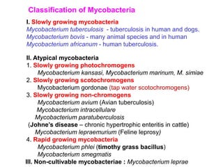
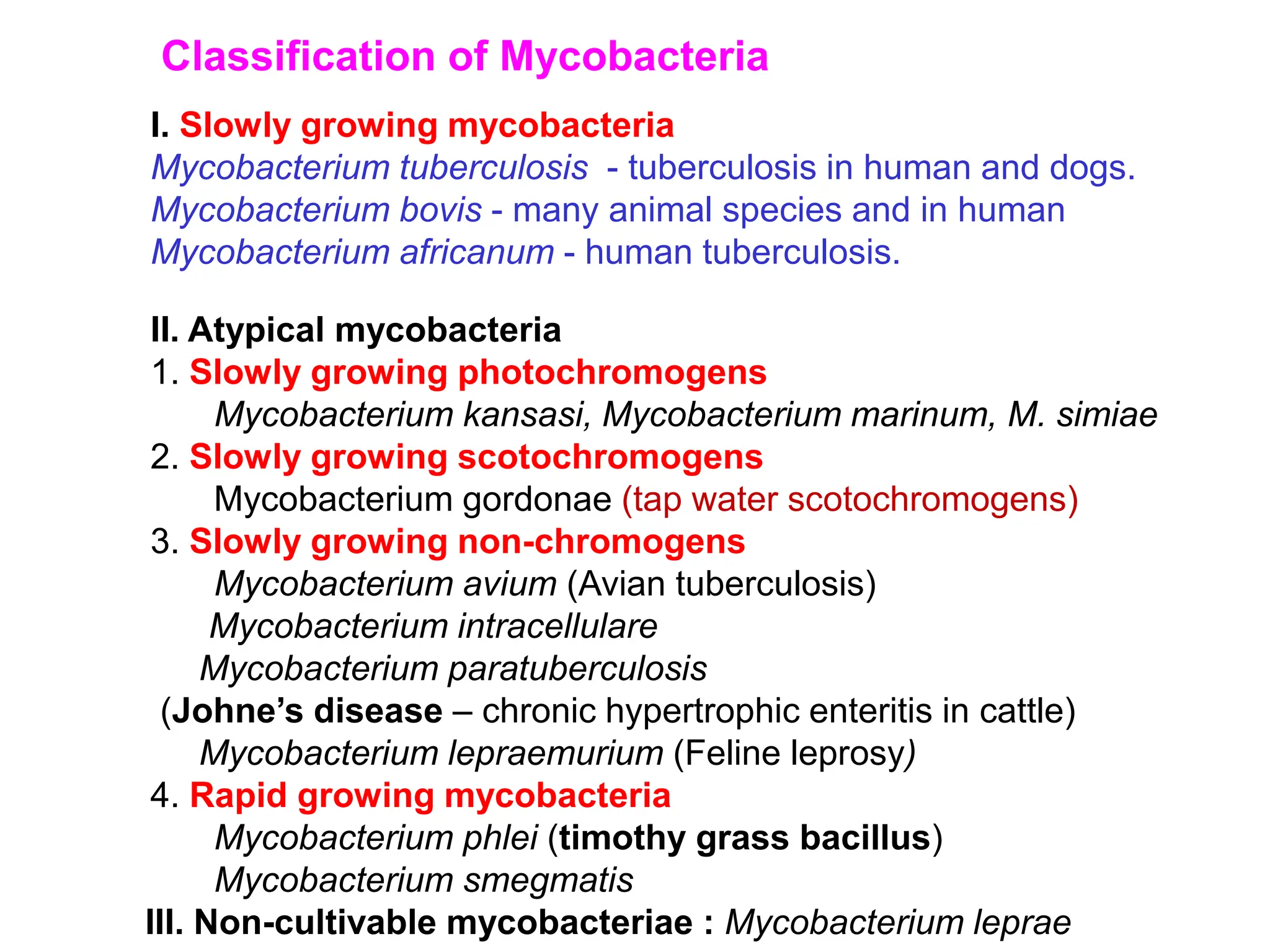
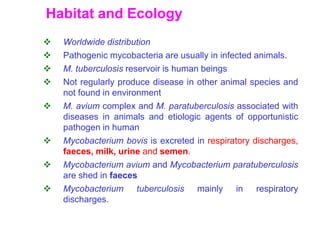
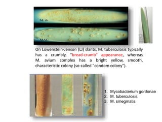
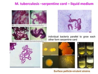
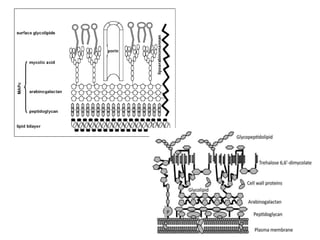
Capitol Tech U Doctoral Presentation - April 2024.pptx
Mycobacterium.ppt bacterial diseases. Tb
1. I. Slowly growing mycobacteria
Mycobacterium tuberculosis - tuberculosis in human and dogs.
Mycobacterium bovis - many animal species and in human
Mycobacterium africanum - human tuberculosis.
Classification of Mycobacteria
II. Atypical mycobacteria
1. Slowly growing photochromogens
Mycobacterium kansasi, Mycobacterium marinum, M. simiae
2. Slowly growing scotochromogens
Mycobacterium gordonae (tap water scotochromogens)
3. Slowly growing non-chromogens
Mycobacterium avium (Avian tuberculosis)
Mycobacterium intracellulare
Mycobacterium paratuberculosis
(Johne’s disease – chronic hypertrophic enteritis in cattle)
Mycobacterium lepraemurium (Feline leprosy)
4. Rapid growing mycobacteria
Mycobacterium phlei (timothy grass bacillus)
Mycobacterium smegmatis
III. Non-cultivable mycobacteriae : Mycobacterium leprae
2. Mycobacteria - History
Lehmann and Neumann (1896) proposed generic name
mycobacterium (fungus bacterium)
First member of this genus - lepra bacillus discovered by
Hansen (1868) – Hansen bacillus.
Koch (1882) isolated the mammalian tubercle bacillus and
proved its causative role in tuberculosis by satisfying
Koch’s postulates.
Acid-fast property of Mycobacterium was discovered by
Ehrlich (1882).
Johne (1895) described Johne’s bacillus - Mycobacterium
paratuberculosis.
4. Habitat and Ecology
Worldwide distribution
Pathogenic mycobacteria are usually in infected animals.
M. tuberculosis reservoir is human beings
Not regularly produce disease in other animal species and
not found in environment
M. avium complex and M. paratuberculosis associated with
diseases in animals and etiologic agents of opportunistic
pathogen in human
Mycobacterium bovis is excreted in respiratory discharges,
faeces, milk, urine and semen.
Mycobacterium avium and Mycobacterium paratuberculosis
are shed in faeces
Mycobacterium tuberculosis mainly in respiratory
discharges.
5. Morphological Characteristics
Acid fast organisms
In tissue straight or slightly curved rod
occurring singly, pairs or in small groups.
On artificial media : coccoid and filamentous forms
Mycobacterium tuberculosis - clinical materials - appear as bundle of
faggots. (serpentine cords in smears from liquid medium)
Mycobacterium kansasi -distinct banded or beaded appearance
Mycobacterium avium - coccoid.
M. paratuberculosis - In clinical materials - appear as bundle of
faggots.
Non-motile, Non-spore farming, Non-capsulated
M. kansasi M. avium M. a. paratuberculosis
6. Cultural characteristics
Slow growth rate, generation time ranges from 14-20hours.
Colonies appear only in about two weeks and sometimes
may be delayed up to 6-8 weeks.
Optimum temperature is 37°C and pH is 6.4 –7.0.
Mycobacterium tuberculosis is an obligate aerobe
Mycobacterium bovis is microaerophilic. Growth is stimulated
by 5-10% CO2.
Susceptible to even traces of toxic substances like fatty acids
in culture media. Toxicity is neutralized by addition of serum,
albumin or charcoal.
Primary isolation : needs complex medium containing egg-
potato base or serum agar base
Colonies : small, dry , scaly
7. Egg based Lowenstein Jensen medium (with potato flour,
glycerol and malachite green) and Stone Brinks medium
(sodium pyruvate instead of glycerol) are most commonly
used.
Malachite green dye (0.025g/100ml) is commonly used as the
selective agent. (inhibitor and pH indicator)
Mycobacterium tuberculosis, Mycobacterium avium and many
of the atypical mycobacteria require glycerol for growth
(eugonic )
Glycerol is inhibitory (dysgonic) to Mycobacterium bovis, while
sodium pyruvate enhances its growth
M. tuberculosis, M. bovis and M .avium are non-chromogenic
Supplementation of media with mycobactin (extracted from
Mycobacteria) is required for M.avium subsp.
paratuberculosis.
Cultural characteristics contd..
8. On Lowenstein-Jenson (LJ) slants, M. tuberculosis typically
has a crumbly, "bread-crumb" appearance, whereas
M. avium complex has a bright yellow, smooth,
characteristic colony (so-called "condom colony").
1. Mycobacterium gordonae
2. M. tuberculosis
3. M. smegmatis
9. M. tuberculosis –serpentine cord – liquid medium
individual bacteria parallel to grow each
other form serpentine card
Surface pellicle-virulent strains
10. Bio-chemical properties
Oxidase positive
Atypical mycobacteria are catalse positive
Tubercle bacilli are peroxidase positive.
Niacin production and nitrate reduction is only by
Mycobacterium tuberculosis.
Urease is reduced by Mycobacterium tuberculosis
and Mycobacterium bovis but not by avian strain
11. Resistance
Resistance to chemical agents due to hydrophobic nature of
cell surface and clumped growth
Resistance to physical influences and will retain their infectivity
in soil and particles of dried faeces for many months.
Not specifically heat resistant; being killed at 60°C in 15-
20mts.
Cultures killed by exposure to direct sunlight for two hours.
Bacilli in sputum may remain alive for 20-30hrs and in droplet
nuclei for 8-10 days and dried sputum for four years
Resistant to disinfectants 5% phenol, 15% H2SO4, 3% nitric
acid, 5% oxalic acid and 4% NaOH
Destroyed by tincture of iodine in 5mts and by 80% ethanol in
2-10mts.
12.
13. Virulence Factors
Do not produce any exotoxins
Surface Mycosides - Peptidoglycolipids or Phenolic
glycolipids- responsible for the control of cellular permeability,
resistance to action of water-soluble enzymes, antibiotics and
disinfectants. Determine colony characters, serologic
specificities and bacteriophage susceptibility.
Subsurface layers – long chain branched mycolic acids.
Linked to peptidoglycan by arabinogalactans – responsible for
acidfastness
Cord factor (Trehalose –6,6’ dimycolate) and Wax D- inhibits
chemotaxis, leukotoxic, responsible for delayed
hypersensitivity
Sulfatides- sulfur containing glycolipids –inhibits
phagolysosome formation and interfere with reactive oxygen
intermediates.
Mycosides, phospholipids and sulpholipids – ensuring
bacterial survival within macrophages.
Ahp – alkyl hydroperoxidase reductase- resistance to
superoxides and nitrogen intermediates
14. Pathogenesis
Inhalation and ingestion.
Mucociliary clearance by mucus and epithelial cilia in the upper
respiratory tract provides defense against infection.
Microorganisms on small particles (1-4 μm in size), such as, dust and
water droplets reach alveolar spaces.
Infected host cells with mycobacteria can reach local lymphnodes
and from there may pass to the thoracic duct with general
dissemination.
After 10-14 days, CMI responses develop and activated
macrophages are able to kill some mycobacteria.
Aggregation of ACTIVATED macrophages contributes to the
formation of a tubercle, and a fibrous layer may encompass the
lesion.
Caseous necrosis due to the cell death and tissue destruction occurs
at the center of the lesion and this may proceed to calcification or
liquefaction.
Once CMI is established, the lymphatic spread is retarded but occurs
via the erosion of bronchi or blood vessels to new area.
Haematogenous spread may produce milliary tuberculosis (in deer).
This involves multifocal tubercle formation in an organ.
15. Tubercle
Avascular granuloma composed of
a caseous necrosis in a central area
encircled by a zone of epitheloid cells,
a peripheral zone of lymphocytes, granulocytes and
fibroblasts. –multinucleated giant cells
Calcification may be present in the necrotic centers.
An outer boundary of fibrous tissue is usually present
between the lesions and normal tissue.
16.
17.
18.
19.
20. Diagnosis of tuberculosis
Microscopical examination
X-ray
Intra dermal caudal fold tuberculin test
Cultures morphology, growth characteristics in
media, biochemical characteristics
Histopathology
ELISA
Polymerase chain amplification (PCR)
Whole-blood gamma interferon (IFN- ) assay
Animal pathogenicity in guinea pigs, rabbits and
chicken.
Inoculated
suspected
material
Mycobacteriu
m tuberculosis
Mycobacterium
bovis
Mycobacterium
avium
Rabbits (I/V) + ++ ++
Guinea pig (S/C) ++ ++ -
Chicken (I/V) - - ++
21.
22.
23. Treatment and control
First effective treatment for TB was developed in
the 1940s – streptomycin
extensive time periods (e.g. 9 months)
organism grows slowly, or dormant
two or more antibiotics
e.g. rifampin, isoniazid and ethambutol
antibiotic resistance minimized
Control
Tuberculin test and slaughter. Not yet eradicated.
Depopulate extensively affected herds.
Disinfect premises with cresylic compounds or
sodium orthophenylphenate.
BCG vaccine (Bacillus of Calmette and Guerin)
effective when given before exposure.
24. • Initial intensive phase - eight weeks of the drugs Isoniazid
(H), Rifampicin (R), Pyrazinamide (Z) and Ethambutol (E).
• continuation phase - three drugs Isoniazid, Rifampicin and
Ethambutol given for another sixteen weeks.
• 2HREZ/4HRE ( Dose : four weight band categories).
• A fixed dose combination (FDC) is when two or more
drugs are combined together in a single pill or tablet.
• Fixed dose combinations of four drugs (Isoniazid,
Rifampicin, Pyrazinamide and Ethambutol), three drugs
(Isoniazid, Rifampicin and Ethambutol) and two drugs
(Isoniazid and Rifampicin) are available - on a daily basis
MDR TB in India is a 6-drug regimen, with an intensive
phase of 6–9 months and a continuation phase of 18
months; the total duration of treatment is about 24–27
months. - kanamycin, levofloxacin, cycloserine,
ethionamide, pyrazinamide, and ethambutol
25. PARATUBERCULOSIS (JOHNE’S DISEASE)
• Mycobacterium avium subsp paratuberculosis
• Crohn’s disease -human
• Herrold’s egg yolk medium -mycobactin - Killed extracts of
M.phelei
• 370C for upto 16 weeks - greyish white colonies
• organism shed in the faeces, milk and semen of infected
animals
• Ingested - engulfed by macrophages - survive and replicate
initially in Peyer’s patches. As the disease progress, an immune
mediated granulomatous reaction develops in the lamina
propria and submucousa
• Diarrhoea- initially intermittent, dark and semisolid. Then
persistent and profuse. Progressive weight loss, emaciation
and eventually death
26. • Mucousa of the terminal small intestine and the
large intestine is usually thickened and folded into
transverse corrugation
• mesentric and ileocaecal lymphnodes are
enlarged and odematous
• live animals - scrapings or pinch biopsies from the
rectum
• Johnin test – i/v or i/d