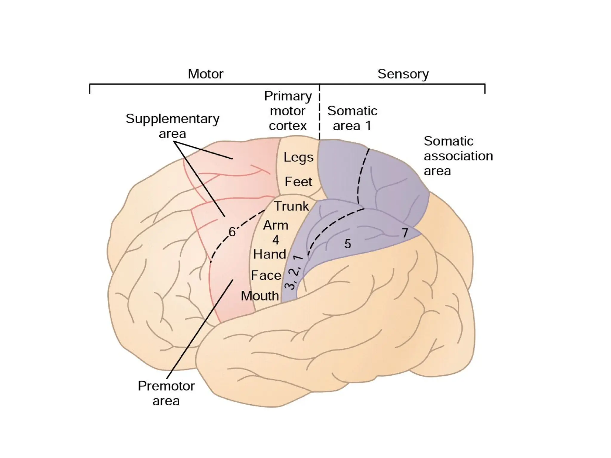
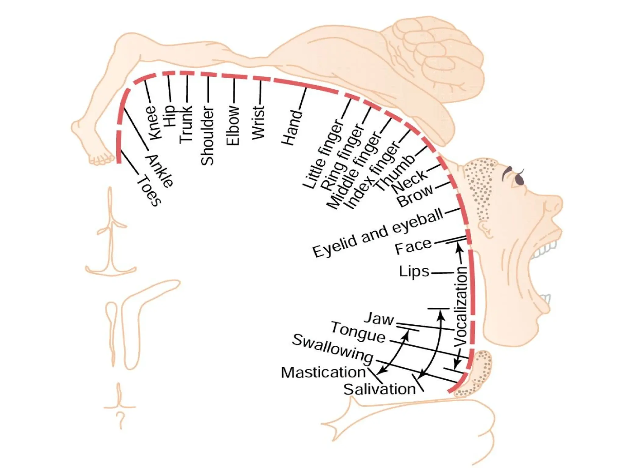
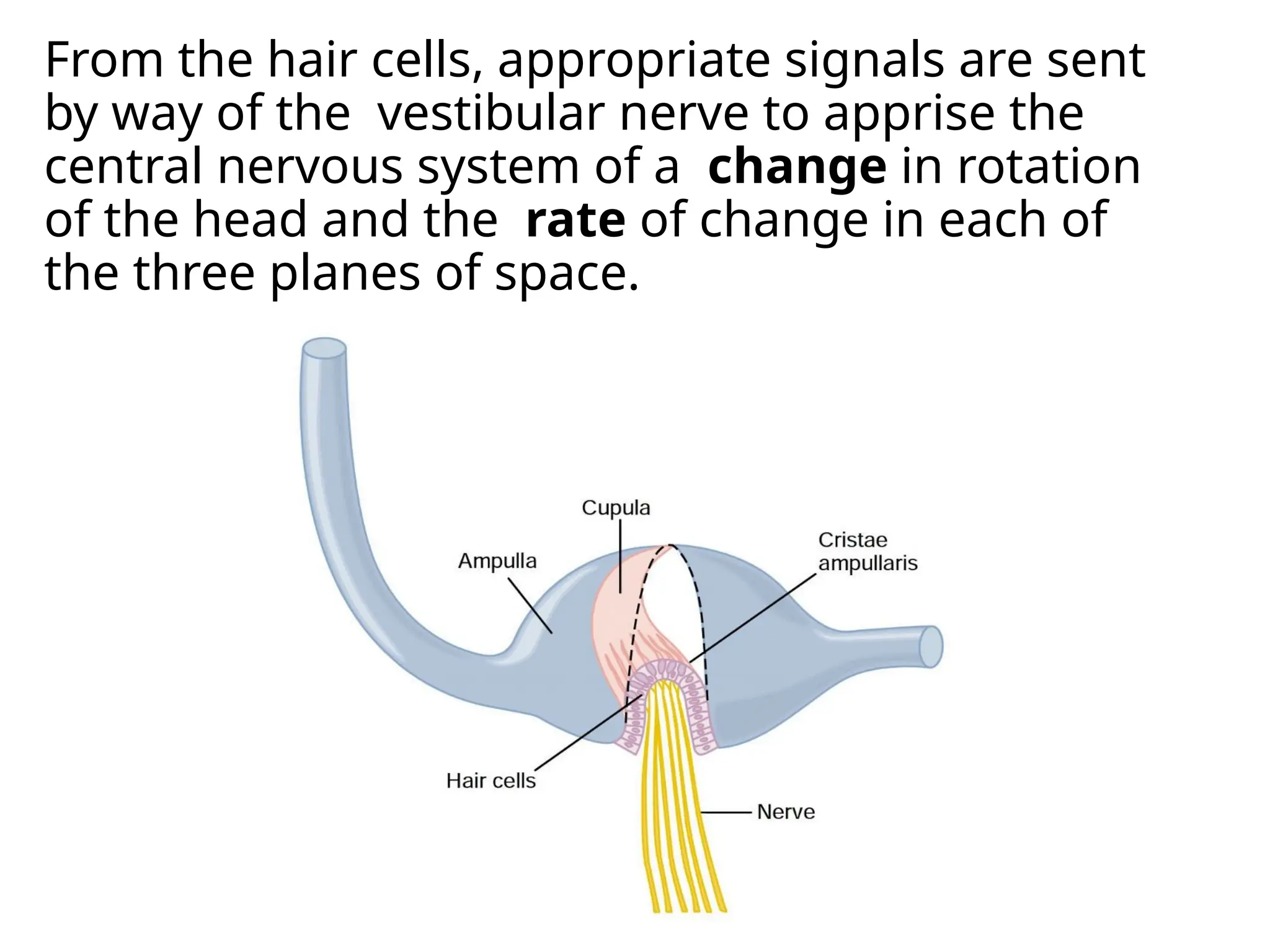
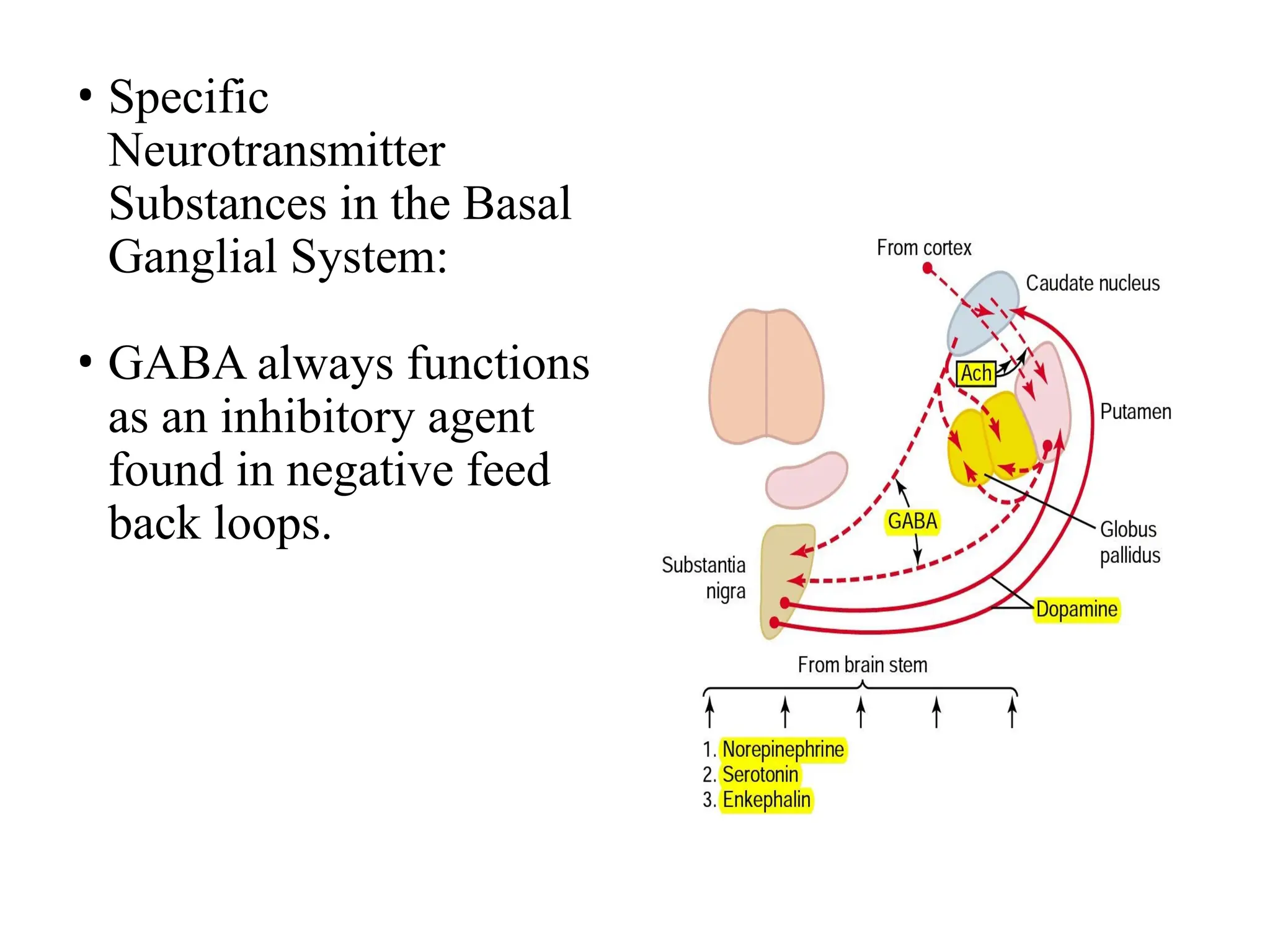
Cortical and Brain Stem
Control of Motor Function
Most “voluntary” movements initiated by the
cerebral cortex are achieved when the cortex activates
“patterns” of function stored in lower brain areas—the
cord,brain stem,basal ganglia, and cerebellum. These
lower centers, in turn, send specific control signals to
the muscles.
MOTOR CORTEX
Anterior to the central cortical sulcus, occupying
approximately the posterior one third of the frontal
lobes.
Divided into:
(1)the primary motor cortex
(2) the premotor area
(3) the supplementary motor area.
Primary motor cortex
 Lies in the first convolution of the frontal lobes
anterior to the central sulcus. It begins laterally in the
sylvian f issure, spreads superiorly to the uppermost
portion of the brain, and then dips deep into the
longitudinal fissure.
 More than one half of the entire primary motor cortex
is concerned with controlling the muscles of the hands
and the muscles of speech.
 Excitation of a single motor cortex neuron usually
excites a specific movement rather than one specific
muscle.
Premotor Area
• Nerve signals generated in the premotor area cause much
more complex “patterns” of movement.
• The anterior part of the premotor area first develops a
“motor image” of the total muscle movement that is to be
performed.Then, in the posterior premotor cortex, this
image excites each successive pattern of muscle activity
required to achieve the image.
• This posterior part of the premotor cortex sends its
signals either directly to the primary motor cortex to
excite specific muscles or, often, by way of the basal
ganglia and thalamus back to the primary motor cortex.
Supplementary Motor Area
 It lies mainly in the longitudinal fissure but extends a
few centimeters onto the superior frontal cortex.
 Contractions elicited by stimulating this area are
often bilateral.
 This area functions in concert with the premotor area
to provide body-wide attitudinal movements,fixation
movements of the different segments of the body,
positional movements of the head and eyes,and so
forth.
Specialized Areas of Motor Control
Broca’s Area and Speech
- “word formation” area.
-Damage to it does not prevent a person from
vocalizing, but it does make it impossible for the person to
speak whole words .
-closely associated cortical area also causes appropriate
respiratory function, so that respiratory activation of the
vocal cords can occur simultaneously with the movements of
the mouth and tongue during speech.
Voluntary Eye Movement Field.
-Damage to this area prevents a person from voluntarily
moving the eyes toward different objects. Instead, the eyes
tend to lock involuntarily onto specific objects.
-This area also controls eyelid movements such as blinking.
Head Rotation.
•This area is closely associated with the eye movement
field; it directs the head toward different objects.
Area for Hand Skills
•Destruction in this area, hand movements become
uncoordinated and nonpurposeful, a condition called
motor apraxia.
Transmission of Signals from the Motor Cortex to the
Muscles :
•Motor signals are transmitted directly from the cortex
to the spinal cord through the corticospinal tract and
indirectly through multiple accessory pathways that
involve the basal ganglia, cerebellum, and various
nuclei of the brain stem.
Corticospinal (Pyramidal) Tract
 The most important output pathway from the motor
cortex .
 It’s fibers originate from giant pyramidal cells, called
Betz cells , these fibers are large myelinated fibers
with a diameter of 16 micrometers.
 The Betz cells are about 60 micrometers in diameter,
and their fibers transmit nerve impulses to the spinal
cord at a velocity of about 70 m/sec, the most rapid
rate of transmission of any signals from the brain to
the cord.
Red nucleus
• Red Nucleus Serves as an alternative pathway for
transmitting cortical signals to the spinal cord
• Red nucleus, located in the mesencephalon, functions
in close association with the corticospinal tract called
“corticorubral tract”
• Fineness of representation of the different muscles is
far less developed than in the motor cortex.
• The corticospinal and rubrospinal tracts together are
called the lateral motor system of the cord.
Extrapyramidal system
• Extrapyramidal motor system is all those portions of
the brain and brain stem that contribute to motor
control but are not part of the direct corticospinal-
pyramidal system.
• These include pathways through the basal ganglia,
the reticular formation of the brain stem, the
vestibular nuclei, and often the red nuclei.
 The cells of the motor cortex are organized in
vertical columns, with thousands of neurons in
each column.
 Each column of cells functions as a unit, usually
stimulating a group of synergistic muscles, but
sometimes stimulating just a single muscle.
 Each column has six distinct layers of cells.
 The pyramidal cells that give rise to the
corticospinal fibers all lie in the fifth layer of
cells from the cortical surface.
 The input signals all enter by way of layers 2
through 4. And the sixth layer gives rise mainly
to fibers that communicate with other regions
of the cerebral cortex itself.
• Each column of cells excites two populations of
pyramidal cell neurons:
1-dynamic neurons : exicted for short
period ,initial rapid development of force.
2-static neurons: fire at a much slower rate, to
maintain the force of contraction as long as the
contraction is required.
• The neurons of the red nucleus have similar
dynamic and static characteristics, except that a
greater percentage of dynamic neurons in it.
• When nerve signals from the motor cortex cause a
muscle to contract, somatosensory signals return all
the way from the activated region of the body to the
neurons in the motor cortex that are initiating the
action.Most of these somatosensory signals arise in :
(1) the muscle spindles
(2) the tendon organs of the muscle tendons
(3) the tactile receptors of the skin overlying the
muscles.
These somatic signals often cause positive feedback
enhancement of the muscle contraction.
 Removal of the Primary Motor Cortex
(Area Pyramidalis)
• Removal of a portion of the primary motor cortex
—area containing the giant Betz pyramidal cells—
causes varying degrees of paralysis of the
represented muscles.
• If the sublying caudate nucleus and adjacent
premotor and supplementary motor are affected
loss of voluntary control of discrete movements of
the distal segments of the limbs, especially of the
hands and fingers.
• The ability to control the fine movements is gone.
• Muscle Spasticity Caused by Lesions That
Damage Large Areas Adjacent to the Motor
Cortex.
• The primary motor cortex normally exerts a
continual tonic stimulatory effect on the motor
neurons of the spinal cord; when this stimulatory
effect is removed, hypotonia results.
• Most lesions of the motor cortex, especially
those caused by a stroke, involve not only the
primary motor cortex but also adjacent parts of
the brain such as the basal ganglia, muscle
spasm occurs on the opposite side, because
inhibitory pathways are affected.
Role of the Brain Stem in Controlling Motor Function
•Brain stem control motor and sensory functions
for the face and head regions , also:
1-Control of respiration
2. Control of the cardiovascular system
3. Partial control of gastrointestinal function
4. Control of many stereotyped movements of the
body
5. Control of equilibrium
6. Control of eye movements
• Reticular nuclei and Vestibular nuclei are
present in the brain stem, they control whole-
body movement and equilibrium.
Excitatory-Inhibitory Antagonism Between
Pontine and Medullary Reticular Nuclei
The reticular nuclei are divided into:
(1) pontine reticular nuclei, located slightly
posteriorly and laterally .
(2) medullary reticular nuclei.
 These two sets of nuclei function mainly
antagonistically to each other,with the pontine
exciting the antigravity muscles and the
medullary relaxing these same muscles.
 when the pontine reticular excitatory system is
unopposed by the medullary reticular system,
it causes powerful excitation of antigravity
muscles.
• The specific role of the vestibular nuclei is to
selectively control the excitatory signals to the
different antigravity muscles to maintain
equilibrium in response to signals from the
vestibular apparatus.
• This control is done by lateral and medial
vestibulospinal tracts.
Maculae
 Sensory Organs of the Utricle and Saccule for
detecting orientation of the head with respect to
gravity.
 The macula of the utricle lies in the horizontal
plane on the inferior surface of the utricle and plays
an important role in determining orientation of the
head when the head is upright.
 Conversely, the macula of the saccule is located
mainly in a vertical plane and signals head
orientation when the person is lying down.
• The calcified statoconia have a specific gravity two
to three times the specific gravity of the
surrounding fluid and tissues.The weight of the
statoconia bends the cilia in the direction of
gravitational pull.
 In each macula, each of the hair cells is oriented in
a different direction so that some of the hair cells
are stimulated when the head bends forward,
some are stimulated when it bends backward,
others are stimulated when it bends to one side,
and so forth.
 Therefore, a different pattern of excitation occurs
in the macular nerve fibers for each orientation of
the head. It is this “pattern” that apprises the brain
of the head’s orientation in space.
Semicircular Ducts.
 When a person’s head begins to rotate in any
direction, the inertia of the fluid in one or
more of the semicircular ducts causes the fluid
to remain stationary while the semicircular
duct rotates with the head. This causes fluid to
flow from the duct and through the ampulla,
bending the cupula to one side.
 Rotation of the head in the opposite direction
causes the cupula to bend to the opposite
side.
 Into the cupula are projected hundreds of cilia
from hair cells located on the ampullary crest.
From the hair cells, appropriate signals are sent
by way of the vestibular nerve to apprise the
central nervous system of a change in rotation
of the head and the rate of change in each of
the three planes of space.
Detection of Head Rotation by the
Semicircular Ducts
The semicircular duct transmits a signal of one
polarity when the head begins to rotate and of
opposite polarity when it stops rotating.
The semicircular duct mechanism predicts that
dysequilibrium is going to occur and thereby
causes the equilibrium centers to make
appropriate anticipatory preventive
adjustments.
“anticipatory correction”.
Other Factors Concerned with
Equilibrium
1-Neck Proprioceptors
joint receptors of the neck:When the head is leaned in one
direction by bending the neck, impulses from the neck
proprioceptors keep the signals originating in the vestibular
apparatus for giving the person a sense of dysequilibrium.
2-Proprioceptive Information from Other Parts of the Body
Ex .pressure sensations from the footpads tell one
(1) whether weight is distributed equally between the two feet
and (2) whether weight on the feet is more forward or backward.
3- A person can still use the visual mechanisms for maintaining
equilibrium.
• The primary pathway for the equilibrium reflexes begins
in the vestibular nerves, where the nerves are excited by
the vestibular apparatus then passes to the vestibular
nuclei and cerebellum. Next, signals are sent into the
reticular nuclei of the brain stem, and down the spinal
cord by way of the vestibulospinal and reticulospinal
tracts. The signals to the cord control the interplay
between facilitation and inhibition of the many antigravity
muscles, thus automatically controlling equilibrium.
• The flocculonodular lobes of the cerebellum are
especially concerned with dynamic equilibrium signals
from the semicircular ducts.
Functions of Brain Stem Nuclei in Controlling
Subconscious, Stereotyped Movements
• Many of the stereotyped motor functions of
the human being are integrated in the brain
stem.
• Example : anencephaly (baby born without
brain structure), they can still cry, yown,
suckling, stretch, move their eye to follow
objects.
• cerebellum and the basal ganglia always function in association with
other systems of motor control.
• Basically, the cerebellum plays major roles in :
• 1-Timing of motor activities and in rapid, smooth progression from one
muscle movement to the next.
• 2- controls intensity of muscle contraction when the muscle load changes.
• 3- controls necessary interplay between agonist and antagonist muscle
groups.
• The basal ganglia help to plan and control complex patterns of muscle
movement.
Contributions of the Cerebellum and
Basal Ganglia to Overall Motor Control
• In the vermis, most cerebellar control functions
for muscle movements of the axial body, neck,
shoulders, and hips are located.
• Cerebellar hemisphere is divided into an
intermediate zone and a lateral zone.
• The intermediate zone of the hemisphere is
concerned with controlling muscle contractions in
the distal portions of the upper and lower limbs.
• Without this lateral zone, most discrete motor
activities of the body lose their appropriate
timing and sequence.
Afferent Pathways from Other Parts of the Brain.
The dorsal spinocerebellar tract and the ventral
spinocerebellar tract.
The signals transmitted in the dorsal spinocerebellar tracts
come mainly from the muscle spindles about muscle
contraction, tension, positions and rates of movement of
the parts of the body, and forces acting on the surfaces
of the body.
the ventral spinocerebellar are excited mainly by motor
signals arriving in the anterior horns of the spinal cord
from the brain and the spinal cord itself.
Deep Cerebellar Nuclei and the Efferent Pathways.
• Located deep in the cerebellar mass on each side
are three deep cerebellar nuclei—the dentate,
interposed, and fastigial.
• Three major layers of the cerebellar cortex are
shown: the molecular layer, Purkinje cell layer, and
granule cell layer.
• Functional Unit of the Cerebellar Cortex is the
Purkinje Cell and the Deep Nuclear Cell.
• The afferent inputs to the cerebellum are
mainly of two types :
A. the climbing fiber type originate from the
inferior olives of the medulla, produce action
potential called the complex spike.
• B. the mossy fiber type originate from the
higher brain, brain stem and spinal cord.
Action potenial produce is called simple spike
(weaker).
• Feedback inhibitory signals from the Purkinje
cell circuit arrive.
• Basket cells and stellate cells are inhibitory
cells with short axons cause lateral inhibition
of adjacent Purkinje cells, thus sharpening the
signals.
Function of the Cerebellum in Motor
Control
• cerebellum coordinate motor control functions
at three levels:
• 1.Vestibulocerebellum
• Equilibrium is far more disturbed during
performance of rapid motions.
• It is presumed that information from both the
body periphery and the vestibular apparatus is
used in a typical feedback control circuit to
provide anticipatory correction of equilibrium.
2.Spinocerebellum
• This part of the cerebellar motor control
system provides smooth, coordinate
movements of the agonist and antagonist
muscles of the distal limbs for performing
acute patterned movements.
• 3.Cerebrocerebellum
• lateral cerebellar zones communicate with the premotor area
and primary motor and associated somatosensory area.
• concerned with two other important but indirect aspects of
motor control:
• (1) the planning of sequential movements:
• Lateral cerebellar zones are concered with what will be
happening during the next sequential movement a fraction of
a second or perhaps even seconds later.
(2) the “timing” of the sequential movements:
• lesions in the lateral zones cause failure of
smooth progression of movements.
Clinical Abnormalities of the
Cerebellum
• 1-Dysmetria: movements ordinarily overshoot
their intended mark then the conscious portion
of the brain overcompensates in the opposite
direction,results in uncoordinated movements
that are called ataxia.
• Caused by cerebellar disease or lesions in the
spinocerebellar tracts.
• 2-Past pointing means that in the absence of the
cerebellum, a person ordinarily moves the hand
or some other moving part of the body
considerably beyond the point of intention.
• 3-Dysdiadochokinesia: failing to predict where the
different parts of the body will be at a given time, it
“loses” perception of the parts during rapid motor
movements.
• Dysarthria:failure of progression during talking.
• 4-intention tremor or an action tremor: results from
cerebellar overshooting and failure of the cerebellar
system to “damp” the motor movements.
• example: Cerebellar nystagmus is tremor of the
eyeballs that occurs usually when one attempts to
fixate the eyes on a scene to one side of the head.
5-hypotonia :
• Results from loss of cerebellar facilitation of
the motor cortex and brain stem motor nuclei
by tonic signals from the deep cerebellar
nuclei.
• Decreased tone of the peripheral body
musculature on the side of the cerebellar
lesion.
Basal ganglia
• Found on each side of the brain, these ganglia
consist of the caudate nucleus, putamen,
globus pallidus, substantia nigra, and
subthalamic nucleus.
• They are located mainly lateral to and
surrounding the thalamus.
• Nerve fibers connecting the cerebral cortex
and spinal cord pass through the space that
lies between the major masses of the basal
ganglia, the caudate nucleus and the
putamen. This space is called the internal
capsule of the brain.
• two major circuits,the putamen circuit and the
caudate circuit.
• The putamen circuit has its inputs mainly from
those parts of the brain adjacent to the
primary motor cortex
• circuits pass from putamen through the
external globus pallidus,the subthalamus, and
the substantia nigra—finally returning to the
motor cortex by way of the thalamus.
• The caudate nucleus plays a major role in
cognitive control of motor activity.
• After the signals pass from the cerebral
cortex to the caudate nucleus, they are next
transmitted to the internal globus pallidus,
then to nuclei of the ventroanterior and
ventrolateral thalamus, and finally back to the
prefrontal, premotor, and supplementary
motor areas of the cerebral cortex.
• In patients with severe lesions of the basal
ganglia, the timing of functions are poor; or
sometimes nonexistent.
• Because the caudate circuit of the basal
ganglial system functions mainly with
association areas of the cerebral cortex such
as the posterior parietal cortex, presumably
the timing and scaling of movements are
functions of this caudate cognitive motor
control circuit.
• Specific
Neurotransmitter
Substances in the Basal
Ganglial System:
• GABA always functions
as an inhibitory agent
found in negative feed
back loops.
• Clinical Syndromes Resulting from damage to the Basal
Ganglia
• 1-Parkinson’s Disease
• known also as paralysis agitans.
• results from widespread destruction of that portion of the
substantia nigra (the pars compacta) that sends dopamine-
secreting nerve fibers to the caudate nucleus and putamen.
• The disease is characterized by:
• (1) rigidity of much of the musculature of the body.
• (2) involuntary tremor of the involved areas even when the
person is resting at a fixed rate of 3 to 6 cycles per second.
• (3) serious difficulty in initiating movement, called akinesia.
Treatment
A. Treatment with l-Dopa:
• L-Dopa passes through blood brain barrier.
• Ameliorates many of the symptoms, especially the
rigidity and akinesia.
• L-dopa is converted in the brain into dopamine, and
the dopamine then restores the normal balance
between inhibition and excitation in the caudate
nucleus and putamen.
B.Treatment with l-Deprenyl:
• This drug inhibits monoamine oxidase, which is
responsible for destruction of most of the dopamine .
• Used in combination with l-Dopa.
c.Treatment with Transplanted Fetal Dopamine Cells.
d.Treatment by Destroying Part of the Feedback Circuitry
in the Basal Ganglia.
2-Huntington’s Disease (Huntington’s Chorea):
• Hereditary disorder that usually begins causing
symptoms at age 30 to 40 years.
• It is characterized at first by flicking movements in
individual muscles and then progressive severe
distortional movements of the entire body.
• The cause is loss of most of the cell bodies of the GABA-
secreting neurons in the caudate nucleus and putamen
and of acetylcholine-secreting neurons in many parts of
the brain(which causes dementia).
Thank you

neurophysiology II. a concise review of brain and spinal cord

  • 1.
    Cortical and BrainStem Control of Motor Function
  • 2.
    Most “voluntary” movementsinitiated by the cerebral cortex are achieved when the cortex activates “patterns” of function stored in lower brain areas—the cord,brain stem,basal ganglia, and cerebellum. These lower centers, in turn, send specific control signals to the muscles.
  • 3.
    MOTOR CORTEX Anterior tothe central cortical sulcus, occupying approximately the posterior one third of the frontal lobes. Divided into: (1)the primary motor cortex (2) the premotor area (3) the supplementary motor area.
  • 6.
    Primary motor cortex Lies in the first convolution of the frontal lobes anterior to the central sulcus. It begins laterally in the sylvian f issure, spreads superiorly to the uppermost portion of the brain, and then dips deep into the longitudinal fissure.  More than one half of the entire primary motor cortex is concerned with controlling the muscles of the hands and the muscles of speech.  Excitation of a single motor cortex neuron usually excites a specific movement rather than one specific muscle.
  • 7.
    Premotor Area • Nervesignals generated in the premotor area cause much more complex “patterns” of movement. • The anterior part of the premotor area first develops a “motor image” of the total muscle movement that is to be performed.Then, in the posterior premotor cortex, this image excites each successive pattern of muscle activity required to achieve the image. • This posterior part of the premotor cortex sends its signals either directly to the primary motor cortex to excite specific muscles or, often, by way of the basal ganglia and thalamus back to the primary motor cortex.
  • 8.
    Supplementary Motor Area It lies mainly in the longitudinal fissure but extends a few centimeters onto the superior frontal cortex.  Contractions elicited by stimulating this area are often bilateral.  This area functions in concert with the premotor area to provide body-wide attitudinal movements,fixation movements of the different segments of the body, positional movements of the head and eyes,and so forth.
  • 9.
    Specialized Areas ofMotor Control Broca’s Area and Speech - “word formation” area. -Damage to it does not prevent a person from vocalizing, but it does make it impossible for the person to speak whole words . -closely associated cortical area also causes appropriate respiratory function, so that respiratory activation of the vocal cords can occur simultaneously with the movements of the mouth and tongue during speech.
  • 11.
    Voluntary Eye MovementField. -Damage to this area prevents a person from voluntarily moving the eyes toward different objects. Instead, the eyes tend to lock involuntarily onto specific objects. -This area also controls eyelid movements such as blinking.
  • 12.
    Head Rotation. •This areais closely associated with the eye movement field; it directs the head toward different objects. Area for Hand Skills •Destruction in this area, hand movements become uncoordinated and nonpurposeful, a condition called motor apraxia.
  • 13.
    Transmission of Signalsfrom the Motor Cortex to the Muscles : •Motor signals are transmitted directly from the cortex to the spinal cord through the corticospinal tract and indirectly through multiple accessory pathways that involve the basal ganglia, cerebellum, and various nuclei of the brain stem.
  • 14.
    Corticospinal (Pyramidal) Tract The most important output pathway from the motor cortex .  It’s fibers originate from giant pyramidal cells, called Betz cells , these fibers are large myelinated fibers with a diameter of 16 micrometers.  The Betz cells are about 60 micrometers in diameter, and their fibers transmit nerve impulses to the spinal cord at a velocity of about 70 m/sec, the most rapid rate of transmission of any signals from the brain to the cord.
  • 16.
    Red nucleus • RedNucleus Serves as an alternative pathway for transmitting cortical signals to the spinal cord • Red nucleus, located in the mesencephalon, functions in close association with the corticospinal tract called “corticorubral tract” • Fineness of representation of the different muscles is far less developed than in the motor cortex. • The corticospinal and rubrospinal tracts together are called the lateral motor system of the cord.
  • 18.
    Extrapyramidal system • Extrapyramidalmotor system is all those portions of the brain and brain stem that contribute to motor control but are not part of the direct corticospinal- pyramidal system. • These include pathways through the basal ganglia, the reticular formation of the brain stem, the vestibular nuclei, and often the red nuclei.
  • 19.
     The cellsof the motor cortex are organized in vertical columns, with thousands of neurons in each column.  Each column of cells functions as a unit, usually stimulating a group of synergistic muscles, but sometimes stimulating just a single muscle.  Each column has six distinct layers of cells.  The pyramidal cells that give rise to the corticospinal fibers all lie in the fifth layer of cells from the cortical surface.  The input signals all enter by way of layers 2 through 4. And the sixth layer gives rise mainly to fibers that communicate with other regions of the cerebral cortex itself.
  • 20.
    • Each columnof cells excites two populations of pyramidal cell neurons: 1-dynamic neurons : exicted for short period ,initial rapid development of force. 2-static neurons: fire at a much slower rate, to maintain the force of contraction as long as the contraction is required. • The neurons of the red nucleus have similar dynamic and static characteristics, except that a greater percentage of dynamic neurons in it.
  • 21.
    • When nervesignals from the motor cortex cause a muscle to contract, somatosensory signals return all the way from the activated region of the body to the neurons in the motor cortex that are initiating the action.Most of these somatosensory signals arise in : (1) the muscle spindles (2) the tendon organs of the muscle tendons (3) the tactile receptors of the skin overlying the muscles. These somatic signals often cause positive feedback enhancement of the muscle contraction.
  • 22.
     Removal ofthe Primary Motor Cortex (Area Pyramidalis) • Removal of a portion of the primary motor cortex —area containing the giant Betz pyramidal cells— causes varying degrees of paralysis of the represented muscles. • If the sublying caudate nucleus and adjacent premotor and supplementary motor are affected loss of voluntary control of discrete movements of the distal segments of the limbs, especially of the hands and fingers. • The ability to control the fine movements is gone.
  • 23.
    • Muscle SpasticityCaused by Lesions That Damage Large Areas Adjacent to the Motor Cortex. • The primary motor cortex normally exerts a continual tonic stimulatory effect on the motor neurons of the spinal cord; when this stimulatory effect is removed, hypotonia results. • Most lesions of the motor cortex, especially those caused by a stroke, involve not only the primary motor cortex but also adjacent parts of the brain such as the basal ganglia, muscle spasm occurs on the opposite side, because inhibitory pathways are affected.
  • 24.
    Role of theBrain Stem in Controlling Motor Function •Brain stem control motor and sensory functions for the face and head regions , also: 1-Control of respiration 2. Control of the cardiovascular system 3. Partial control of gastrointestinal function 4. Control of many stereotyped movements of the body 5. Control of equilibrium 6. Control of eye movements • Reticular nuclei and Vestibular nuclei are present in the brain stem, they control whole- body movement and equilibrium.
  • 25.
    Excitatory-Inhibitory Antagonism Between Pontineand Medullary Reticular Nuclei The reticular nuclei are divided into: (1) pontine reticular nuclei, located slightly posteriorly and laterally . (2) medullary reticular nuclei.  These two sets of nuclei function mainly antagonistically to each other,with the pontine exciting the antigravity muscles and the medullary relaxing these same muscles.  when the pontine reticular excitatory system is unopposed by the medullary reticular system, it causes powerful excitation of antigravity muscles.
  • 27.
    • The specificrole of the vestibular nuclei is to selectively control the excitatory signals to the different antigravity muscles to maintain equilibrium in response to signals from the vestibular apparatus. • This control is done by lateral and medial vestibulospinal tracts.
  • 29.
    Maculae  Sensory Organsof the Utricle and Saccule for detecting orientation of the head with respect to gravity.  The macula of the utricle lies in the horizontal plane on the inferior surface of the utricle and plays an important role in determining orientation of the head when the head is upright.  Conversely, the macula of the saccule is located mainly in a vertical plane and signals head orientation when the person is lying down.
  • 30.
    • The calcifiedstatoconia have a specific gravity two to three times the specific gravity of the surrounding fluid and tissues.The weight of the statoconia bends the cilia in the direction of gravitational pull.  In each macula, each of the hair cells is oriented in a different direction so that some of the hair cells are stimulated when the head bends forward, some are stimulated when it bends backward, others are stimulated when it bends to one side, and so forth.  Therefore, a different pattern of excitation occurs in the macular nerve fibers for each orientation of the head. It is this “pattern” that apprises the brain of the head’s orientation in space.
  • 31.
    Semicircular Ducts.  Whena person’s head begins to rotate in any direction, the inertia of the fluid in one or more of the semicircular ducts causes the fluid to remain stationary while the semicircular duct rotates with the head. This causes fluid to flow from the duct and through the ampulla, bending the cupula to one side.  Rotation of the head in the opposite direction causes the cupula to bend to the opposite side.  Into the cupula are projected hundreds of cilia from hair cells located on the ampullary crest.
  • 32.
    From the haircells, appropriate signals are sent by way of the vestibular nerve to apprise the central nervous system of a change in rotation of the head and the rate of change in each of the three planes of space.
  • 33.
    Detection of HeadRotation by the Semicircular Ducts The semicircular duct transmits a signal of one polarity when the head begins to rotate and of opposite polarity when it stops rotating. The semicircular duct mechanism predicts that dysequilibrium is going to occur and thereby causes the equilibrium centers to make appropriate anticipatory preventive adjustments. “anticipatory correction”.
  • 34.
    Other Factors Concernedwith Equilibrium 1-Neck Proprioceptors joint receptors of the neck:When the head is leaned in one direction by bending the neck, impulses from the neck proprioceptors keep the signals originating in the vestibular apparatus for giving the person a sense of dysequilibrium. 2-Proprioceptive Information from Other Parts of the Body Ex .pressure sensations from the footpads tell one (1) whether weight is distributed equally between the two feet and (2) whether weight on the feet is more forward or backward. 3- A person can still use the visual mechanisms for maintaining equilibrium.
  • 35.
    • The primarypathway for the equilibrium reflexes begins in the vestibular nerves, where the nerves are excited by the vestibular apparatus then passes to the vestibular nuclei and cerebellum. Next, signals are sent into the reticular nuclei of the brain stem, and down the spinal cord by way of the vestibulospinal and reticulospinal tracts. The signals to the cord control the interplay between facilitation and inhibition of the many antigravity muscles, thus automatically controlling equilibrium. • The flocculonodular lobes of the cerebellum are especially concerned with dynamic equilibrium signals from the semicircular ducts.
  • 37.
    Functions of BrainStem Nuclei in Controlling Subconscious, Stereotyped Movements • Many of the stereotyped motor functions of the human being are integrated in the brain stem. • Example : anencephaly (baby born without brain structure), they can still cry, yown, suckling, stretch, move their eye to follow objects.
  • 38.
    • cerebellum andthe basal ganglia always function in association with other systems of motor control. • Basically, the cerebellum plays major roles in : • 1-Timing of motor activities and in rapid, smooth progression from one muscle movement to the next. • 2- controls intensity of muscle contraction when the muscle load changes. • 3- controls necessary interplay between agonist and antagonist muscle groups. • The basal ganglia help to plan and control complex patterns of muscle movement. Contributions of the Cerebellum and Basal Ganglia to Overall Motor Control
  • 40.
    • In thevermis, most cerebellar control functions for muscle movements of the axial body, neck, shoulders, and hips are located. • Cerebellar hemisphere is divided into an intermediate zone and a lateral zone. • The intermediate zone of the hemisphere is concerned with controlling muscle contractions in the distal portions of the upper and lower limbs. • Without this lateral zone, most discrete motor activities of the body lose their appropriate timing and sequence.
  • 41.
    Afferent Pathways fromOther Parts of the Brain. The dorsal spinocerebellar tract and the ventral spinocerebellar tract. The signals transmitted in the dorsal spinocerebellar tracts come mainly from the muscle spindles about muscle contraction, tension, positions and rates of movement of the parts of the body, and forces acting on the surfaces of the body. the ventral spinocerebellar are excited mainly by motor signals arriving in the anterior horns of the spinal cord from the brain and the spinal cord itself.
  • 42.
    Deep Cerebellar Nucleiand the Efferent Pathways. • Located deep in the cerebellar mass on each side are three deep cerebellar nuclei—the dentate, interposed, and fastigial. • Three major layers of the cerebellar cortex are shown: the molecular layer, Purkinje cell layer, and granule cell layer. • Functional Unit of the Cerebellar Cortex is the Purkinje Cell and the Deep Nuclear Cell.
  • 43.
    • The afferentinputs to the cerebellum are mainly of two types : A. the climbing fiber type originate from the inferior olives of the medulla, produce action potential called the complex spike. • B. the mossy fiber type originate from the higher brain, brain stem and spinal cord. Action potenial produce is called simple spike (weaker).
  • 44.
    • Feedback inhibitorysignals from the Purkinje cell circuit arrive. • Basket cells and stellate cells are inhibitory cells with short axons cause lateral inhibition of adjacent Purkinje cells, thus sharpening the signals.
  • 45.
    Function of theCerebellum in Motor Control • cerebellum coordinate motor control functions at three levels: • 1.Vestibulocerebellum • Equilibrium is far more disturbed during performance of rapid motions. • It is presumed that information from both the body periphery and the vestibular apparatus is used in a typical feedback control circuit to provide anticipatory correction of equilibrium.
  • 46.
    2.Spinocerebellum • This partof the cerebellar motor control system provides smooth, coordinate movements of the agonist and antagonist muscles of the distal limbs for performing acute patterned movements.
  • 47.
    • 3.Cerebrocerebellum • lateralcerebellar zones communicate with the premotor area and primary motor and associated somatosensory area. • concerned with two other important but indirect aspects of motor control: • (1) the planning of sequential movements: • Lateral cerebellar zones are concered with what will be happening during the next sequential movement a fraction of a second or perhaps even seconds later.
  • 48.
    (2) the “timing”of the sequential movements: • lesions in the lateral zones cause failure of smooth progression of movements.
  • 49.
    Clinical Abnormalities ofthe Cerebellum • 1-Dysmetria: movements ordinarily overshoot their intended mark then the conscious portion of the brain overcompensates in the opposite direction,results in uncoordinated movements that are called ataxia. • Caused by cerebellar disease or lesions in the spinocerebellar tracts. • 2-Past pointing means that in the absence of the cerebellum, a person ordinarily moves the hand or some other moving part of the body considerably beyond the point of intention.
  • 50.
    • 3-Dysdiadochokinesia: failingto predict where the different parts of the body will be at a given time, it “loses” perception of the parts during rapid motor movements. • Dysarthria:failure of progression during talking. • 4-intention tremor or an action tremor: results from cerebellar overshooting and failure of the cerebellar system to “damp” the motor movements. • example: Cerebellar nystagmus is tremor of the eyeballs that occurs usually when one attempts to fixate the eyes on a scene to one side of the head.
  • 51.
    5-hypotonia : • Resultsfrom loss of cerebellar facilitation of the motor cortex and brain stem motor nuclei by tonic signals from the deep cerebellar nuclei. • Decreased tone of the peripheral body musculature on the side of the cerebellar lesion.
  • 52.
    Basal ganglia • Foundon each side of the brain, these ganglia consist of the caudate nucleus, putamen, globus pallidus, substantia nigra, and subthalamic nucleus. • They are located mainly lateral to and surrounding the thalamus. • Nerve fibers connecting the cerebral cortex and spinal cord pass through the space that lies between the major masses of the basal ganglia, the caudate nucleus and the putamen. This space is called the internal capsule of the brain.
  • 54.
    • two majorcircuits,the putamen circuit and the caudate circuit. • The putamen circuit has its inputs mainly from those parts of the brain adjacent to the primary motor cortex • circuits pass from putamen through the external globus pallidus,the subthalamus, and the substantia nigra—finally returning to the motor cortex by way of the thalamus.
  • 56.
    • The caudatenucleus plays a major role in cognitive control of motor activity. • After the signals pass from the cerebral cortex to the caudate nucleus, they are next transmitted to the internal globus pallidus, then to nuclei of the ventroanterior and ventrolateral thalamus, and finally back to the prefrontal, premotor, and supplementary motor areas of the cerebral cortex.
  • 58.
    • In patientswith severe lesions of the basal ganglia, the timing of functions are poor; or sometimes nonexistent. • Because the caudate circuit of the basal ganglial system functions mainly with association areas of the cerebral cortex such as the posterior parietal cortex, presumably the timing and scaling of movements are functions of this caudate cognitive motor control circuit.
  • 59.
    • Specific Neurotransmitter Substances inthe Basal Ganglial System: • GABA always functions as an inhibitory agent found in negative feed back loops.
  • 60.
    • Clinical SyndromesResulting from damage to the Basal Ganglia • 1-Parkinson’s Disease • known also as paralysis agitans. • results from widespread destruction of that portion of the substantia nigra (the pars compacta) that sends dopamine- secreting nerve fibers to the caudate nucleus and putamen. • The disease is characterized by: • (1) rigidity of much of the musculature of the body. • (2) involuntary tremor of the involved areas even when the person is resting at a fixed rate of 3 to 6 cycles per second. • (3) serious difficulty in initiating movement, called akinesia.
  • 61.
    Treatment A. Treatment withl-Dopa: • L-Dopa passes through blood brain barrier. • Ameliorates many of the symptoms, especially the rigidity and akinesia. • L-dopa is converted in the brain into dopamine, and the dopamine then restores the normal balance between inhibition and excitation in the caudate nucleus and putamen. B.Treatment with l-Deprenyl: • This drug inhibits monoamine oxidase, which is responsible for destruction of most of the dopamine . • Used in combination with l-Dopa.
  • 62.
    c.Treatment with TransplantedFetal Dopamine Cells. d.Treatment by Destroying Part of the Feedback Circuitry in the Basal Ganglia. 2-Huntington’s Disease (Huntington’s Chorea): • Hereditary disorder that usually begins causing symptoms at age 30 to 40 years. • It is characterized at first by flicking movements in individual muscles and then progressive severe distortional movements of the entire body. • The cause is loss of most of the cell bodies of the GABA- secreting neurons in the caudate nucleus and putamen and of acetylcholine-secreting neurons in many parts of the brain(which causes dementia).
  • 63.

Editor's Notes

  • #5 large representation of handand speech muscles usually conraction of group of muscles rather than one muscle.
  • #15 fibers go after leaving the cortex to the internal capsule (between caudate and putamen nucleas in the basal ganglia) then through the brain stem forming the pyramidsof the medulla , majority of the pyramids pass to the opposite side in the lower part of medualla and descend through lateral corticosinal tract, then terminate on the interneurons in the intermediate region of gray matter, few terminate on the sensory neurons and others terminate on the motor neurons.
  • #55 premotor and supplementary area then to putamen nucleus then to globus pallidus then to ventroanteriorand ventrolateral relay nuclei of the thalamus then back to the motor cortex( premotor and supplementary areas) and little to primary motor area.
  • #57 signals pass from cerebral cortex to caudate nucleas then to the internal globbus pallidus then to ventroanterior and ventrolateral of the thalamus then back to premotor prefrontal and supplementary motor area with almost none reaching primary motor area.