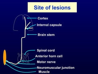
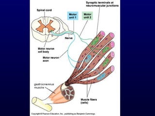
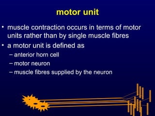
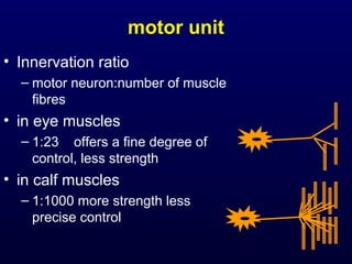
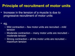
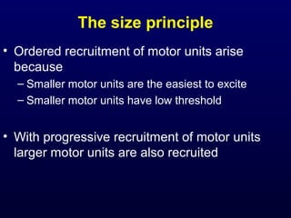
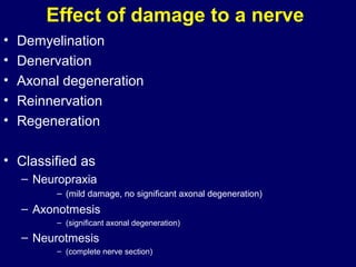
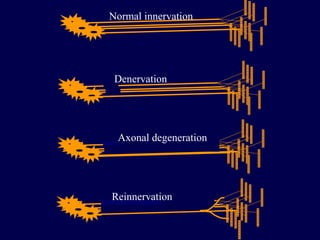
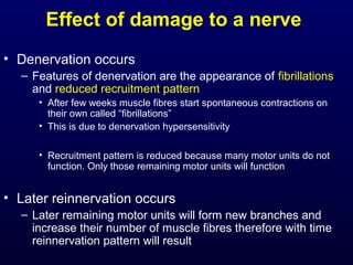
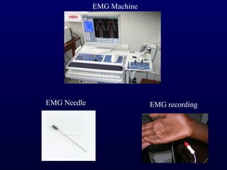
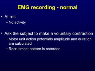
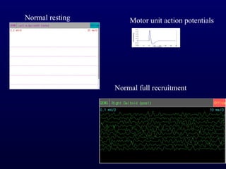
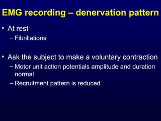
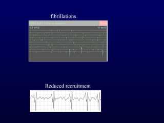
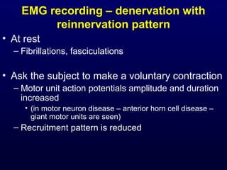
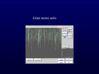
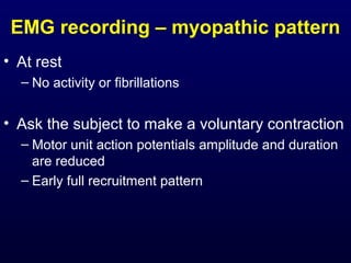
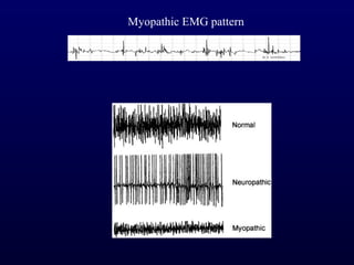
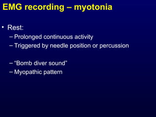
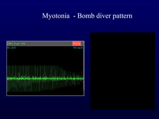
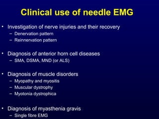
The document outlines the physiology of motor units and their neural control, how disorders at different levels can affect muscle function including lesions in the cortex, brainstem, spinal cord, nerve, neuromuscular junction or muscle, and how electromyography can be used to investigate nerve injuries, anterior horn cell diseases, muscle disorders, and diagnose conditions like myasthenia gravis.
This PPT share the principles used in exercise prescription and the parameters which should be kept in mind while prescribing and progressing the exercise regimen
Biomechanics of fracture and fracture healingsara kowsar
A bone fracture is a broken bone that
occurs when a force impels against a
bone that is stronger than it can structurally withstand.Bone healing refers to complex and sequential events that occur to restore injured bone to pre-injury state.
Walking is a phenomenon that is taken for granted by healthy individuals, but requires a complex control of the neuromusculoskeletal system. Walking is mainly a result of an automatic process, involving the spinal cord and brainstem mechanisms. Hemiplegic type of gait of a person who has had a brain insult and depends on which area of the brain is affected. Hemiplegic gait usually has:
Decreased stance phase and prolonged swing phase of the paretic side.
Decreased walking speed and shorter stride length.
Fitness testing is a way of gaining information about the health-related and skill related components of an athletes fitness. Testing can take place in a number of environments, with laboratory testing being the most accurate.
CENTRAL NERVOUS SYSTEM : EXERCISE PHYSIOLOGY J. Priyanka
Nervous System : What is it?
Divisions
Functions of Nervous System
Neuron
Neuron Types
Brain
Spinal Cord
Peripheral Nervous System
Somatic Neural System
Autonomic Neural System
skeletal, cardiac & smooth Muscles by Thiru Murugan.pptxthiru murugan
Unit III – The Muscular System - Anatomy
Types and structure of muscles
Muscle groups
Alterations in disease
Applications and implications in nursing
Muscle:
Muscle is a soft tissue and it is one of the 4 basic tissues, along with nervous tissue, epithelium, and connective tissue.
Muscles helps in movement, support and protection of internal organs.
Muscles can perform variety of functions
Muscles tissue is made up of cells called “MYOCYTES” or muscle fibers.
There are more than 600 muscles in the human body. A kind of elastic tissue makes up each muscle, which consists of thousands, or tens of thousands, of small muscle fibers.
Types of Muscles: There are 3 main types of muscles
Skeletal muscle
Cardiac muscle
Smooth muscle
Skeletal muscle:
These are having close relationship to the bone or skeleton, so called Skeletal muscles
It present in limbs and related body parts & It form about 40% of body weight.
Under microscope the skeletal muscles fibers shows prominent striations, so called “Striated Muscles” & It is also known as “Voluntary Muscles” (movements are under our control)
Structure of Skeletal muscle:
Muscle fibers shows transverse striations under light microscope so it is called “striated muscles”
The nucleus is located peripherally.
Each skeletal muscle is an organ that consists of numerous cells called muscle fibers.
Each muscle fibers surrounded by “ Endomysium”
Inside each skeletal muscle, muscle fibers are organized into bundles, called fascicles, each fascicle surrounded by perimysium.
The whole muscle is covered by “epimysium”
Each skeletal muscle has three layers: endomysium, perimysium and epimysium
Muscle fibers:
Muscle is composed of many long cylindrical-shaped elongated fibres called muscle fibers
Length varies according to the size and shape of the muscles.
The actual arrangement of the fibres depending on the function of the muscle.
Each muscle fibers covered by a membrane is called the sarcolemma.
The cytoplasm of a muscle fiber is called Sarcoplasm
In sarcoplasm there are many mitochondria and bundles of fine longitudinal thread like part is called “myofibrils”
Microscopic structure of myofibrils:
A myofibril (also known as a muscle fibril or sarcostyle) is a basic rod-like part of a muscle cell.
Muscles are composed of tubular cells called myocytes, known as muscle fibres in striated muscle, and these cells in turn contain many chains of myofibrils.
They are created during embryonic development in a process known as myogenesis.
Under light microscope each myofibril consist of 2 bands:
Light band or “I” Band and Dark band or “A” Band
The alternating pattern of these bands results in the striated appearance of skeletal muscle.
Light band or “I” Band:
The I-bands (isotropic in polarized light) appear light in color.
I band divided into 2 portions by a narrow dark line called “Z” line or “Z” Disc.
This “Z” line is formed by protein which does not permit the light.
The part in between 2 “Z” lines called “sarc
2. Unit - 7-Muscles Anatomy, Thiru Murugan, Msc Professorthiru murugan
The Muscular System
By Thiru murugan. M
The Muscular system:
Types and structure of muscles
Muscle groups: muscles of the head, neck, thorax, abdomen, pelvis, upper limb and lower limbs
Principal muscles: deltoid, biceps, triceps, respiratory, abdominal, pelvic floor, pelvic floor muscles, gluteal muscles and vastus lateralis
Major muscles involved in nursing procedures
Muscle:
Muscle is a soft tissue and it is one of the 4 basic tissues, along with nervous tissue, epithelium, and connective tissue.
Muscles helps in movement, support and protection of internal organs.
Muscles can perform variety of functions
Muscles tissue is made up of cells called “MYOCYTES” or muscle fibers.
There are more than 600 muscles in the human body. A kind of elastic tissue makes up each muscle, which consists of thousands, or tens of thousands, of small muscle fibers.
Types of Muscles: There are 3 main types of muscles
Skeletal muscle
Cardiac muscle
Smooth muscle
Skeletal muscle:
These are having close relationship to the bone or skeleton, so called Skeletal muscles
It present in limbs and related body parts & It form about 40% of body weight.
Under microscope the skeletal muscles fibers shows prominent striations, so called “Striated Muscles” & It is also known as “Voluntary Muscles” (movements are under our control)
Structure of Skeletal muscle:
Muscle fibers shows transverse striations under light microscope so it is called “striated muscles”
The nucleus is located peripherally.
Each skeletal muscle is an organ that consists of numerous cells called muscle fibers.
Each muscle fibers surrounded by “ Endomysium”
Inside each skeletal muscle, muscle fibers are organized into bundles, called fascicles, each fascicle surrounded by perimysium.
The whole muscle is covered by “epimysium”
Each skeletal muscle has three layers: endomysium, perimysium and epimysium
Muscle fibers:
Muscle is composed of many long cylindrical-shaped elongated fibres called muscle fibers
Length varies according to the size and shape of the muscles.
The actual arrangement of the fibres depending on the function of the muscle.
Each muscle fibers covered by a membrane is called the sarcolemma.
The cytoplasm of a muscle fiber is called Sarcoplasm
In sarcoplasm there are many mitochondria and bundles of fine longitudinal thread like part is called “myofibrils”
Microscopic structure of myofibrils:
A myofibril (also known as a muscle fibril or sarcostyle) is a basic rod-like part of a muscle cell.
Muscles are composed of tubular cells called myocytes, known as muscle fibres in striated muscle, and these cells in turn contain many chains of myofibrils.
They are created during embryonic development in a process known as myogenesis.
Under light microscope each myofibril consist of 2 bands:
Light band or “I” Band & Dark band or “A” Band
The alternating pattern of these bands results in the striated appearance of skeletal muscle.
Light band or “I” Band:
The I-bands (isotropic in polarize
1. Mechanisms of
dysfunction of
muscles
Prof. Vajira Weerasinghe
Department of Physiology
Lecture is available at
www.slideshare.net/vajira54
2. Objectives
1. Recall the physiology of the motor unit and its
neural control
2. Outline how disorders at different levels in the
control mechanisms affect muscle function
3. Site of lesions
Cortex
Internal capsule
Brain stem
Spinal cord
Anterior horn cell
Motor nerve
Neuromuscular junction
Muscle
4. Motor unit
• A single motor neuron and the group muscle
fibres supplied by the branches of the axon
5.
6. motor unit
• muscle contraction occurs in terms of motor
units rather than by single muscle fibres
• a motor unit is defined as
– anterior horn cell
– motor neuron
– muscle fibres supplied by the neuron
7. motor unit
• Innervation ratio
– motor neuron:number of muscle
fibres
• in eye muscles
– 1:23 offers a fine degree of
control, less strength
• in calf muscles
– 1:1000 more strength less
precise control
8. Principle of recruitment of motor units
• Increase in the tension of a muscle is due to
progressive recruitment of motor units
• eg.
– Mild contraction – few motor units are recruited – mild
tension
– Moderate contraction – many motor units are recruited –
moderate tension
– Strong contraction – all the motor units are recruited –
maximum tension
9. The size principle
• Ordered recruitment of motor units arise
because
– Smaller motor units are the easiest to excite
– Smaller motor units have low threshold
• With progressive recruitment of motor units
larger motor units are also recruited
10. Effect of damage to a nerve
• Demyelination
• Denervation
• Axonal degeneration
• Reinnervation
• Regeneration
• Classified as
– Neuropraxia
– (mild damage, no significant axonal degeneration)
– Axonotmesis
– (significant axonal degeneration)
– Neurotmesis
– (complete nerve section)
12. Effect of damage to a nerve
• Denervation occurs
– Features of denervation are the appearance of fibrillations
and reduced recruitment pattern
• After few weeks muscle fibres start spontaneous contractions on
their own called “fibrillations”
• This is due to denervation hypersensitivity
• Recruitment pattern is reduced because many motor units do not
function. Only those remaining motor units will function
• Later reinnervation occurs
– Later remaining motor units will form new branches and
increase their number of muscle fibres therefore with time
reinnervation pattern will result
18. slow & fast fibres
• Slow twitch fibre (type I fibre)
• Fast twitch fibre (type II fibre)
19. Slow twitch fibre (type I fibre)
– Slow cross-bridge cycling
– slow rate of shortening (eg. soleus muscle in calf)
– high resistance to fatigue
– high myoglobin content
– high capillary density
– many mitochondria
– low glycolytic enzyme content
– They are red muscle fibres
20. Fast twitch fibre (type II fibre)
– rapid cross-bridge cycling,
– rapid rate of shortening (eg. extra-ocular
muscles)
– low resistance to fatigue
– low myoglobin content
– low capillary density
– few mitochondria
– high glycolytic enzyme content
– fast twitch fibers use anaerobic metabolism
to create fuel, they are much better at
generating short bursts of strength or speed
than slow muscles
21. fast slow
fibres fibres
Sprinters 63% 37%
Marathon
runners 18% 82%
Average man 55% 45%
26. Duchenne Muscular dystrophy
• Duchenne muscular
dystrophy is a lethal
degenerative disease of
muscles in which the
protein dystrophin is
absent
• Gower’s sign is seen
• Dystrophic muscles are
more susceptible to
stretch-induced muscle
damage
27. Dystrophin
• Dystrophin is a rod-shaped cytoplasmic protein, and a
vital part of a protein complex that connects the
cytoskeleton of a muscle fiber to the surrounding
extracellular matrix through the cell membrane
• It provides an anchoring function to the muscle
proteins
28.
29. Myotonia
• Some muscle disorders could be due to derangement
of electrical activity in the muscle membrane
– Na+, K+ Cl- channel derangements
– Called channelopathies
– Myotonia dystrophica, myotonia congenita
• Lack of K+ or Cl- channels
• Depolarisation is normal
• Repolarisation will not take place normally
30.
31. Anterior horn cell diseases
• SMA (Spinal muscular atrophy)
• Affect infants
• Poor prognosis
• Several types are present SMA type I, II etc
• DSMA (Distal spinal muscular atrophy)
• Affect adolesecents
• Main feature is a wasting of small muscles of the hand
• Non-progressive and benign
• MND (Motor neuron disease)
or ALS (amyotrophic lateral sclerosis)
• Affect adults (after 40 years)
• Features include weakness and wasting of limb muscles, tongue fasciculations,
dysarthria, dysphagia
• Slowly progressive and poor prognosis
34. Electromyography
• This is a neurophysiological test done in order to
detect muscle disorders
• Recording electrodes are needles (EMG needles)
• They contain cathode and anode in the form of a
needle
• This is inserted into the muscle
• Motor unit recording pattern is recorded visually in the
screen and sound pattern is recorded from a
loudspeaker
36. EMG recording - normal
• At rest
– No activity
• Ask the subject to make a voluntary contraction
– Motor unit action potentials amplitude and duration
are calculated
– Recruitment pattern is recorded
37. Normal resting Motor unit action potentials
Normal full recruitment
38. EMG recording – denervation pattern
• At rest
– Fibrillations
• Ask the subject to make a voluntary contraction
– Motor unit action potentials amplitude and duration
normal
– Recruitment pattern is reduced
40. EMG recording – denervation with
reinnervation pattern
• At rest
– Fibrillations, fasciculations
• Ask the subject to make a voluntary contraction
– Motor unit action potentials amplitude and duration
increased
• (in motor neuron disease – anterior horn cell disease –
giant motor units are seen)
– Recruitment pattern is reduced
42. EMG recording – myopathic pattern
• At rest
– No activity or fibrillations
• Ask the subject to make a voluntary contraction
– Motor unit action potentials amplitude and duration
are reduced
– Early full recruitment pattern