Introduction to MotorCortex
• The motor cortex is the region of the cerebral cortex
involved in planning, control, and execution of voluntary
movements. It is located in the frontal lobe, anterior to the
central sulcus. It includes:
1.Primary motor cortex (M1)
2.Premotor cortex
3.Supplementary motor area (SMA)
• Each area contributes uniquely to the generation and
coordination of movement.
3.
a) Primary motorcortex
b) Premotor cortex
c) Supplementary motor cortex
5.
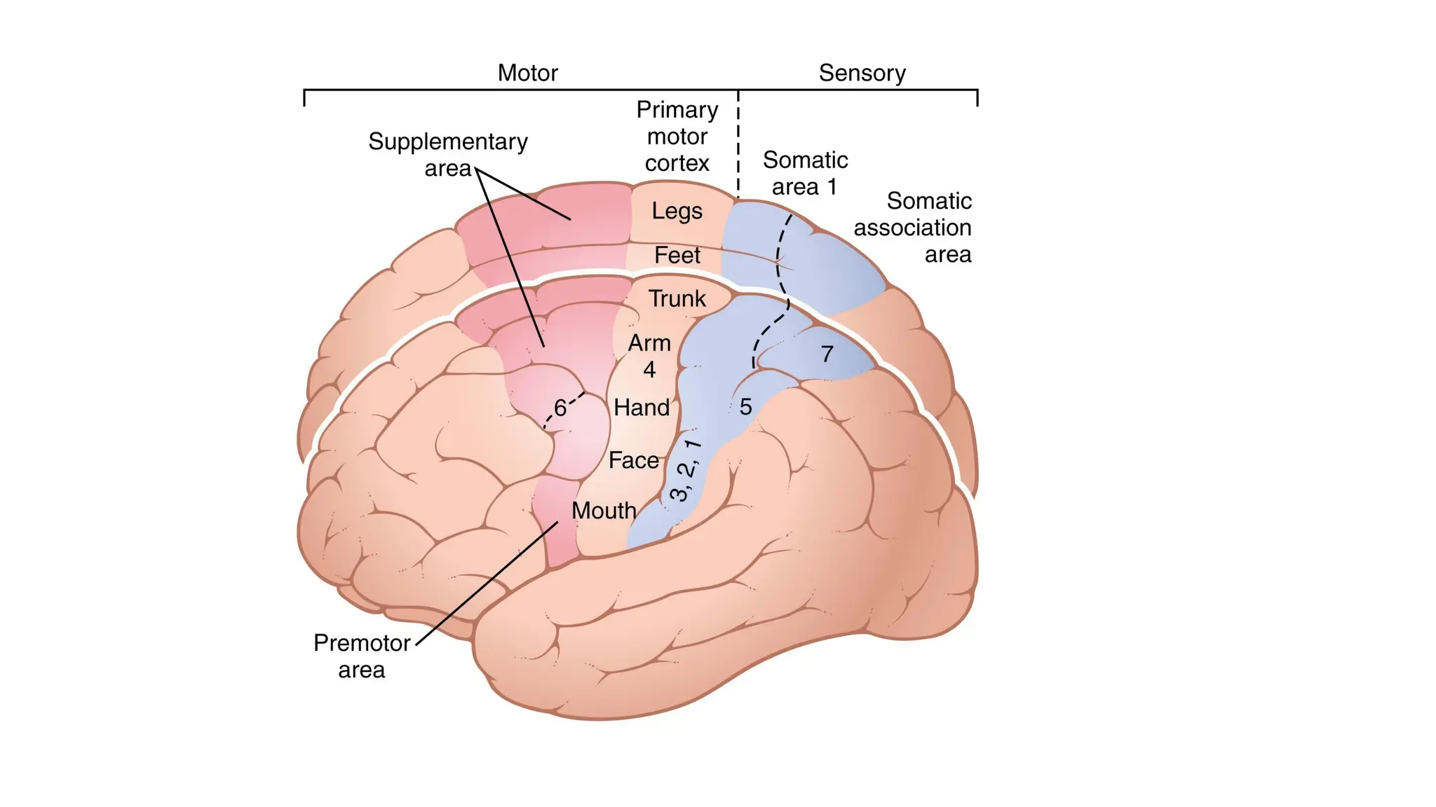
Primary Motor Cortex(M1)
Location:
• Lies in the precentral gyrus of the frontal lobe.
• Brodmann area 4.
Functions:
• Directly executes voluntary movements, especially fine, skilled
movements (e.g., fingers, hands, face).
• Controls contralateral body muscles (opposite side).
• Sends output signals via corticospinal and corticobulbar tracts to
lower motor neurons.
6.
Characteristics:
• Has asomatotopic organization (motor homunculus).
• Produces individual and discrete muscle contractions.
• Responsible for simple movements (e.g., flexing a finger)
7.
Premotor Cortex
Location:
• Anteriorto the primary motor cortex.
• Brodmann area 6 (lateral part).
• Functions:
• Responsible for planning and coordination of complex movements.
• Controls postural adjustments needed for voluntary movements.
• Active during motor learning and movement guided by external stimuli
(e.g., seeing a ball and reaching for it).
• Coordinates proximal muscles and trunk muscles.
8.
Connections:
• Receives sensoryinput from parietal lobe and visual cortex.
• Sends outputs to primary motor cortex and directly to the spinal
cord.
9.
Supplementary Motor Area(SMA)
Location:
• Medial part of Brodmann area 6 (on the medial surface of the
hemisphere, superior to the premotor cortex).
Functions:
• Controls bilateral movements (e.g., buttoning a shirt).
• Involved in motor planning, especially sequential and complex
movements.
• Important for movement initiated from memory (internal cues),
rather than external stimuli.
• Active during mental rehearsal of movement.
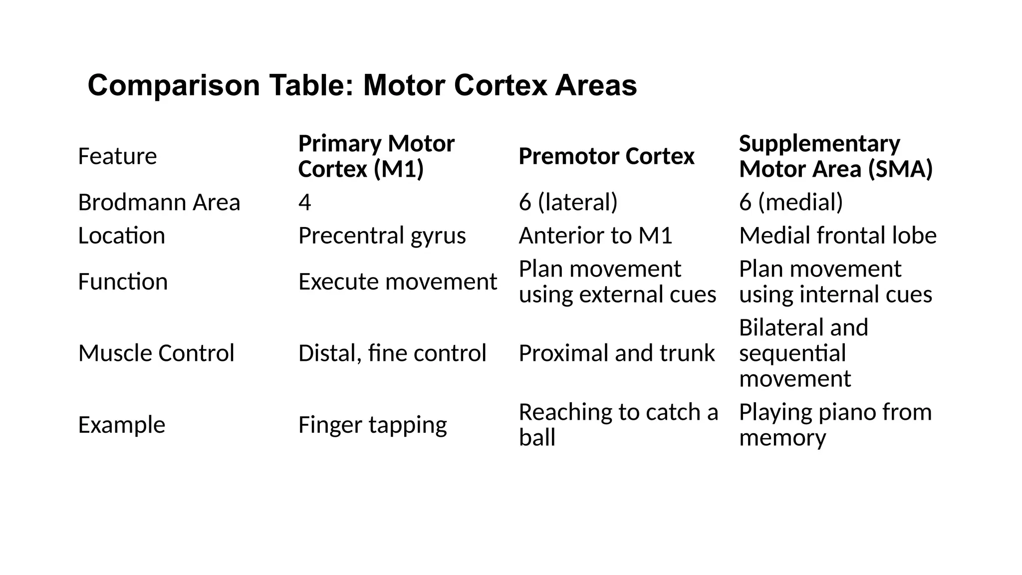
Feature Primary Motor
Cortex(M1)
Premotor Cortex Supplementary
Motor Area (SMA)
Brodmann Area 4 6 (lateral) 6 (medial)
Location Precentral gyrus Anterior to M1 Medial frontal lobe
Function Execute movement Plan movement
using external cues
Plan movement
using internal cues
Muscle Control Distal, fine control Proximal and trunk
Bilateral and
sequential
movement
Example Finger tapping Reaching to catch a
ball
Playing piano from
memory
Comparison Table: Motor Cortex Areas
12.
The motor cortexis an intricately organized system where:
• Primary motor cortex executes specific voluntary movements.
• Premotor cortex helps in externally guided movements.
• Supplementary motor area plans internally generated, complex, and
bilateral movements.
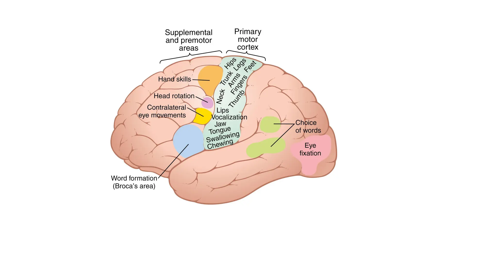
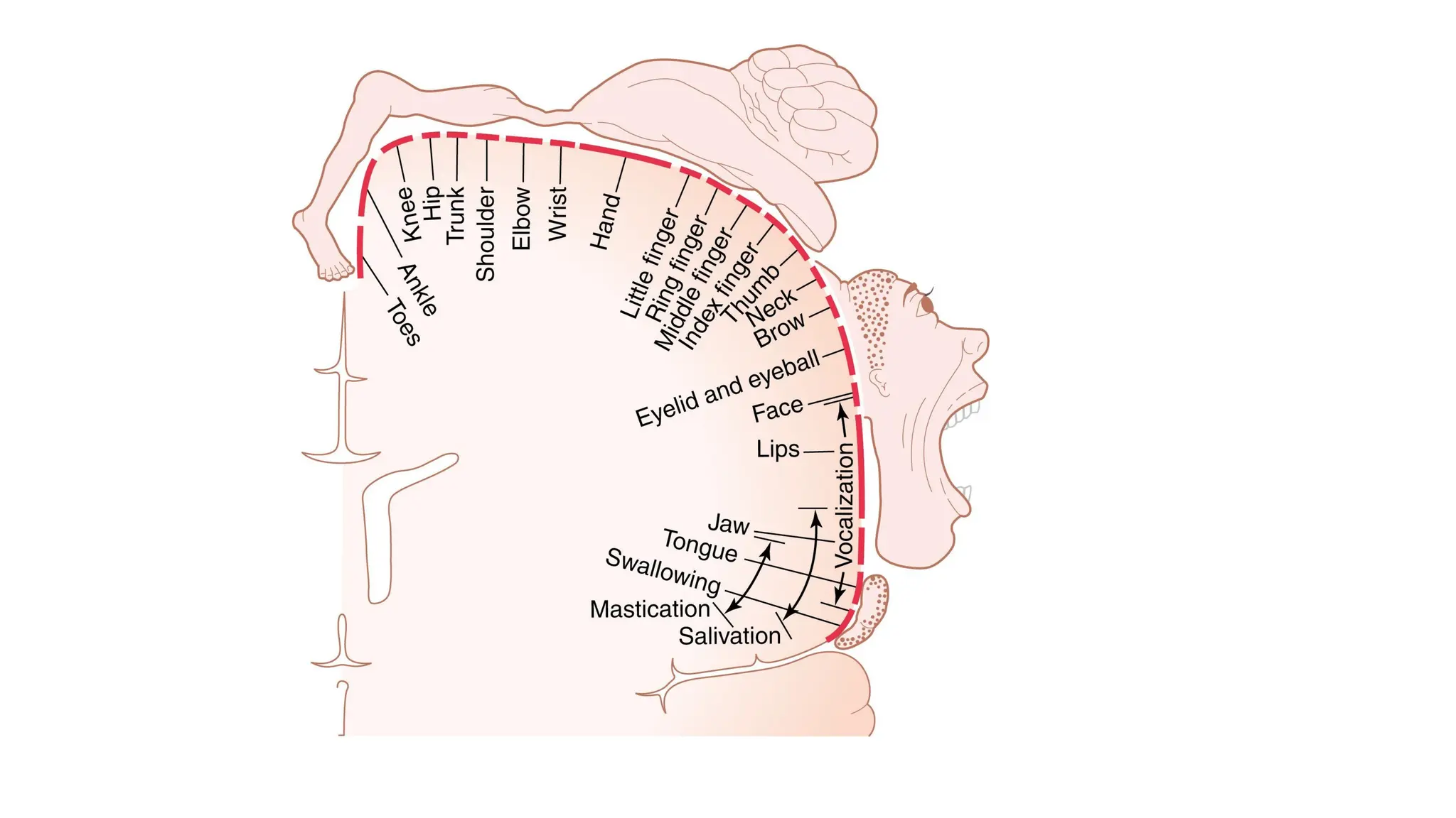
Definition:
• The motorhomunculus is a somatotopic map representing how
different parts of the body are controlled by specific regions of the
primary motor cortex.
• Key Features:
• Located in the primary motor cortex (precentral gyrus).
• The body is represented upside down:
• Lower limbs = medial cortex
• Face and tongue = lateral cortex
• The size of each body part on the homunculus is proportional to the
precision of motor control, not its physical size.
17.
Examples:
• Large areas:hands, face, lips (fine motor control).
• Small areas: trunk, thighs (gross movements).
Clinical Relevance:
• Helps localize brain lesions affecting motor function.
• Explains focal motor deficits (e.g., stroke in medial cortex affects leg
more than face).
18.
The motor homunculusprovides a visual map that reflects the
importance of precision and control in different body parts, making it a
key concept in neuroanatomy and clinical neurology.
19.
The transmission ofsignals from the motor cortex to skeletal muscles
is primarily carried out by descending motor tracts.
These are divided into two major systems:
1. Corticospinal Tract (Pyramidal Tract)
2. Extrapyramidal Tracts
20.
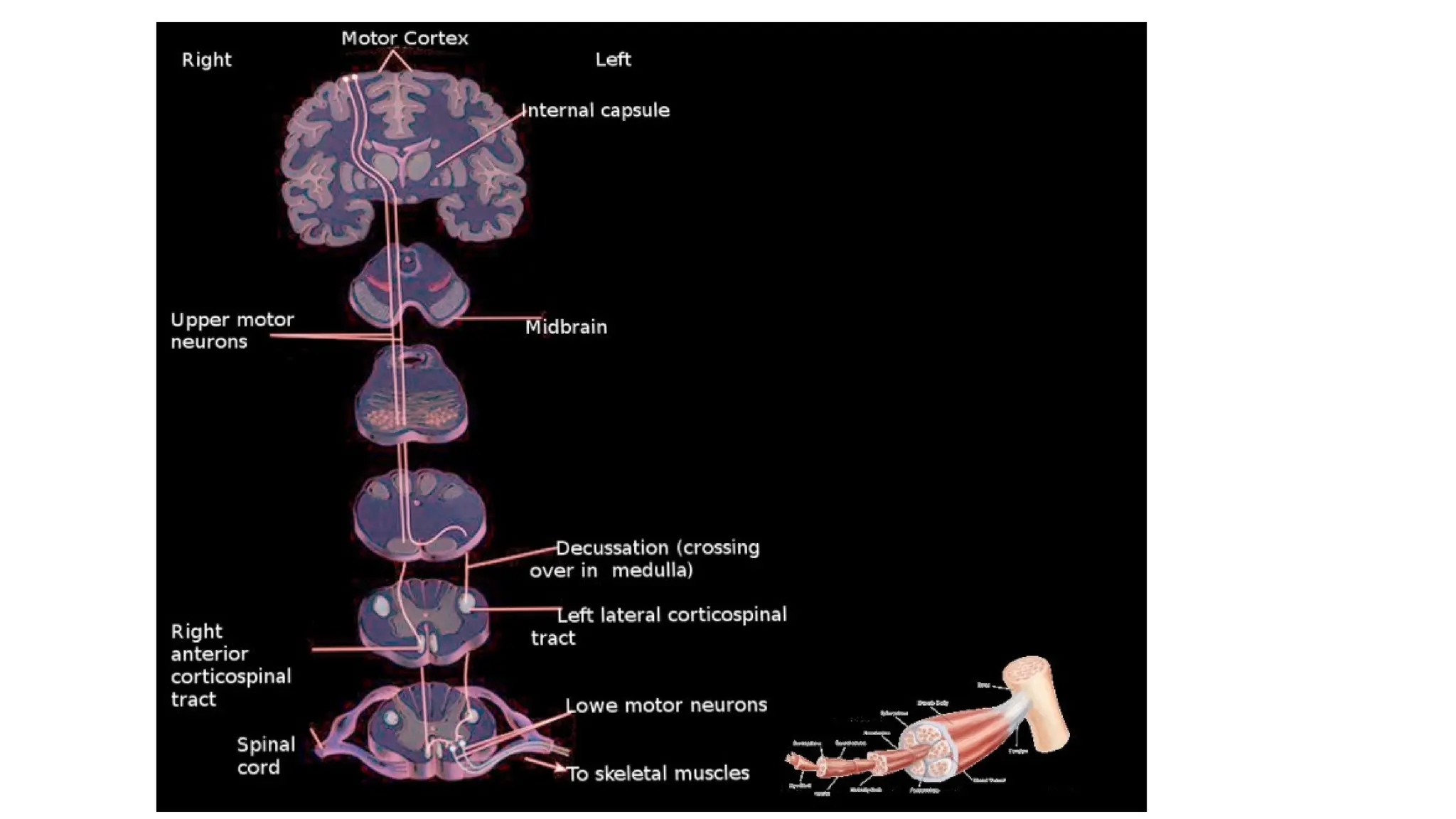
Corticospinal Tract (PyramidalTract)
Origin:
• Arises from the primary motor cortex (area 4), premotor cortex,
supplementary motor areas and somatosensory cortex.
22.
Pathway:
• Fibers descendthrough the internal capsule, cerebral peduncles
(midbrain), pons, and medulla.
• In the medulla, most fibers form the pyramids and about 75–90%
decussate (cross over) to the opposite side in the pyramidal
decussation.
• These crossed fibers form the lateral corticospinal tract, which
descends in the spinal cord.
• The remaining uncrossed fibers form the anterior corticospinal tract
(some cross at the spinal segment level).
Function:
• Responsible forvoluntary movements, especially fine, skilled
movements of distal limbs (e.g., fingers, hands).
25.
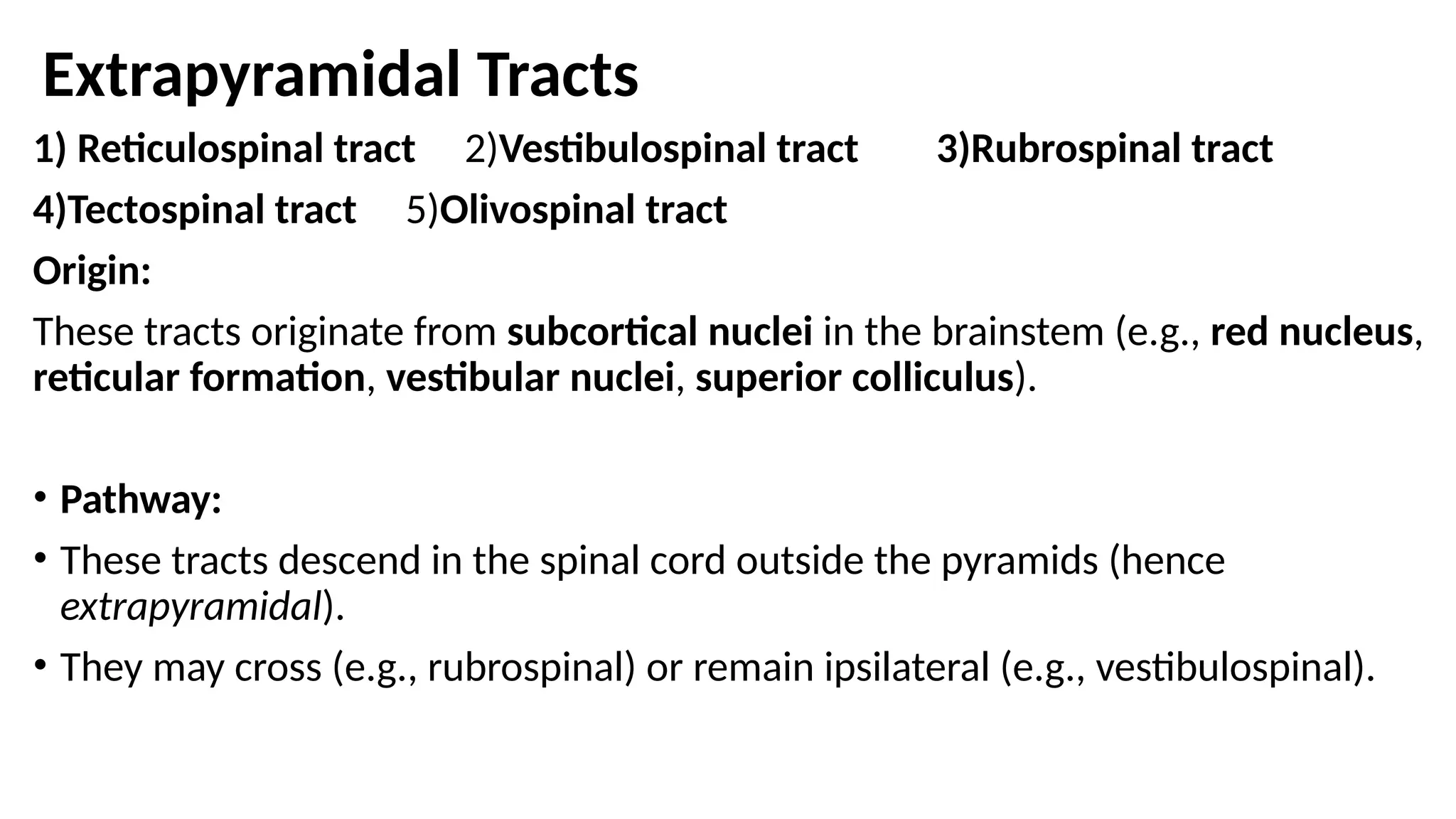
Extrapyramidal Tracts
1) Reticulospinaltract 2)Vestibulospinal tract 3)Rubrospinal tract
4)Tectospinal tract 5)Olivospinal tract
Origin:
These tracts originate from subcortical nuclei in the brainstem (e.g., red nucleus,
reticular formation, vestibular nuclei, superior colliculus).
• Pathway:
• These tracts descend in the spinal cord outside the pyramids (hence
extrapyramidal).
• They may cross (e.g., rubrospinal) or remain ipsilateral (e.g., vestibulospinal).
26.
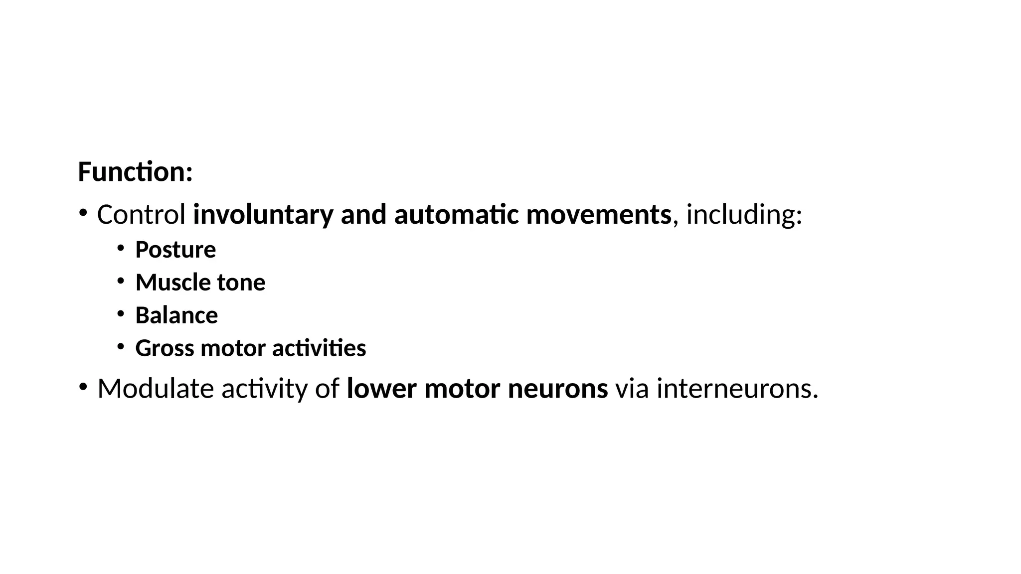
Function:
• Control involuntaryand automatic movements, including:
• Posture
• Muscle tone
• Balance
• Gross motor activities
• Modulate activity of lower motor neurons via interneurons.
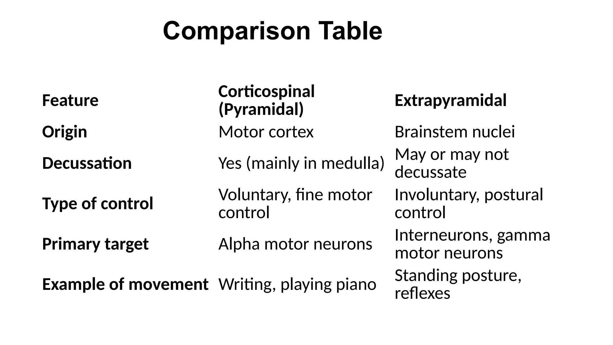
27.
Feature Corticospinal
(Pyramidal)
Extrapyramidal
Origin Motorcortex Brainstem nuclei
Decussation Yes (mainly in medulla) May or may not
decussate
Type of control
Voluntary, fine motor
control
Involuntary, postural
control
Primary target Alpha motor neurons Interneurons, gamma
motor neurons
Example of movement Writing, playing piano Standing posture,
reflexes
Comparison Table
28.
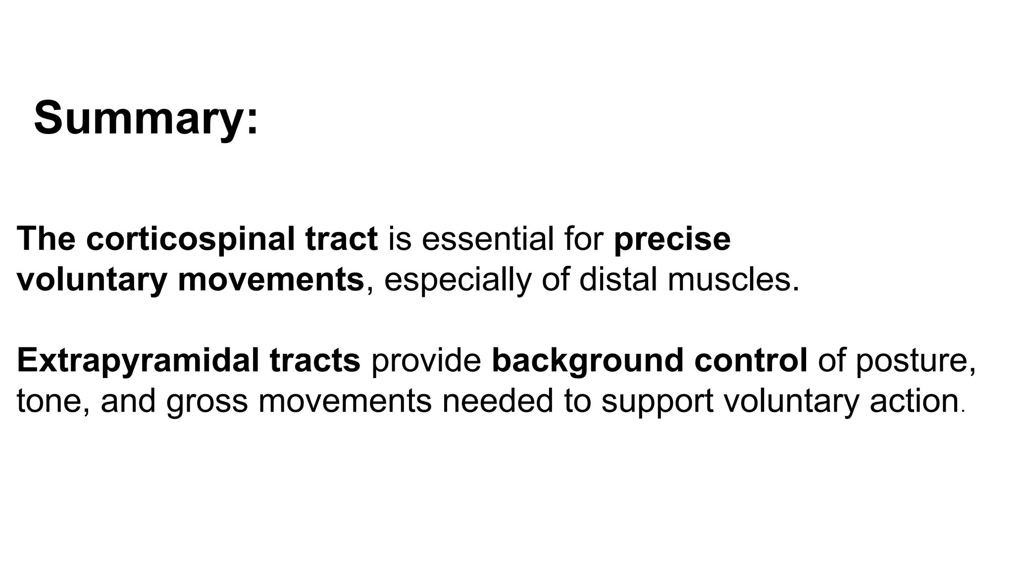
Summary:
The corticospinal tractis essential for precise
voluntary movements, especially of distal muscles.
Extrapyramidal tracts provide background control of posture,
tone, and gross movements needed to support voluntary action.