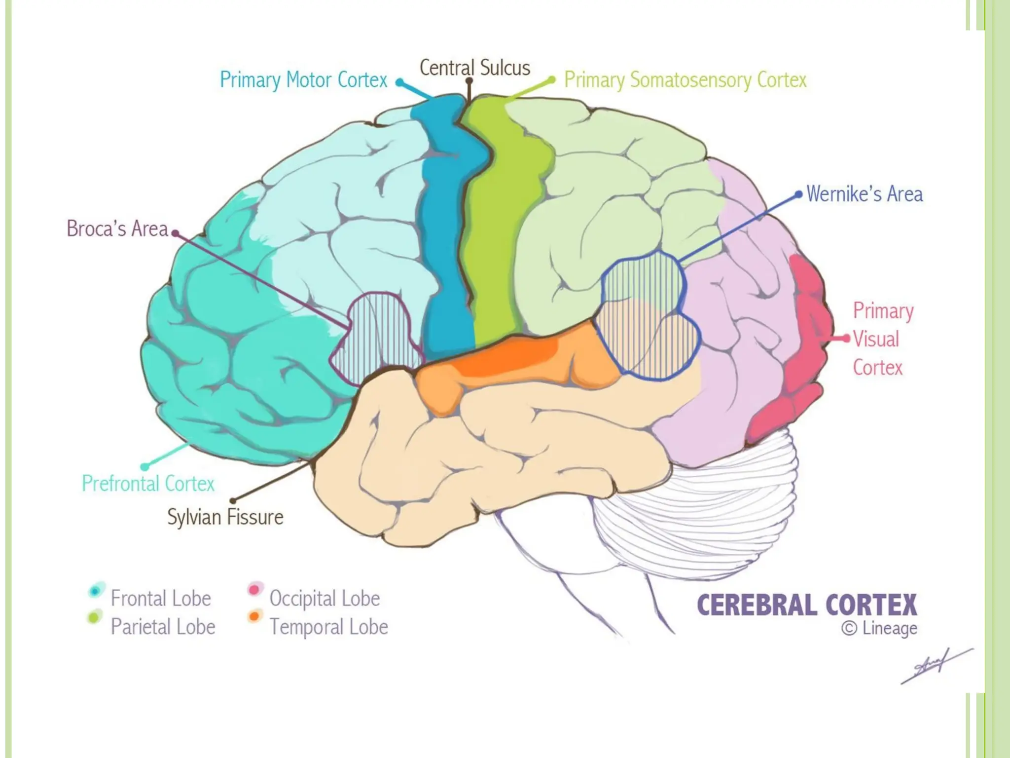
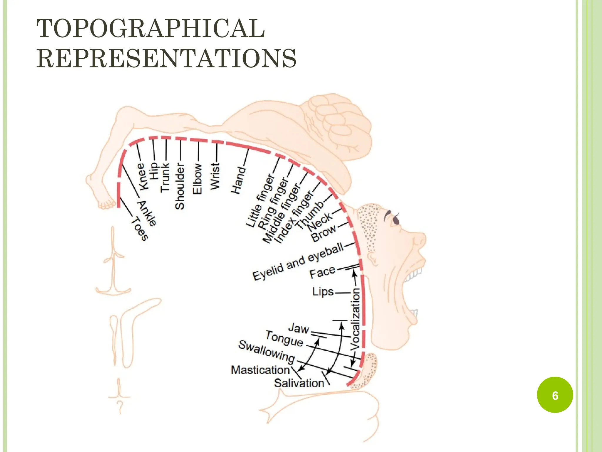
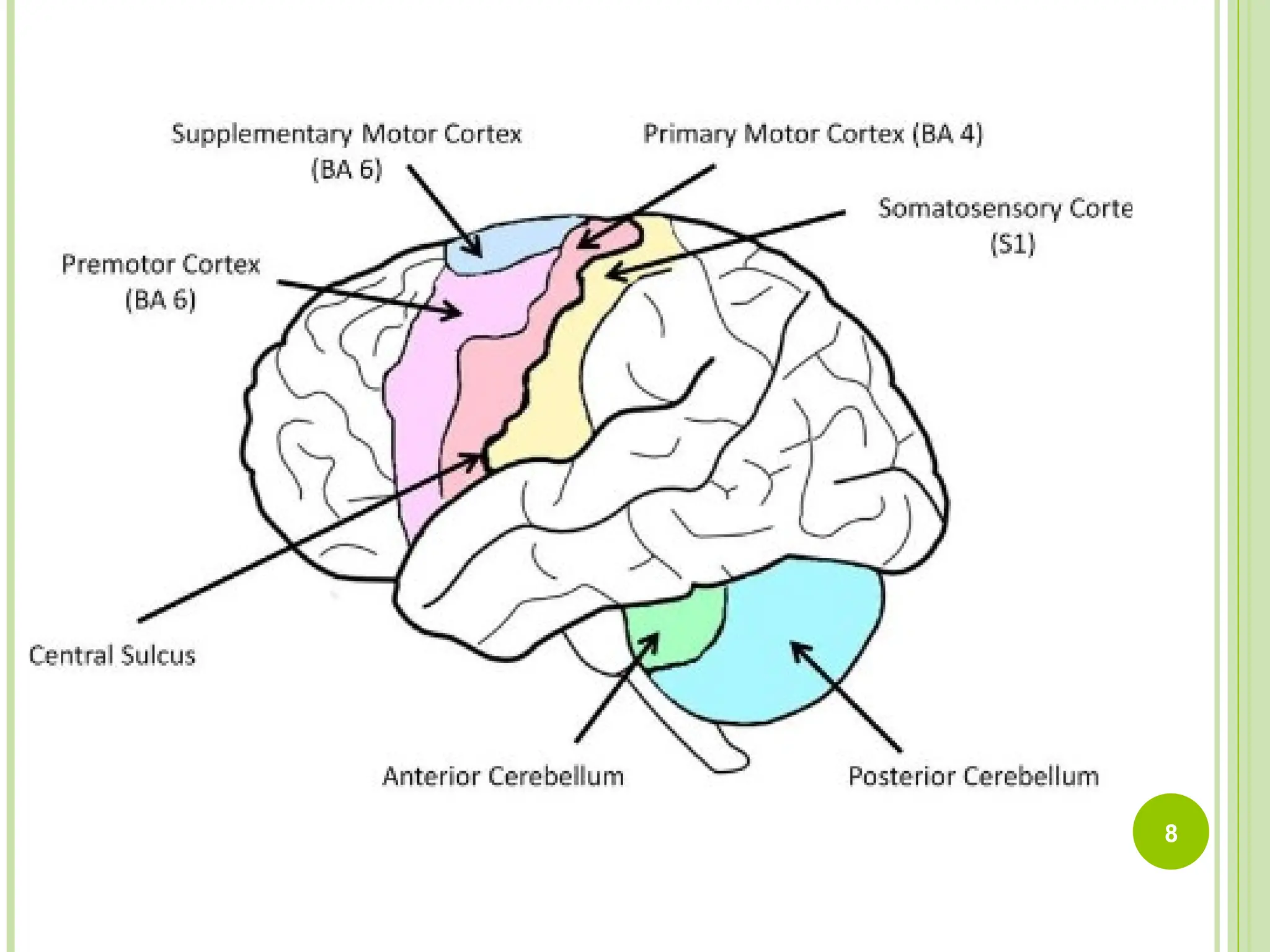
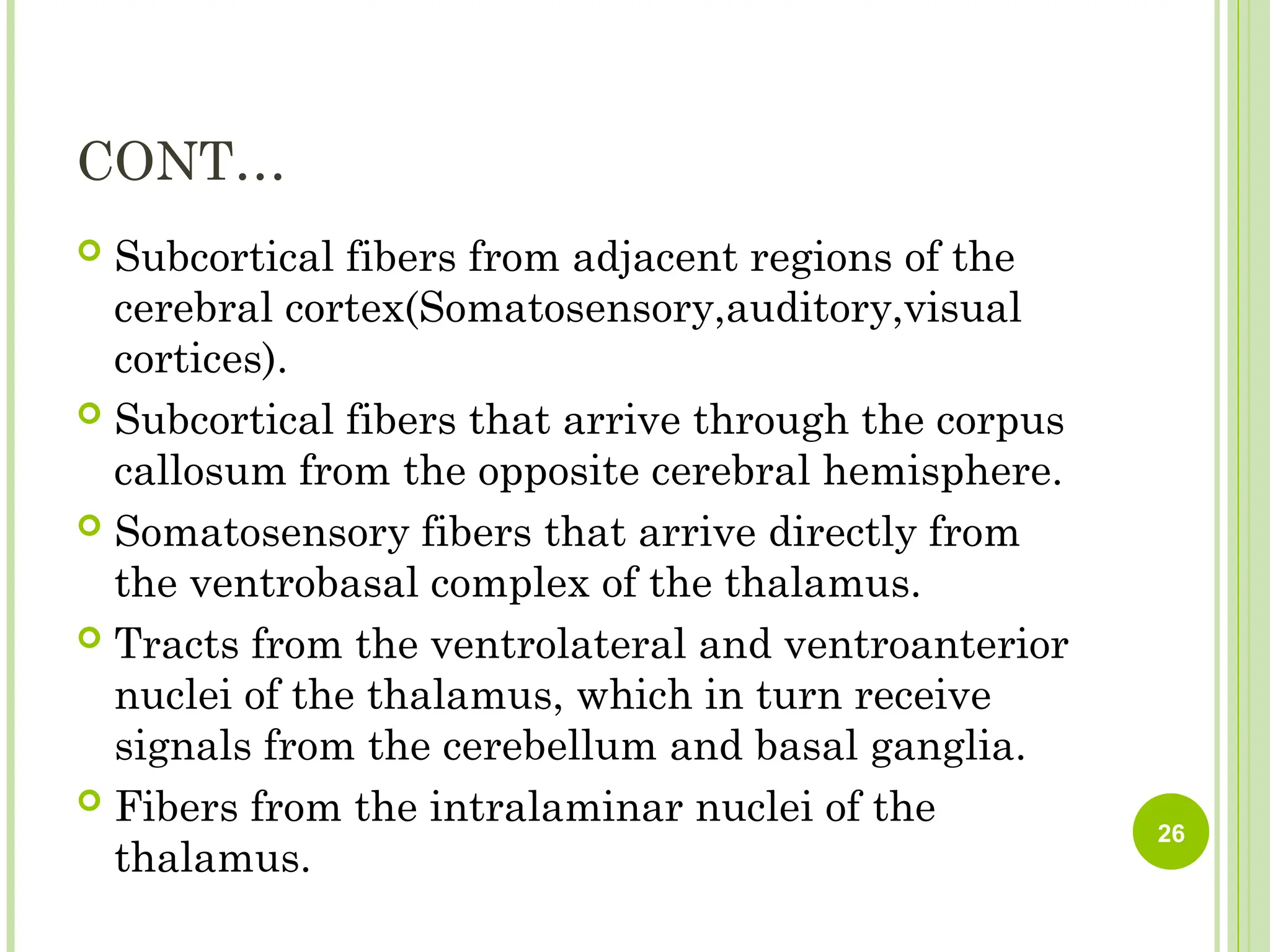
This presentation provides a concise and high-yield overview of the cerebellum and brainstem, focusing on essential anatomical structures, functional divisions, and clinical correlations. The cerebellum is introduced with its major components, including the hemispheres, vermis, cerebellar cortex, white matter, and deep nuclei. The functional organization is explained through its three major divisions: the vestibulocerebellum, which maintains balance and coordinates eye movements; the spinocerebellum, which regulates posture, muscle tone, and gait; and the cerebrocerebellum, which is involved in planning skilled voluntary movements and motor learning. Key clinical signs of cerebellar lesions—such as ataxia, intention tremor, dysmetria, dysdiadochokinesia, nystagmus, and a wide-based gait—are clearly highlighted to help learners connect structure with function.
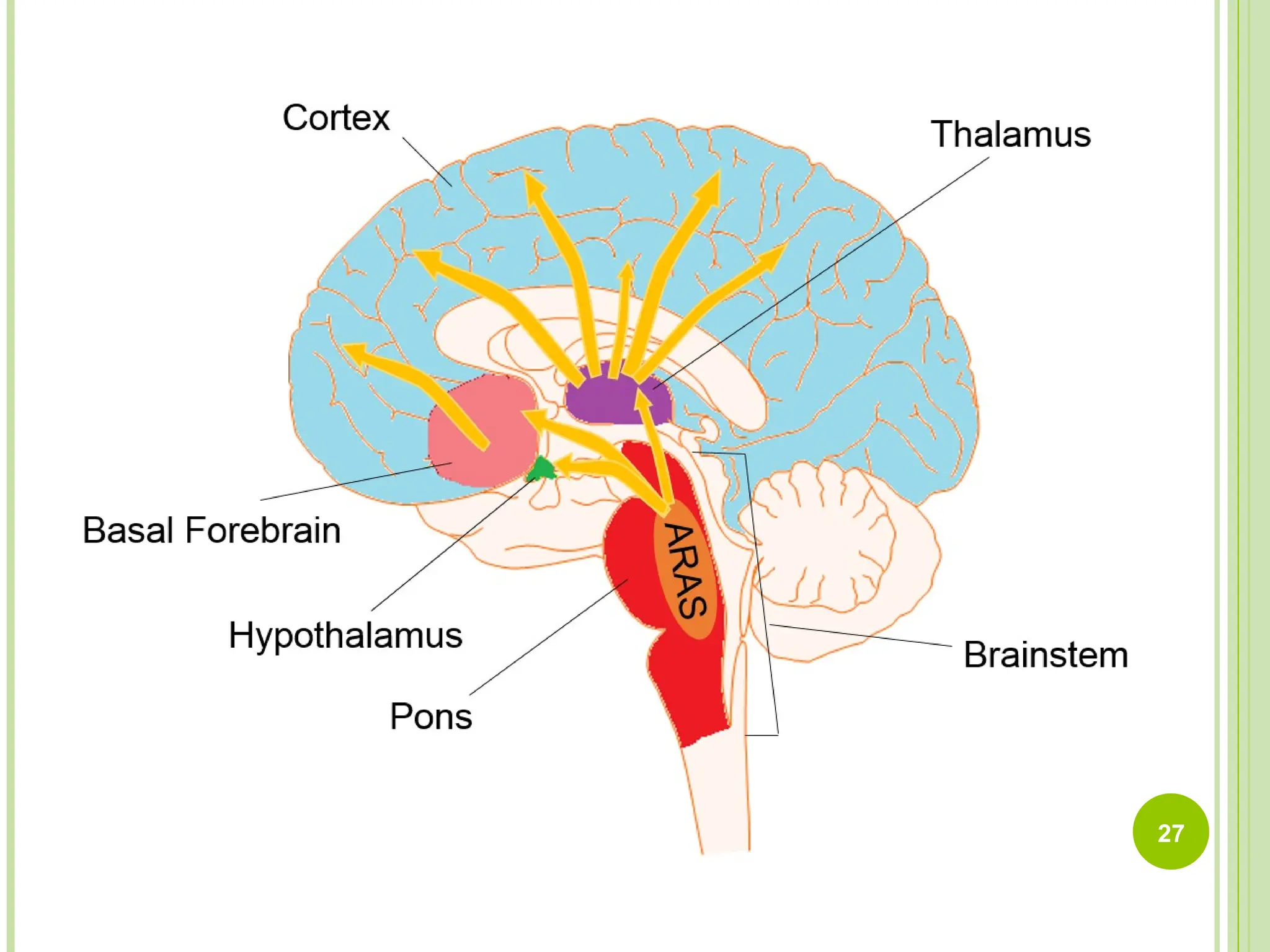
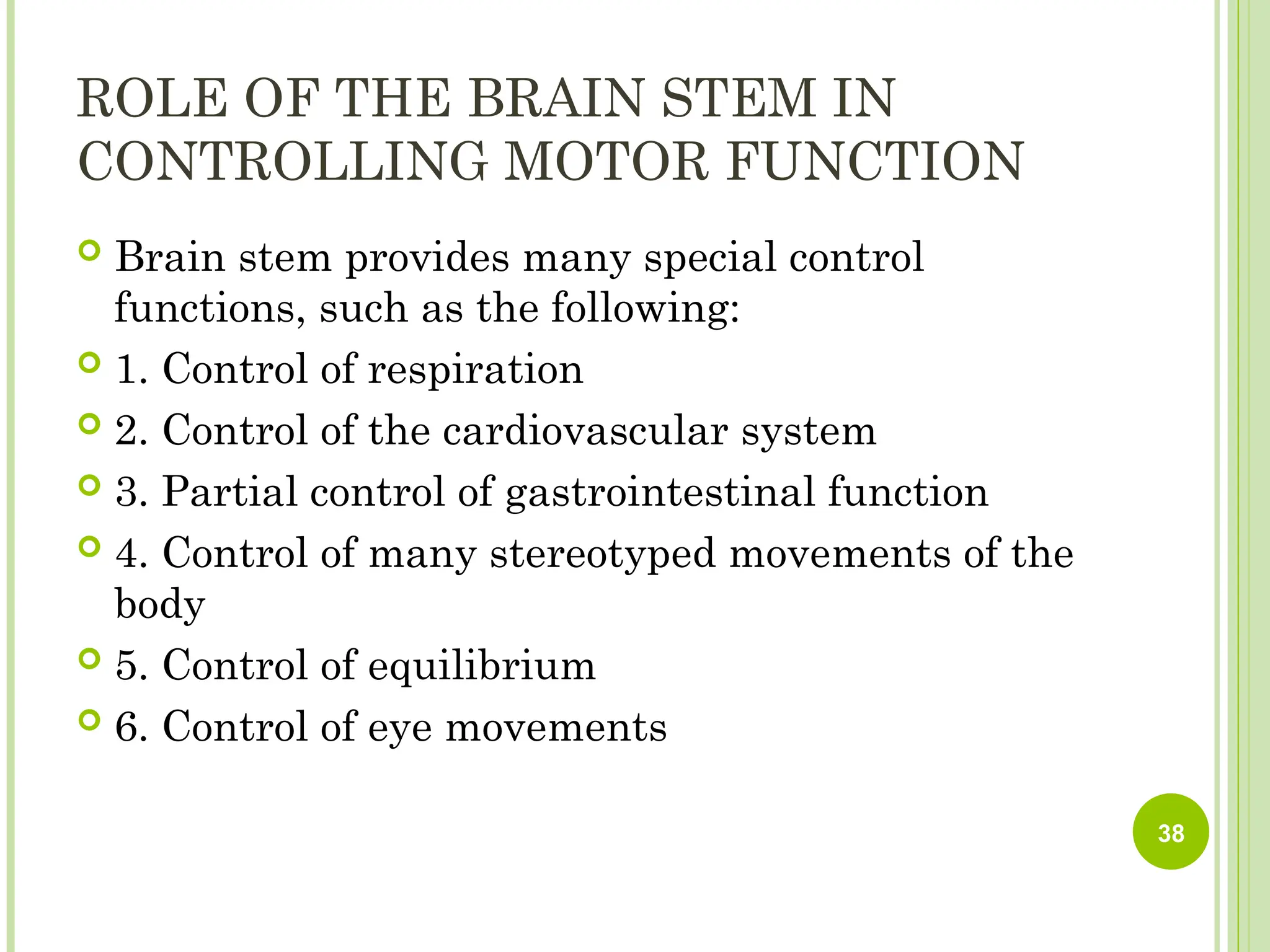
The brainstem section covers the midbrain, pons, and medulla in an easy-to-follow format. Each region is described with its important external and internal landmarks, major tracts, and cranial nerve nuclei. The midbrain is emphasized for its roles in eye movement control, auditory and visual reflexes, and its association with structures like the superior and inferior colliculi. The pons is explained as a bridge for motor pathways and home to cranial nerve nuclei that control facial expression, chewing, and aspects of respiration. The medulla is presented as the vital center for cardiovascular and respiratory regulation and includes nuclei involved in swallowing, gag reflex, and other primitive functions necessary for survival.
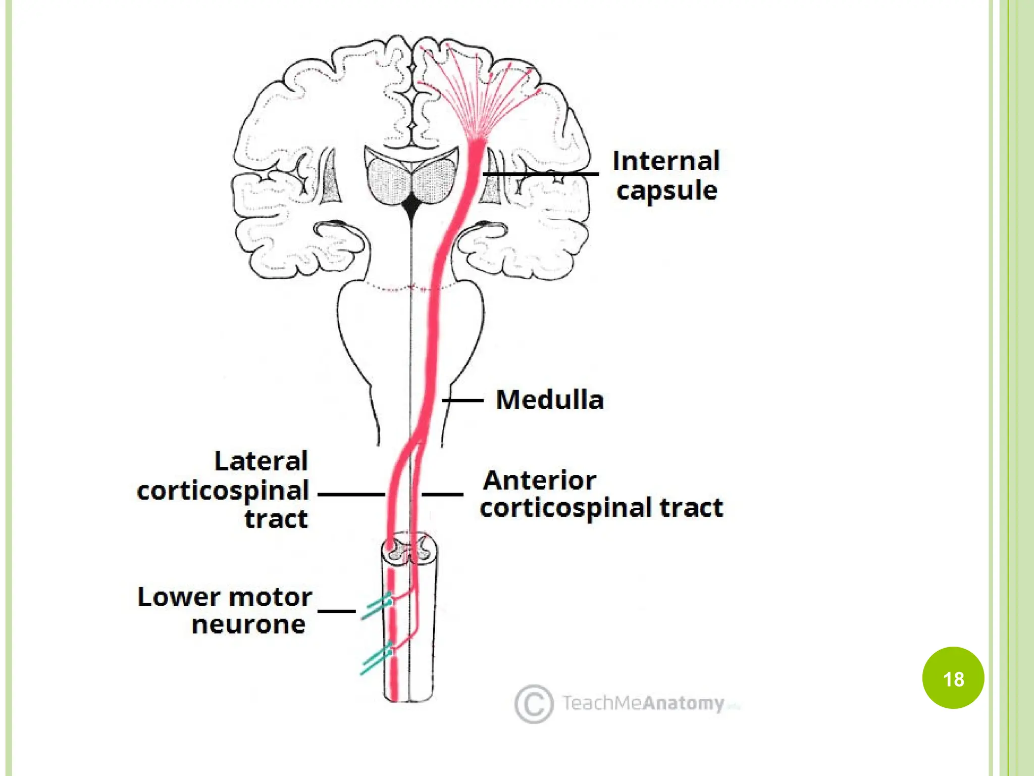
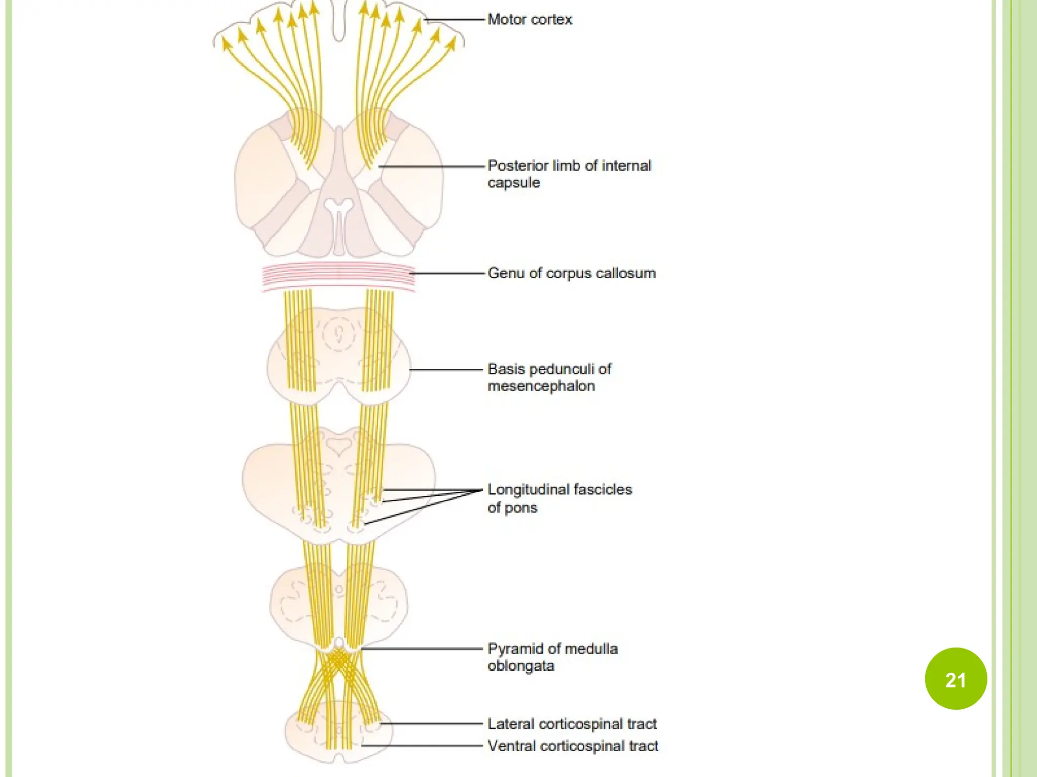
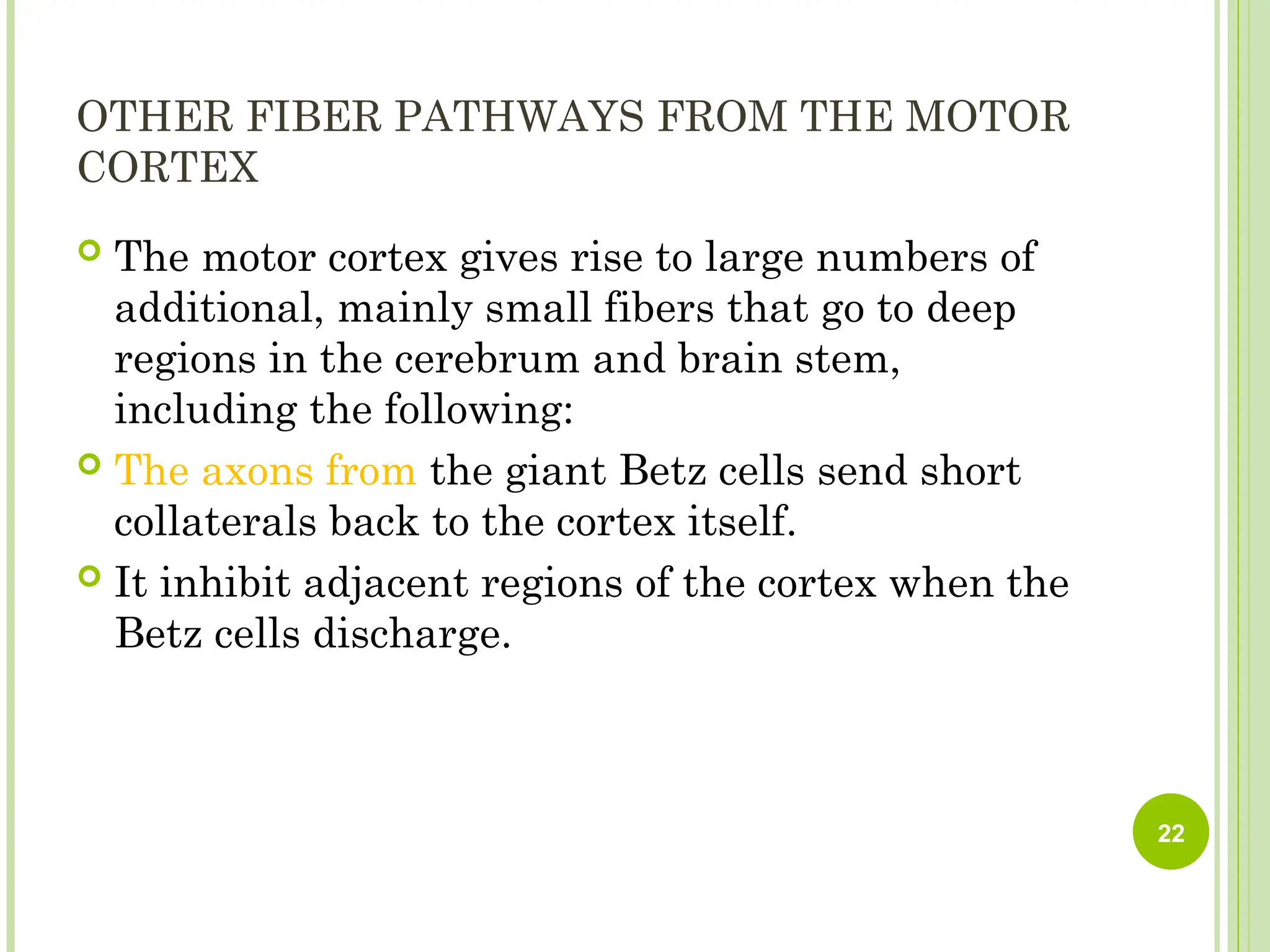
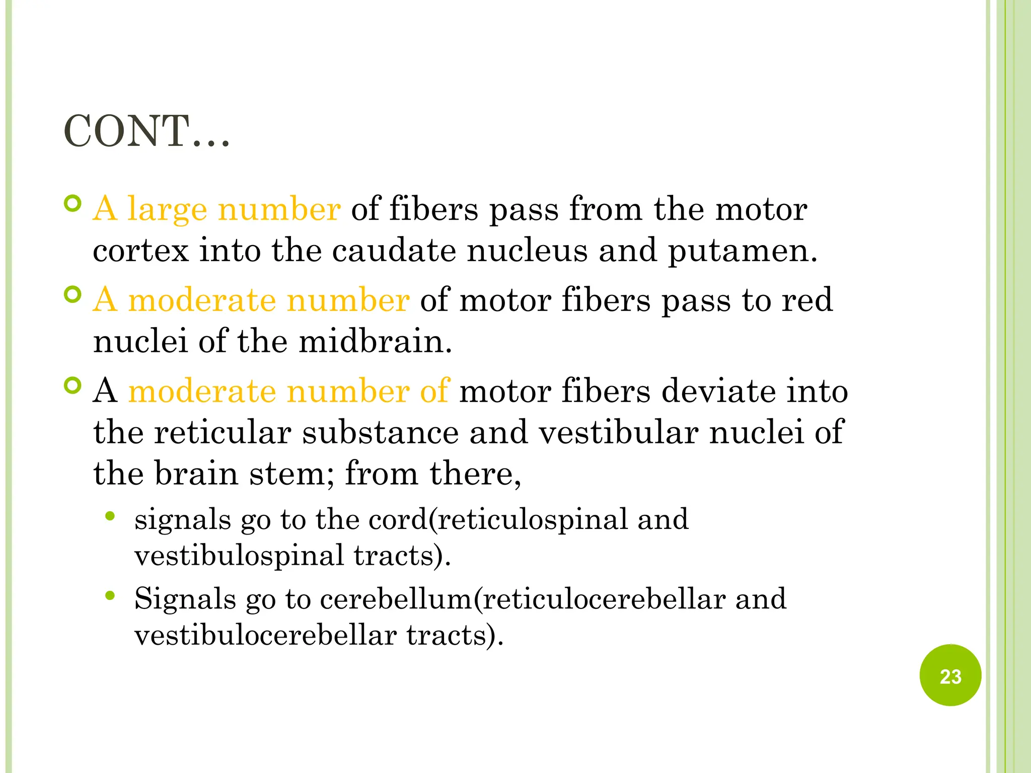
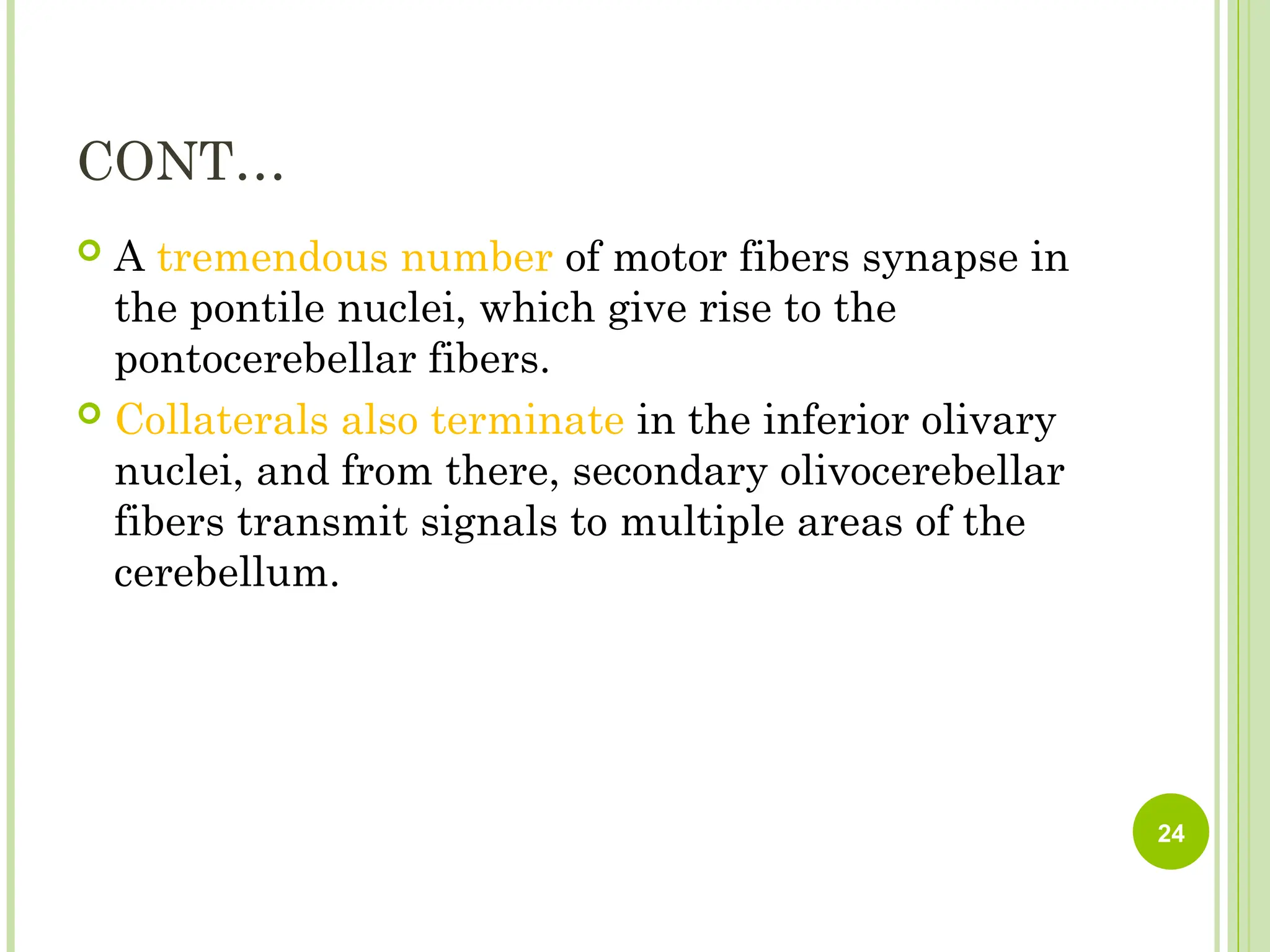
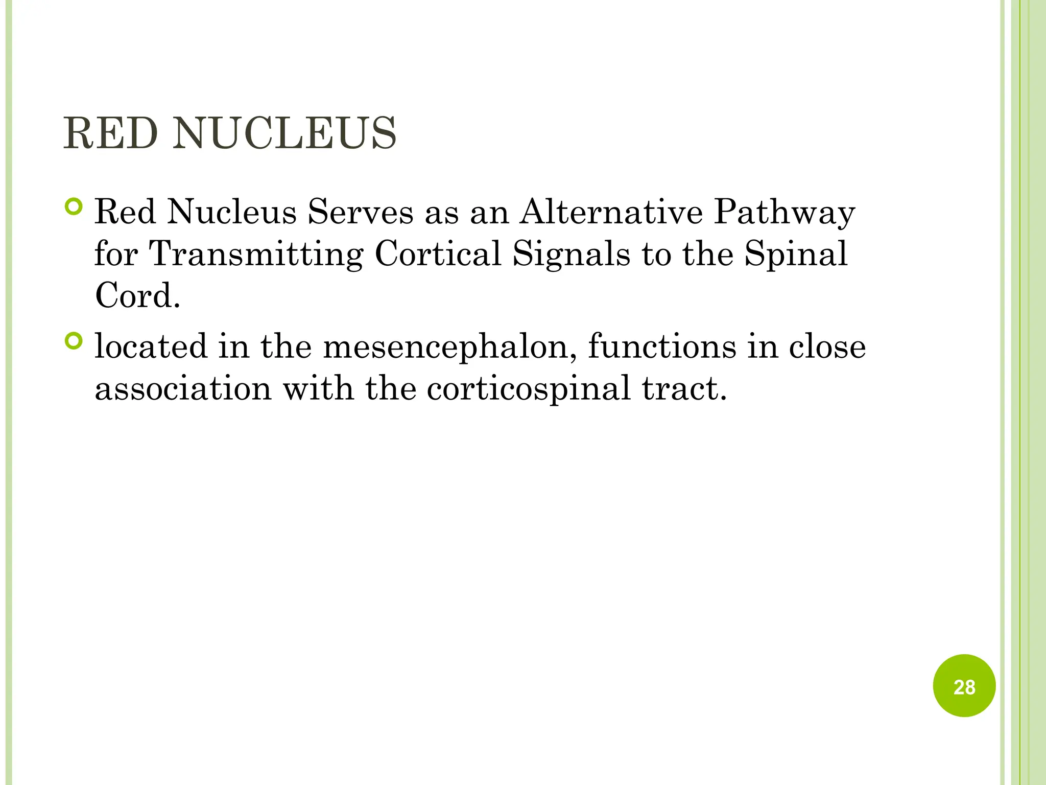
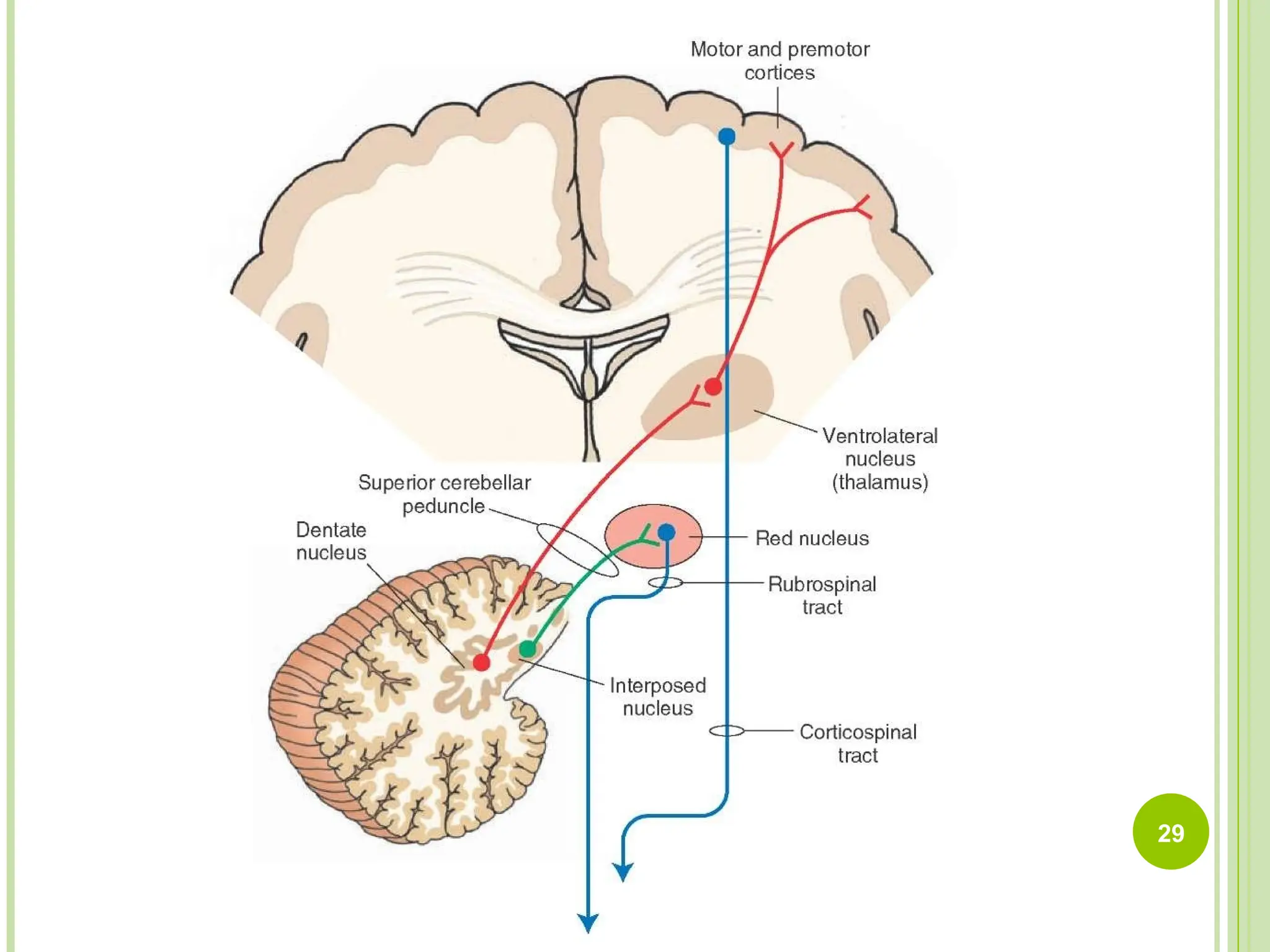
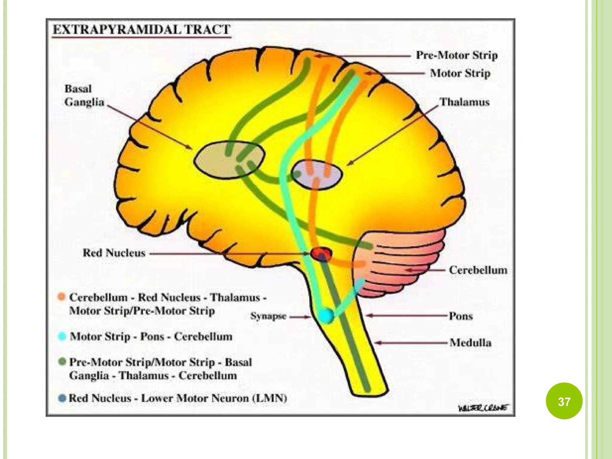
The presentation also underscores the importance of the brainstem as a conduit for ascending sensory pathways and descending motor pathways and explains how lesions at different levels can produce characteristic “crossed findings,” with ipsilateral cranial nerve deficits and contralateral body weakness. The relationship between cerebellar peduncles and brainstem connections is also addressed, clarifying how information travels between the cerebellum, spinal cord, and cerebral cortex.
Overall, this PPT is designed to provide medical and health-science students with a clear, structured, and clinically relevant summary of two critical parts of the central nervous system. It serves as a helpful visual guide for exam preparation, course review, and understanding neurological disorders involving the cerebellum and brainstem.