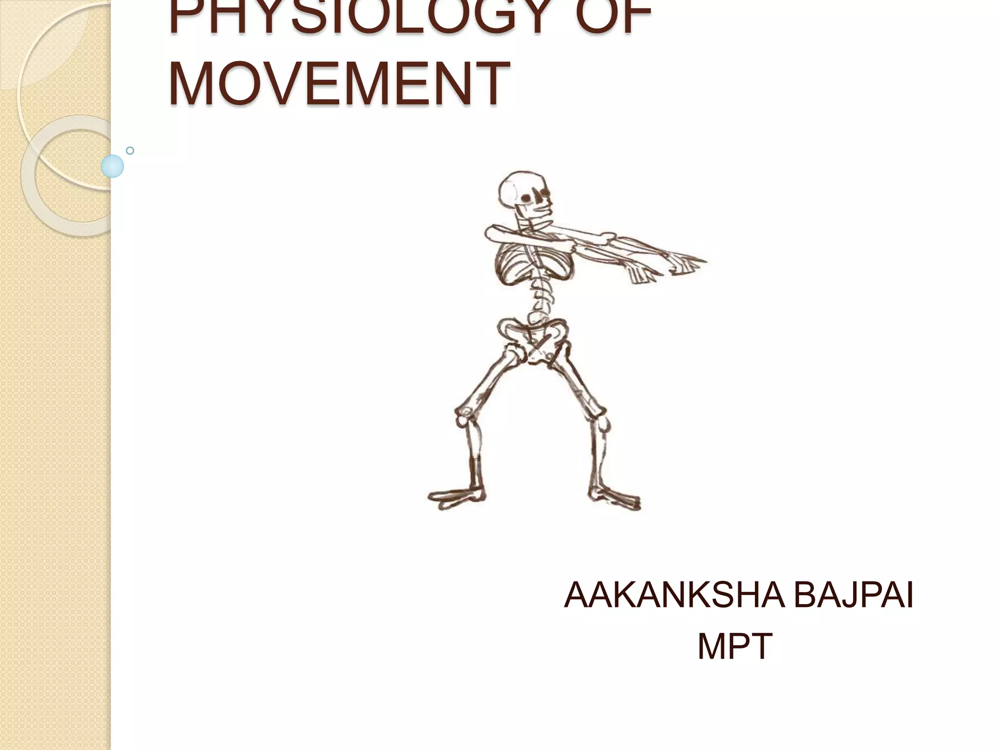
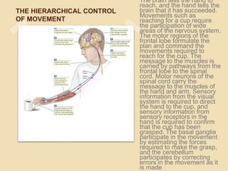
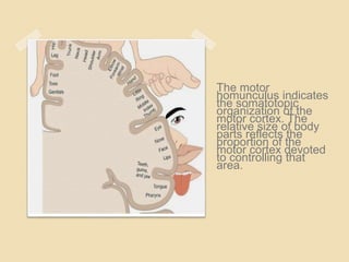
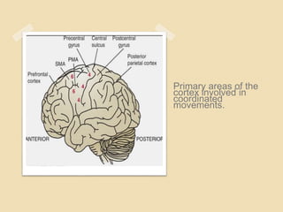
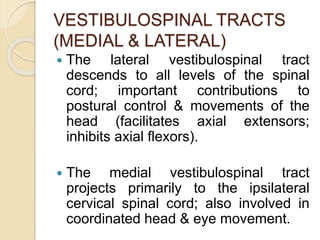
PHYSIOLOGY OF
MOVEMENT
AAKANKSHA BAJPAI
MPT
 There are distinct areas in the brain for
planning and execution and feedback of
the movements.
 The basic requisite for any movement is
the state of preparedness(muscle tone) in
the groups of muscles where movement is
to be executed.
 MOTOR CONTROL has been defined as
an area of study dealing with the
understanding of the neural, physical and
behavioral aspects of biological (e.g.
humans) movements.
SUMMARY OF NORMAL
MOTOR CONTROL
 For normal movement, the movement
planning areas , control circuits and
descending tracts must act in contact
with sensory information to provide
instruction to lower motor neuron. The
basal ganglia receive most of their
input from the cerebral cortex.
 In contrast, the cerebellum receives
copious information from the spinal
cord, vestibular system and brainstem
the cerebellum influences movement
via motor areas of cerebral cortex and
extensive connections with upper
motor neuron that arise in the
brainstem only lower motor neuron
deliver the signals from the CNS to the
skeletal muscle that generate
movement.
Three fundamental types of
movement
Postural
Primarily
Controlled by
brain stem
mechanisms
Ambulatory
Controlled by
brainstem &
spinal
regions
Reaching/
grasping
Controlled by
the cerebral
cortex
However, all regions of the nervous
system contribute to each type of
movement.
POSTURAL CONTROL
 Postural control provides orientation and
balance(equilibrium).
 Postural control is achieved by central
commands to lower motor neuron; the
central output is adjusted to the
environmental contact by sensory input.
 The central commands are mediated by
the tectospinal, medial reticulospinal,
vestibulospinal and medial corticospinal
tracts.
 To orient the world we use 3 senses:-
a) Somatosensation
b) Vision
c) Vestibular
 Somatosensation provides information about
weight bearing and the relative position of body
parts.
 Vision provides information about movement
and cues for judging upright.
 Vestibular input from receptors in the inner ear
informs us about head position relative to
gravity and about head movement.
AMBULATION
 All region of the nervous system are
required for normal human ambulation
the cerebral cortex provides goal
orientation, the basal ganglia govern
generation of force, and the
cerebellum provides timing, inter limbs
coordination and error correction.
REACHING & GRASPING
 Vision and somatosensation are essential
for normal reaching and grasping.
 The post parietal cortex contains neurons
associated with both sensation and
movement; these neurons project to pre
motor cortical areas controlling reaching,
grasping and eye movement.
 The premotor cortical areas for each action
are somewhat distinct, with the result that
reaching, grasping and eye movement are
controlled separately but coordinated by
connection among the areas.
THE HIERARCHICAL CONTROL
OF MOVEMENT
The brain tells the hand to
reach, and the hand tells the
brain that it has succeeded.
Movements such as
reaching for a cup require
the participation of wide
areas of the nervous system.
The motor regions of the
frontal lobe formulate the
plan and command the
movements required to
reach for the cup. The
message to the muscles is
carried by pathways from the
frontal lobe to the spinal
cord. Motor neurons of the
spinal cord carry the
message to the muscles of
the hand and arm. Sensory
information from the visual
system is required to direct
the hand to the cup, and
sensory information from
sensory receptors in the
hand is required to confirm
that the cup has been
grasped. The basal ganglia
participate in the movement
by estimating the forces
required to make the grasp,
and the cerebellum
participates by correcting
errors in the movement as it
is made
THE FOREBRAIN &
MOVEMENT INITIATION
 The frontal lobe of each hemisphere is
responsible for planning and initiating
sequences of behaviour. The frontal
lobe is divided into a number of
different regions from front to back,
they are the prefrontal cortex,
premotor cortex and primary motor
cortex.
The prefrontal cortex
of the frontal lobe
plans movements.
The premotor cortex
organizes sequences
of movements. The
motor cortex
executes specific
movements.
Information flow is
from prefrontal to
premotor cortex and
then to motor cortex.
THE BRAINSTEM – TYPICAL
MOVEMENT
 The brainstem is important for
posture, for the ability to stand upright
& to make coordinated movement of
the limbs, for swimming & walking.
 Grooming provides an example of a
particularly complex movement
pattern that is coordinated mainly by
the brainstem.
 The effect of damage to regions of the
brainstem that organize sequences of
movement can be seen in a person
with cerebral palsy. Damage to the
brainstem can also cause changes in
cognitive functions such as occurs in
Autism.
THE SPINAL CORD &
MOVEMENT EXECUTION-
 In view of the complex behaviors that
the brain produces, the spinal cord is
sometimes considered simply a
pathway for conveying information
between the brain & the rest of the
body.
THE ORGANIZATION OF THE
MOTOR SYSTEM-
 MOTOR CORTEX-
The principle brain area involved in motor
fucntion is the motor cortex, which
comprises cortical (broadmann’s) area 4 and
6 located in a demarcated area of the frontal
lobe called the precentral gyrus.
 However, planning coordinated
movement to accomplish a task involves
many areas of the neocortex as it
requires knowledge of the body’s
position in space, the location of the
intended target selection of an optimum
movement strategy (i.e., which joints,
muscle or body segments will be used)
memory storage until time of execution
and specific instructions to implement
the movement strategy selected (where
to move or what to do).
The motor
homunculus indicates
the somatotopic
organization of the
motor cortex. The
relative size of body
parts reflects the
proportion of the
motor cortex devoted
to controlling that
area.
Primary areas of the
cortex involved in
coordinated
movements.
 Broadmann’ area 4 is designated the
primary motor cortex (PMC) as it is the
most specific cortical motor area
containing the largest concentration of
corticospinal neurons. This area is
electrically excitable and stimuli of low
intensity evoke a motor response. It lies
anterior to the central sulcus on the
precentral gyrus and controls
contralateral voluntary movements.
 Broadmann’s area 6 is also electrically
excitable but requires stimuli of higher
intensities to cause a motor response.
It lies just anterior to area 4 and is
subdivided into the the superiorly
placed supplementary motor area
(SMA) and the inferiorly positioned
premotor area (PMA).
DESCENDING MOTOR
PATHWAYS
 The most important descending
pathway of the motor system is the
CORTICOSPINAL (pyramidal) tract
that transmits signals from the motor
cortex directly to the spinal cord. It is
among the longest & largest CNS
tracts.
 The other major descending motor
pathways that control neurons
innervating muscle include the
CORTICOBULBAR TRACT
 Some fibers project directly to motor
cranial nerve (CN) nuclei (e.g.
trigeminal, facial, hypoglossal) &
others to the reticular formation before
reaching cranial nerve nuclei.
TECTOSPINAL TRACT
 This relatively small tract projects to
motor neurons in the cervical cord;
fibers influence neurons innervating
neck muscles as well as the spinal
accessory nucleus (CN X1) ; important
in guiding head movements during
visual motor tasks.
RETICULOSPINAL TRACT
(MEDIAL & LATERAL)-
 Important influence on muscle tone &
reflex activity via influence on muscle
spindle activity (increasing or
decreasing sensitivity).
 The pontine (medial) reticulospinal
tract facilitates extension of the lower
extremities (excitation of extensor
motor neurons) augmenting antigravity
reflexes of the spinal cord; important
influence on posture & gait.
 The medullary (lateral) reticulospinal
tract has the reverse effect (excitation
of flexor motor neuron).
VESTIBULOSPINAL TRACTS
(MEDIAL & LATERAL)
 The lateral vestibulospinal tract
descends to all levels of the spinal
cord; important contributions to
postural control & movements of the
head (facilitates axial extensors;
inhibits axial flexors).
 The medial vestibulospinal tract
projects primarily to the ipsilateral
cervical spinal cord; also involved in
coordinated head & eye movement.
RUBROSPINAL TRACT
 This tract merges with the
corticospinal tract in the cervical
region.
CEREBELLUM
 The primary function of the cerebellum
is regulation of movement, postural
control, & muscle tone.
BASAL GANGLIA
 Basal ganglia play an important role in
maintaining normal background
muscle tone. The basal ganglia also
are believed to influence some
aspects of both perceptual & cognitive
functions.
DORSAL COLUMN- MEDIAL
LEMNISCAL PATHWAY
 Regulation of movement is dependent
on sensory afferent information.
 The dorsal column- medial lemniscal
pathway is particularly important to
coordinated movement, as it is
responsible for the afferent
transmission of discriminative
sensations.
 Sensations transmitted by the dorsal
column- medial lemniscal pathway
include discriminative touch,
stereognosis, tactile pressure,
baragnosis, graphesthesia,
recognition of texture, kinesthesia,
two-point discrimination,
proprioception & vibration.
THANK YOU

Physiology of movement

  • 1.
  • 2.
     There aredistinct areas in the brain for planning and execution and feedback of the movements.  The basic requisite for any movement is the state of preparedness(muscle tone) in the groups of muscles where movement is to be executed.  MOTOR CONTROL has been defined as an area of study dealing with the understanding of the neural, physical and behavioral aspects of biological (e.g. humans) movements.
  • 3.
    SUMMARY OF NORMAL MOTORCONTROL  For normal movement, the movement planning areas , control circuits and descending tracts must act in contact with sensory information to provide instruction to lower motor neuron. The basal ganglia receive most of their input from the cerebral cortex.
  • 4.
     In contrast,the cerebellum receives copious information from the spinal cord, vestibular system and brainstem the cerebellum influences movement via motor areas of cerebral cortex and extensive connections with upper motor neuron that arise in the brainstem only lower motor neuron deliver the signals from the CNS to the skeletal muscle that generate movement.
  • 5.
    Three fundamental typesof movement Postural Primarily Controlled by brain stem mechanisms Ambulatory Controlled by brainstem & spinal regions Reaching/ grasping Controlled by the cerebral cortex However, all regions of the nervous system contribute to each type of movement.
  • 6.
    POSTURAL CONTROL  Posturalcontrol provides orientation and balance(equilibrium).  Postural control is achieved by central commands to lower motor neuron; the central output is adjusted to the environmental contact by sensory input.  The central commands are mediated by the tectospinal, medial reticulospinal, vestibulospinal and medial corticospinal tracts.
  • 7.
     To orientthe world we use 3 senses:- a) Somatosensation b) Vision c) Vestibular  Somatosensation provides information about weight bearing and the relative position of body parts.  Vision provides information about movement and cues for judging upright.  Vestibular input from receptors in the inner ear informs us about head position relative to gravity and about head movement.
  • 8.
    AMBULATION  All regionof the nervous system are required for normal human ambulation the cerebral cortex provides goal orientation, the basal ganglia govern generation of force, and the cerebellum provides timing, inter limbs coordination and error correction.
  • 9.
    REACHING & GRASPING Vision and somatosensation are essential for normal reaching and grasping.  The post parietal cortex contains neurons associated with both sensation and movement; these neurons project to pre motor cortical areas controlling reaching, grasping and eye movement.  The premotor cortical areas for each action are somewhat distinct, with the result that reaching, grasping and eye movement are controlled separately but coordinated by connection among the areas.
  • 10.
    THE HIERARCHICAL CONTROL OFMOVEMENT The brain tells the hand to reach, and the hand tells the brain that it has succeeded. Movements such as reaching for a cup require the participation of wide areas of the nervous system. The motor regions of the frontal lobe formulate the plan and command the movements required to reach for the cup. The message to the muscles is carried by pathways from the frontal lobe to the spinal cord. Motor neurons of the spinal cord carry the message to the muscles of the hand and arm. Sensory information from the visual system is required to direct the hand to the cup, and sensory information from sensory receptors in the hand is required to confirm that the cup has been grasped. The basal ganglia participate in the movement by estimating the forces required to make the grasp, and the cerebellum participates by correcting errors in the movement as it is made
  • 11.
    THE FOREBRAIN & MOVEMENTINITIATION  The frontal lobe of each hemisphere is responsible for planning and initiating sequences of behaviour. The frontal lobe is divided into a number of different regions from front to back, they are the prefrontal cortex, premotor cortex and primary motor cortex.
  • 12.
    The prefrontal cortex ofthe frontal lobe plans movements. The premotor cortex organizes sequences of movements. The motor cortex executes specific movements. Information flow is from prefrontal to premotor cortex and then to motor cortex.
  • 13.
    THE BRAINSTEM –TYPICAL MOVEMENT  The brainstem is important for posture, for the ability to stand upright & to make coordinated movement of the limbs, for swimming & walking.  Grooming provides an example of a particularly complex movement pattern that is coordinated mainly by the brainstem.
  • 14.
     The effectof damage to regions of the brainstem that organize sequences of movement can be seen in a person with cerebral palsy. Damage to the brainstem can also cause changes in cognitive functions such as occurs in Autism.
  • 15.
    THE SPINAL CORD& MOVEMENT EXECUTION-  In view of the complex behaviors that the brain produces, the spinal cord is sometimes considered simply a pathway for conveying information between the brain & the rest of the body.
  • 16.
    THE ORGANIZATION OFTHE MOTOR SYSTEM-  MOTOR CORTEX- The principle brain area involved in motor fucntion is the motor cortex, which comprises cortical (broadmann’s) area 4 and 6 located in a demarcated area of the frontal lobe called the precentral gyrus.
  • 17.
     However, planningcoordinated movement to accomplish a task involves many areas of the neocortex as it requires knowledge of the body’s position in space, the location of the intended target selection of an optimum movement strategy (i.e., which joints, muscle or body segments will be used) memory storage until time of execution and specific instructions to implement the movement strategy selected (where to move or what to do).
  • 18.
    The motor homunculus indicates thesomatotopic organization of the motor cortex. The relative size of body parts reflects the proportion of the motor cortex devoted to controlling that area.
  • 19.
    Primary areas ofthe cortex involved in coordinated movements.
  • 20.
     Broadmann’ area4 is designated the primary motor cortex (PMC) as it is the most specific cortical motor area containing the largest concentration of corticospinal neurons. This area is electrically excitable and stimuli of low intensity evoke a motor response. It lies anterior to the central sulcus on the precentral gyrus and controls contralateral voluntary movements.
  • 21.
     Broadmann’s area6 is also electrically excitable but requires stimuli of higher intensities to cause a motor response. It lies just anterior to area 4 and is subdivided into the the superiorly placed supplementary motor area (SMA) and the inferiorly positioned premotor area (PMA).
  • 22.
    DESCENDING MOTOR PATHWAYS  Themost important descending pathway of the motor system is the CORTICOSPINAL (pyramidal) tract that transmits signals from the motor cortex directly to the spinal cord. It is among the longest & largest CNS tracts.  The other major descending motor pathways that control neurons innervating muscle include the
  • 23.
    CORTICOBULBAR TRACT  Somefibers project directly to motor cranial nerve (CN) nuclei (e.g. trigeminal, facial, hypoglossal) & others to the reticular formation before reaching cranial nerve nuclei.
  • 24.
    TECTOSPINAL TRACT  Thisrelatively small tract projects to motor neurons in the cervical cord; fibers influence neurons innervating neck muscles as well as the spinal accessory nucleus (CN X1) ; important in guiding head movements during visual motor tasks.
  • 25.
    RETICULOSPINAL TRACT (MEDIAL &LATERAL)-  Important influence on muscle tone & reflex activity via influence on muscle spindle activity (increasing or decreasing sensitivity).
  • 26.
     The pontine(medial) reticulospinal tract facilitates extension of the lower extremities (excitation of extensor motor neurons) augmenting antigravity reflexes of the spinal cord; important influence on posture & gait.  The medullary (lateral) reticulospinal tract has the reverse effect (excitation of flexor motor neuron).
  • 27.
    VESTIBULOSPINAL TRACTS (MEDIAL &LATERAL)  The lateral vestibulospinal tract descends to all levels of the spinal cord; important contributions to postural control & movements of the head (facilitates axial extensors; inhibits axial flexors).  The medial vestibulospinal tract projects primarily to the ipsilateral cervical spinal cord; also involved in coordinated head & eye movement.
  • 28.
    RUBROSPINAL TRACT  Thistract merges with the corticospinal tract in the cervical region.
  • 29.
    CEREBELLUM  The primaryfunction of the cerebellum is regulation of movement, postural control, & muscle tone.
  • 30.
    BASAL GANGLIA  Basalganglia play an important role in maintaining normal background muscle tone. The basal ganglia also are believed to influence some aspects of both perceptual & cognitive functions.
  • 31.
    DORSAL COLUMN- MEDIAL LEMNISCALPATHWAY  Regulation of movement is dependent on sensory afferent information.  The dorsal column- medial lemniscal pathway is particularly important to coordinated movement, as it is responsible for the afferent transmission of discriminative sensations.
  • 32.
     Sensations transmittedby the dorsal column- medial lemniscal pathway include discriminative touch, stereognosis, tactile pressure, baragnosis, graphesthesia, recognition of texture, kinesthesia, two-point discrimination, proprioception & vibration.
  • 33.