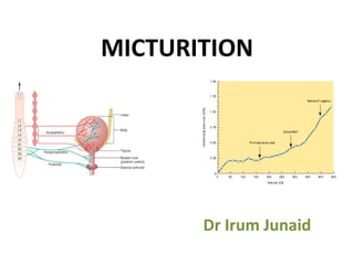
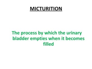
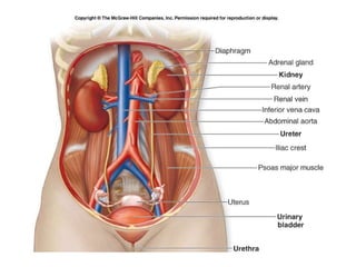
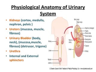
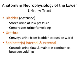
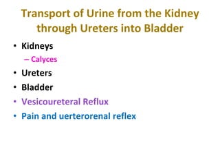
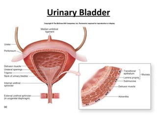
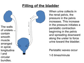
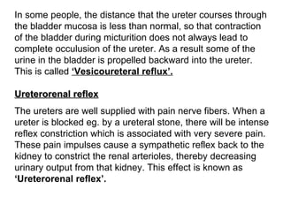
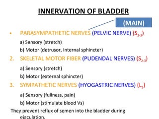
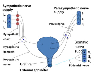
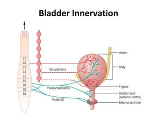
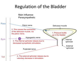
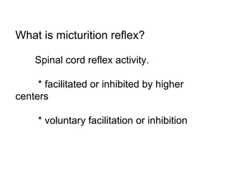
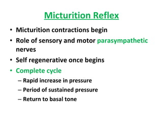
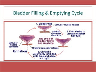
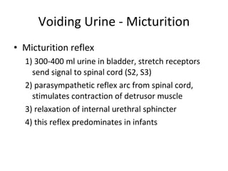
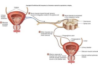
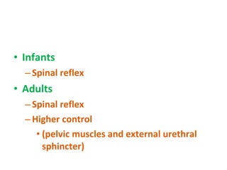
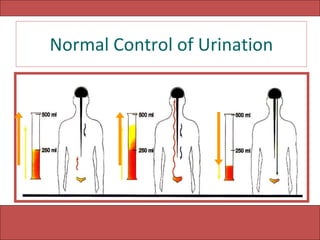
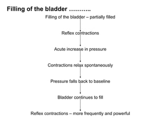
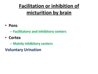
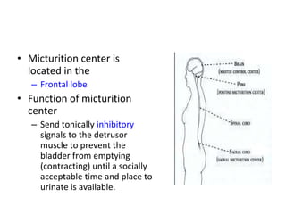
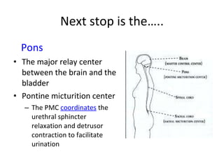
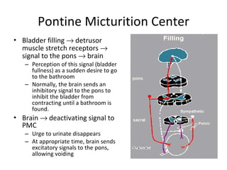
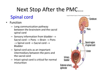
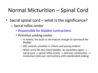
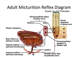
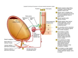
MICTURITION Dr Irum Junaid
MICTURITION The process by which the urinary bladder empties when it becomes filled
 
Physiological Anatomy of Urinary System Kidneys  (cortex, medulla, nephron, pelvis ) Ureters  (mucosa, muscle, fibrous) Urinary Bladder  (body, neck), (mucosa,muscle, fibrous) (detrusor, trigone) Urethra   Internal and External  sphincters
Anatomy & Neurophysiology of the Lower Urinary Tract Bladder  (detrusor) Stores urine at low pressure Compresses urine for voiding Urethra Conveys urine from bladder to outside world Sphincter(s) internal & external Controls urine flow & maintain continence between voidings
Transport of Urine from the Kidney through Ureters into Bladder Kidneys Calyces Ureters Bladder Vesicoureteral Reflux Pain and uerterorenal reflex
Urinary Bladder
Filling of the bladder When urine collects in the renal pelvis, the pressure in the pelvis increases. This increase in the pressure initiates a peristaltic contraction beginning in the pelvis and spreading downward along the ureter to force urine toward the bladder.  Peristaltic waves occur 1-5 times/minute The walls of ureters contain smooth muscle arranged in spiral, longitudinal and circular bundles.
In some people, the distance that the ureter courses through the bladder mucosa is less than normal, so that contraction of the bladder during micturition does not always lead to complete occulusion of the ureter. As a result some of the urine in the bladder is propelled backward into the ureter. This is called  ‘Vesicoureteral reflux’.   Ureterorenal reflex The ureters are well supplied with pain nerve fibers. When a ureter is blocked eg. by a ureteral stone, there will be intense reflex constriction which is associated with very severe pain. These pain impulses cause a sympathetic reflex back to the kidney to constrict the renal arterioles, thereby decreasing urinary output from that kidney. This effect is known as  ‘Ureterorenal reflex’.
INNERVATION OF BLADDER PARASYMPATHETIC NERVES  (PELVIC NERVE)   (S 2-3 ) a) Sensory (stretch) b) Motor (detrusor, Internal sphincter) 2.  SKELETAL MOTOR FIBER  (PUDENDAL NERVES)   (S 2-3 )   a) Sensory (stretch) b) Motor (external sphincter) 3.  SYMPATHETIC NERVES  (HYOGASTRIC NERVES)   (L 2 )   a) Sensory (fullness, pain) b) Motor (stimulate blood Vs) They prevent reflux of semen into the bladder during ejaculation.  ( MAIN )
L 1 L 2 L 3 Sympathetic nerve supply Sympathetic  chain Hypogastric  ganglion Hypogastric  nerve Urethra External sphincter Parasympathetic nerve supply S 2 S 3 S 4 S 2 S 3 S 4 Pelvic nerve Pudendal nerve Somatic nerve supply
Bladder Innervation
Regulation of the Bladder Main Influence: Parasympathetic Pelvic nerve Detrussor muscle Hypogastric nerve Pudendal nerve Internal sphincter External sphincter Para Sym Vol 1. Pressure builds up in the bladder 2. This causes the contraction of the detrussor muscle, via the pelvic nerve. 3. The internal sphincter relaxes due to decreased sympathetic stimulation. 4. The external sphincter relaxes due to  voluntary decrease in stimulation.
What is micturition reflex? Spinal cord reflex activity. * facilitated or inhibited by higher centers * voluntary facilitation or inhibition
Micturition Reflex Micturition contractions begin Role of sensory and motor  parasympathetic  nerves Self regenerative once begins Complete cycle Rapid increase in pressure Period of sustained pressure Return to basal tone
Bladder Filling & Emptying Cycle The cycle of bladder filling and emptying 1. Bladder fills 2. First desire to urinate (bladder half full) Urination 3. Urination voluntarily inhibited until time and place are right Detrusor  muscle contracts Detrusor muscle relaxes Urethral sphincter contracts Urethral sphincter relaxes
Voiding Urine - Micturition Micturition reflex 1) 300-400 ml urine in bladder, stretch receptors send signal to spinal cord (S2, S3) 2) parasympathetic reflex arc from spinal cord, stimulates contraction of detrusor muscle  3) relaxation of internal urethral sphincter 4) this reflex predominates in infants
 
Infants Spinal reflex Adults Spinal reflex  Higher control (pelvic muscles and external urethral sphincter)
Normal Control of Urination
Cystometrogram 100  200  300  400 Intravesical volume (mL) Intravesical pressure (cm of  Water) 80 60 40 20 0 0 Ia Ib
Bladder filling – cystometrogram Relation between bladder volume & pressure. Empty bladder……P zero 30-50 ml urine……P 5-10 cm H 2 O 50 – 300 ml urine…. P 5-10 cm H 2 O  More than 400 ml…..rapid rise in P
LAW of LAPLACE This is in accordance with law of Laplace. In the bladder tension increases as the urine is filled. At the same time, the radius also increases due to relaxation of the detrusor muscle. Because of this, the pressure rise is almost nil.
When bladder wall stretches during filling it will initiate a reflex contraction which has lower threshold. That does not trigger micturition reflex. When bladder is filled about 300 – 400 mL of urine, there will be sharp rise in the intravesical pressure as the micturition reflex is triggered. At this point also voluntary control is possible. Beyond 600 – 700 mL of urine voluntary control starts failing.
Filling of the bladder – partially filled Reflex contractions Acute increase in pressure Contractions relax spontaneously Pressure falls back to baseline Bladder continues to fill Reflex contractions – more frequently and powerful Filling of the bladder ………..
Facilitation or inhibition of micturition by brain Pons Facilitatory and inhibitory centers Cortex Mainly inhibitory centers Voluntary Urination
Micturition center is located in the  Frontal lobe Function of micturition center  Send tonically  inhibitory  signals to the detrusor muscle to prevent the bladder from emptying (contracting) until a socially acceptable time and place to urinate is available.
Next stop is the….. Pons The major relay center between the brain and the bladder  Pontine micturition center The PMC  coordinates  the urethral sphincter relaxation and detrusor contraction to facilitate urination
Pontine Micturition Center Bladder filling    detrusor muscle stretch receptors    signal to the pons    brain  Perception of this signal (bladder fullness) as a sudden desire to go to the bathroom Normally, the brain sends an inhibitory signal to the pons to inhibit the bladder from contracting until a bathroom is found.  Brain    deactivating signal to PMC Urge to urinate disappears At appropriate time, brain sends excitatory signals to the pons, allowing voiding
Spinal cord   Function  Long communication pathway between the brainstem and the sacral spinal cord Sensory information from bladder    Sacral cord    Pons    Brain    Pons    Spinal cord    Sacral cord    Bladder Spinal cord acts as an important intermediary between the pons and the sacral cord Intact spinal cord is critical for normal micturition Next Stop After the PMC….
Normal Micturition – Spinal Cord Sacral spinal cord – what is the significance? Sacral reflex center Responsible for bladder contractions Primitive voiding center In infants, the brain is not mature enough to command the bladder SRC controls urination in infants and young children  When urine fills the infant bladder, an excitatory signal    sacral cord    spinal reflex center    detrusor contraction    involuntary detrusor contractions with coordinated voiding
Adult Micturition Reflex Diagram
 
Cystometrogram
Abnormalities Atonic bladder Sensory nerve fibers from the bladder to the spinal cord are destroyed Overflow incontinence Crush injury, syphilis,
Automatic bladder Above the sacral region with intact sacral cord segments No longer controlled by the brain Spinal shock contd.
T he urinary bladder looses its tone and becomes flaccid and unresponsive. So, the bladder is completely filled, and later urine overflows by dribbling. After the spinal shock has passed, the voiding reflex returns although there is no voluntary and higher centre control.  Whenever, the bladder is filled with some amount of urine, there is automatic evacuation of the bladder. (Spastic neurogenic bladder)
Uninhibited Neurogenic Bladder Partial damage in the spinal cord or the brain stem interrupting most of the inhibitory signals Slight quantity of urine elicits an uncontrollable micturition reflex
Nocturnal micturition (Bed wetting) This is normal in infants and children below 3 years. It occurs due to incomplete myelination of motor nerve fibers of the bladder resulting loss of voluntary control of micturition .
Incontinence from impaired sphincter function  Of lesser degree In response to sudden rise in intravesical pressure (coughing, sneezing) After multiple child births in women After prostatic surgery involving damage to sphincter
 

Micturation reflex by Dr Irum

  • 1.
  • 2.
  • 3.
    MICTURITION The processby which the urinary bladder empties when it becomes filled
  • 4.
  • 5.
    Physiological Anatomy ofUrinary System Kidneys (cortex, medulla, nephron, pelvis ) Ureters (mucosa, muscle, fibrous) Urinary Bladder (body, neck), (mucosa,muscle, fibrous) (detrusor, trigone) Urethra Internal and External sphincters
  • 6.
    Anatomy & Neurophysiologyof the Lower Urinary Tract Bladder (detrusor) Stores urine at low pressure Compresses urine for voiding Urethra Conveys urine from bladder to outside world Sphincter(s) internal & external Controls urine flow & maintain continence between voidings
  • 7.
    Transport of Urinefrom the Kidney through Ureters into Bladder Kidneys Calyces Ureters Bladder Vesicoureteral Reflux Pain and uerterorenal reflex
  • 8.
  • 9.
    Filling of thebladder When urine collects in the renal pelvis, the pressure in the pelvis increases. This increase in the pressure initiates a peristaltic contraction beginning in the pelvis and spreading downward along the ureter to force urine toward the bladder. Peristaltic waves occur 1-5 times/minute The walls of ureters contain smooth muscle arranged in spiral, longitudinal and circular bundles.
  • 10.
    In some people,the distance that the ureter courses through the bladder mucosa is less than normal, so that contraction of the bladder during micturition does not always lead to complete occulusion of the ureter. As a result some of the urine in the bladder is propelled backward into the ureter. This is called ‘Vesicoureteral reflux’. Ureterorenal reflex The ureters are well supplied with pain nerve fibers. When a ureter is blocked eg. by a ureteral stone, there will be intense reflex constriction which is associated with very severe pain. These pain impulses cause a sympathetic reflex back to the kidney to constrict the renal arterioles, thereby decreasing urinary output from that kidney. This effect is known as ‘Ureterorenal reflex’.
  • 11.
    INNERVATION OF BLADDERPARASYMPATHETIC NERVES (PELVIC NERVE) (S 2-3 ) a) Sensory (stretch) b) Motor (detrusor, Internal sphincter) 2. SKELETAL MOTOR FIBER (PUDENDAL NERVES) (S 2-3 ) a) Sensory (stretch) b) Motor (external sphincter) 3. SYMPATHETIC NERVES (HYOGASTRIC NERVES) (L 2 ) a) Sensory (fullness, pain) b) Motor (stimulate blood Vs) They prevent reflux of semen into the bladder during ejaculation. ( MAIN )
  • 12.
    L 1 L2 L 3 Sympathetic nerve supply Sympathetic chain Hypogastric ganglion Hypogastric nerve Urethra External sphincter Parasympathetic nerve supply S 2 S 3 S 4 S 2 S 3 S 4 Pelvic nerve Pudendal nerve Somatic nerve supply
  • 13.
  • 14.
    Regulation of theBladder Main Influence: Parasympathetic Pelvic nerve Detrussor muscle Hypogastric nerve Pudendal nerve Internal sphincter External sphincter Para Sym Vol 1. Pressure builds up in the bladder 2. This causes the contraction of the detrussor muscle, via the pelvic nerve. 3. The internal sphincter relaxes due to decreased sympathetic stimulation. 4. The external sphincter relaxes due to voluntary decrease in stimulation.
  • 15.
    What is micturitionreflex? Spinal cord reflex activity. * facilitated or inhibited by higher centers * voluntary facilitation or inhibition
  • 16.
    Micturition Reflex Micturitioncontractions begin Role of sensory and motor parasympathetic nerves Self regenerative once begins Complete cycle Rapid increase in pressure Period of sustained pressure Return to basal tone
  • 17.
    Bladder Filling &Emptying Cycle The cycle of bladder filling and emptying 1. Bladder fills 2. First desire to urinate (bladder half full) Urination 3. Urination voluntarily inhibited until time and place are right Detrusor muscle contracts Detrusor muscle relaxes Urethral sphincter contracts Urethral sphincter relaxes
  • 18.
    Voiding Urine -Micturition Micturition reflex 1) 300-400 ml urine in bladder, stretch receptors send signal to spinal cord (S2, S3) 2) parasympathetic reflex arc from spinal cord, stimulates contraction of detrusor muscle 3) relaxation of internal urethral sphincter 4) this reflex predominates in infants
  • 19.
  • 20.
    Infants Spinal reflexAdults Spinal reflex Higher control (pelvic muscles and external urethral sphincter)
  • 21.
  • 22.
    Cystometrogram 100 200 300 400 Intravesical volume (mL) Intravesical pressure (cm of Water) 80 60 40 20 0 0 Ia Ib
  • 23.
    Bladder filling –cystometrogram Relation between bladder volume & pressure. Empty bladder……P zero 30-50 ml urine……P 5-10 cm H 2 O 50 – 300 ml urine…. P 5-10 cm H 2 O More than 400 ml…..rapid rise in P
  • 24.
    LAW of LAPLACEThis is in accordance with law of Laplace. In the bladder tension increases as the urine is filled. At the same time, the radius also increases due to relaxation of the detrusor muscle. Because of this, the pressure rise is almost nil.
  • 25.
    When bladder wallstretches during filling it will initiate a reflex contraction which has lower threshold. That does not trigger micturition reflex. When bladder is filled about 300 – 400 mL of urine, there will be sharp rise in the intravesical pressure as the micturition reflex is triggered. At this point also voluntary control is possible. Beyond 600 – 700 mL of urine voluntary control starts failing.
  • 26.
    Filling of thebladder – partially filled Reflex contractions Acute increase in pressure Contractions relax spontaneously Pressure falls back to baseline Bladder continues to fill Reflex contractions – more frequently and powerful Filling of the bladder ………..
  • 27.
    Facilitation or inhibitionof micturition by brain Pons Facilitatory and inhibitory centers Cortex Mainly inhibitory centers Voluntary Urination
  • 28.
    Micturition center islocated in the Frontal lobe Function of micturition center Send tonically inhibitory signals to the detrusor muscle to prevent the bladder from emptying (contracting) until a socially acceptable time and place to urinate is available.
  • 29.
    Next stop isthe….. Pons The major relay center between the brain and the bladder Pontine micturition center The PMC coordinates the urethral sphincter relaxation and detrusor contraction to facilitate urination
  • 30.
    Pontine Micturition CenterBladder filling  detrusor muscle stretch receptors  signal to the pons  brain Perception of this signal (bladder fullness) as a sudden desire to go to the bathroom Normally, the brain sends an inhibitory signal to the pons to inhibit the bladder from contracting until a bathroom is found. Brain  deactivating signal to PMC Urge to urinate disappears At appropriate time, brain sends excitatory signals to the pons, allowing voiding
  • 31.
    Spinal cord Function Long communication pathway between the brainstem and the sacral spinal cord Sensory information from bladder  Sacral cord  Pons  Brain  Pons  Spinal cord  Sacral cord  Bladder Spinal cord acts as an important intermediary between the pons and the sacral cord Intact spinal cord is critical for normal micturition Next Stop After the PMC….
  • 32.
    Normal Micturition –Spinal Cord Sacral spinal cord – what is the significance? Sacral reflex center Responsible for bladder contractions Primitive voiding center In infants, the brain is not mature enough to command the bladder SRC controls urination in infants and young children When urine fills the infant bladder, an excitatory signal  sacral cord  spinal reflex center  detrusor contraction  involuntary detrusor contractions with coordinated voiding
  • 33.
  • 34.
  • 35.
  • 36.
    Abnormalities Atonic bladderSensory nerve fibers from the bladder to the spinal cord are destroyed Overflow incontinence Crush injury, syphilis,
  • 37.
    Automatic bladder Abovethe sacral region with intact sacral cord segments No longer controlled by the brain Spinal shock contd.
  • 38.
    T he urinarybladder looses its tone and becomes flaccid and unresponsive. So, the bladder is completely filled, and later urine overflows by dribbling. After the spinal shock has passed, the voiding reflex returns although there is no voluntary and higher centre control. Whenever, the bladder is filled with some amount of urine, there is automatic evacuation of the bladder. (Spastic neurogenic bladder)
  • 39.
    Uninhibited Neurogenic BladderPartial damage in the spinal cord or the brain stem interrupting most of the inhibitory signals Slight quantity of urine elicits an uncontrollable micturition reflex
  • 40.
    Nocturnal micturition (Bedwetting) This is normal in infants and children below 3 years. It occurs due to incomplete myelination of motor nerve fibers of the bladder resulting loss of voluntary control of micturition .
  • 41.
    Incontinence from impairedsphincter function Of lesser degree In response to sudden rise in intravesical pressure (coughing, sneezing) After multiple child births in women After prostatic surgery involving damage to sphincter
  • 42.